 E-submission
Articles
- Page Path
- HOME > J Pathol Transl Med > Volume 48(2); 2014 > Article
-
Brief Case Report
An Unusual Case of Pulmonary Mucous Gland Adenoma with Fibromyxoid Stroma and Cartilage Islands in 68-Year-Old Woman - Boram Lee, In Ho Choi, Joungho Han, Kyung Soo Lee1, Young Mog Shim2
-
Korean Journal of Pathology 2014;48(2):167-169.
DOI: https://doi.org/10.4132/KoreanJPathol.2014.48.2.167
Published online: April 28, 2014
Department of Pathology, Samsung Medical Center, Sungkyunkwan University School of Medicine, Seoul, Korea.
1Department Radiology, Samsung Medical Center, Sungkyunkwan University School of Medicine, Seoul, Korea.
2Department Thoracic Surgery, Samsung Medical Center, Sungkyunkwan University School of Medicine, Seoul, Korea.
- Corresponding Author: Joungho Han, M.D. Department of Pathology, Samsung Medical Center, Sungkyunkwan University School of Medicine, 81 Irwon-ro, Gangnam-gu, Seoul 135-710, Korea. Tel: +82-2-3410-2800, Fax: +82-2-3410-0025, hanjho@skku.edu
• Received: May 20, 2013 • Revised: June 11, 2013 • Accepted: June 12, 2013
© 2014 The Korean Society of Pathologists/The Korean Society for Cytopathology
This is an Open Access article distributed under the terms of the Creative Commons Attribution Non-Commercial License (http://creativecommons.org/licenses/by-nc/3.0/) which permits unrestricted non-commercial use, distribution, and reproduction in any medium, provided the original work is properly cited.
- 68-year-old woman, who had no specific medical history or history of smoking, was transferred to our hospital due to an incidentally identified mass at the peripheral territory of the right upper lobe of the lung (Fig. 1A). The mass measured about 3 cm in diameter with no definite change of size over 18 months. In spite of bronchoscopic biopsy, no definite diagnosis could be made. One year later, positron emission tomography revealed a mild hypermetabolic state of the mass with no other remarkable findings, and wedge resection of the lung was performed for histological confirmation.
- Macroscopically, the mass was a 3×1.9-cm-sized, solid ovoid mass that had a grayish and weakly glistening cut surface and disclosed several small cystic spaces and fragments of cartilage. There was no identifiable connection to the bronchus on any of multiple sections. Microscopically, the tumor was well circumscribed and composed of multiple cystic glands and myxoid stroma containing lobules of cartilage (Fig. 1B). Mucous and serous glands were haphazardly scattered on the myxoid background, and some of them were cystically dilated; however, no invasive foci were found (Fig. 1C). Irregularly-shaped cartilaginous islands were noted on the myxoid stroma, and some of the dilated glands were filled with proteinaceous or secretory material (Fig. 1D). The lining epithelial cells of the glands had basally located hyperchromatic nuclei and abundant clear or eosinophilic cytoplasm (Fig. 1E). No mitotic figure was observed. Immunohistochemical staining for p63 (1:200, 4A4, Dako, Glostrup, Denmark) (Fig. 1F) and calponin (1:200, CALP, Bio-Genex, San Ramon, CA, USA) showed positivity in the myoepithelial cells in glands but negativity in the stromal cells. The patient is alive and well five months after the operation.
CASE REPORT
- Pulmonary MGA with lack of bronchial relation is extremely unusual.4 Moreover, MGA of the peripheral lung with cartilage islands has not been reported.
- The histologic findings of a benign mucous glandular proliferation mixed with cartilage/mesenchymal component suggest several possible scenarios. First, it is reasonable that MGA with myxoid stroma develops chondroid metaplasia, as occurs in other organs. The stroma of MGAs varies, and hyalinized sclerosis can be seen.2 However, no report describing myxoid stroma and cartilage islands in MGA could be found in the English literature.
- We also considered coincidental combination of MGA and pulmonary hamartoma. There was a case report of a collision tumor of pulmonary hamartoma and a carcinoid.5 However, this was likely an extremely extraordinary situation, considering the rarity of mucus gland adenoma.
- There have been two reported cases of salivary gland-type tumors developed in hamartoma; myoepithelioma and malignant mixed tumor.6 The myoepithelial cells composing these tumors can arise from stromal cells of hamartoma. Additionally, there was no glandular component like our case of MGA.
- Pleomorphic adenoma of the lung also can be considered, showing varying admixtures of epithelial elements and a stromal/mesenchymal component showing abundant myxoid or focally chondroid matrix. In a literature review of 24 cases of salivary gland-type mixed tumor of the lung, Matsumoto et al.7 suggested that peripherally developed tumors might be derived from primitive stem cells with the ability to differentiate toward ductal structures, myoepithelium, and chondroid and myxoid matrix components. However, our case is morphologically different from pleomorphic adenoma, and immunohistochemical staining of myoepithelial cells showed positivity for normal myoepithelial cells within glands and negative for the stromal cells, while pleomorphic adenoma shows positivity for the stromal cells.
- Although none of the above possibilities sufficiently explains the histologic finding of our case, it is a very interesting case. Herein, we report a rare case of MGA with fibromyxoid stroma and cartilage islands.
DISCUSSION
- 1. Kwon JE, Kim GY, Han J, Kim TS, Kim K. Mucous gland adenoma presenting as a peripheral lung mass: a brief case report. Korean J Pathol 2004; 38: 126-128.
- 2. England DM, Hochholzer L. Truly benign "bronchial adenoma". Report of 10 cases of mucous gland adenoma with immunohistochemical and ultrastructural findings. Am J Surg Pathol 1995; 19: 887-899. ArticlePubMed
- 3. Jin SY, Lee KG. Mucous gland adenoma of the bronchus: a case report. Korean J Pathol 1985; 19: 368-373.
- 4. Karpathiou G, Sivridis E, Mikroulis D, Froudarakis M, Giatromanolaki A. Pulmonary mucus gland adenomas: are they always of endobronchial localization? Case Rep Pathol 2013; 2013: 239173.ArticlePubMedPMCPDF
- 5. Cavazza A, Paci M, Rossi G. Pulmonary hamartoma associated with typical carcinoid/tumorlet. Virchows Arch 2006; 449: 392-393. ArticlePubMedPDF
- 6. Pelosi G, Rodriguez J, Viale G, Rosai J. Salivary gland-type tumors with myoepithelial differentiation arising in pulmonary hamartoma: report of 2 cases of a hitherto unrecognized association. Am J Surg Pathol 2006; 30: 375-387. PubMed
- 7. Matsumoto M, Sonobe H, Furihata M, Nonami Y, Ohmori Y, Ohtsuki Y. A case of salivary gland-type mixed tumor of the lung differentiating toward type II alveolar epithelial cells in glandular components with a literature review. Virchows Arch 2002; 441: 618-621. ArticlePubMedPDF
REFERENCES
Fig. 1Mucous gland adenoma with fibromyxoid stroma and cartilage islands of lung. (A) Computed tomography shows a 3-cm-sized, well circumscribed mass at the posterior segment of the right upper lobe of lung. (B) The tumor is well circumscribed and shows multiple varying cysts surrounded by myxoid stroma and several separated islands of cartilage. (C) Irregularly-shaped mucous glands are noted on the myxoid background. (D) Some of the glands are cystically dilated and filled with proteinaceous material and separated islands of cartilage. (E) Mucoserous glands are lined by columnar or flat cuboidal epithelial cells with clear or eosinophilic cytoplasm and basally located small nuclei with no atypia. (F) Myoepithelial cells of glands show positivity for p63; however, stromal cells are negative.
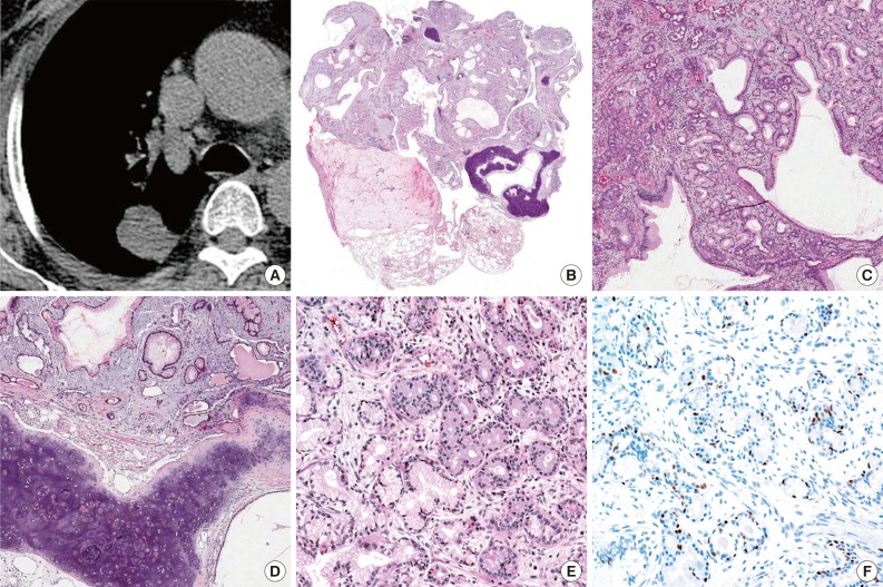
Figure & Data
References
Citations
Citations to this article as recorded by 
- Case report: Bronchoscopic intervention for rare benign airway tumors: a report of 4 cases and literature review
Zhifang Cui, Jinhong Wang, Hongwu Wang, Lei Li, Xiaohui Si, Yanbin Zhang, Heng Zou
Frontiers in Oncology.2024;[Epub] CrossRef - Extremely Rare Coexistence of Peripherally Located Mucous Gland Adenoma and Pulmonary Chondroid Hamartoma
Sevilay Özmen, Funda Demirağ, Harika Derya Tamer, Adem Karaman, Yener Aydın
British Journal of Hospital Medicine.2024; : 1. CrossRef - Peripheral pulmonary mucous gland adenoma with a cavity mimicking lung cancer
Hiroaki Komatsu, Nobuhiro Izumi, Takuma Tsukioka, Hidetoshi Inoue, Ryuichi Ito, Yumi Matsuda, Noritoshi Nishiyama
General Thoracic and Cardiovascular Surgery.2022; 70(1): 92. CrossRef - Un cas d’adénome muqueux glandulaire bronchique : un diagnostic rare à ne pas méconnaître !
Arnault Tauziède-Espariat, Bertrand Grand, Odette Georges, Ali Benali, Philippe Viehl, Laura Bitton, Gérard Antin, Albane Gareton, Raphaël Saffroy, René Jancovici, Diane Damotte
Annales de Pathologie.2021; 41(2): 192. CrossRef - Mucous Gland Adenoma: The Spectrum of Growth Patterns and the Diagnostic Challenges
Michael P. Zaleski, Neda Kalhor, Cesar A. Moran
Advances in Anatomic Pathology.2020; 27(6): 371. CrossRef - Peripheral mucous gland adenoma of the lung with parenchymal involvement and smooth muscle in the stroma
Xue-Ting Zhang, Man Yang, Xiao-Fang Liu, Xu-Yong Lin
Medicine.2018; 97(3): e9597. CrossRef - An unusual case of pulmonary hamartoma with predominant bronchial mucous glands in the peripheral lung
Rin Yamada, Akiko Tonooka, Shin‐ichiro Horiguchi, Toru Motoi, Hirotoshi Horio, Tsunekazu Hishima
Pathology International.2018; 68(7): 431. CrossRef
 PubReader
 ePub Link
-
 Cite this Article
- Cite this Article
-
- Close
- Download Citation
- Close
- Figure
-

An Unusual Case of Pulmonary Mucous Gland Adenoma with Fibromyxoid Stroma and Cartilage Islands in 68-Year-Old Woman

Fig. 1 Mucous gland adenoma with fibromyxoid stroma and cartilage islands of lung. (A) Computed tomography shows a 3-cm-sized, well circumscribed mass at the posterior segment of the right upper lobe of lung. (B) The tumor is well circumscribed and shows multiple varying cysts surrounded by myxoid stroma and several separated islands of cartilage. (C) Irregularly-shaped mucous glands are noted on the myxoid background. (D) Some of the glands are cystically dilated and filled with proteinaceous material and separated islands of cartilage. (E) Mucoserous glands are lined by columnar or flat cuboidal epithelial cells with clear or eosinophilic cytoplasm and basally located small nuclei with no atypia. (F) Myoepithelial cells of glands show positivity for p63; however, stromal cells are negative.
Fig. 1
An Unusual Case of Pulmonary Mucous Gland Adenoma with Fibromyxoid Stroma and Cartilage Islands in 68-Year-Old Woman

