
 E-submission
Articles
- Page Path
- HOME > J Pathol Transl Med > Forthcoming articles > Article
-
Case Report
Clinicopathological characteristics of primary malignant cutaneous perivascular epithelioid cell tumor: case report and literature review -
Huaming Li
 , Dan Zhang, Shiwu Zhang
 -
DOI: https://doi.org/10.4132/jptm.2026.05.10
Published online: July 20, 2026
Department of Pathology, Tianjin Union Medical Center, The First Affiliated Hospital of Nankai University, Nankai University, Tianjin, China
- Corresponding Author Shiwu Zhang, MD, PhD Department of Pathology, Tianjin Union Medical Center, The First Affiliated Hospital of Nankai University, 190# Jieyuan Road, Hongqiao, Tianjin 300121, China Tel: +86-15620876109, Fax: +86-022-27557193, E-mail: zhangshiwu666@aliyun.com
© The Korean Society of Pathologists/The Korean Society for Cytopathology
This is an Open Access article distributed under the terms of the Creative Commons Attribution Non-Commercial License (https://creativecommons.org/licenses/by-nc/4.0) which permits unrestricted non-commercial use, distribution, and reproduction in any medium, provided the original work is properly cited.
- 113 Views
- 7 Download


Abstract
- Primary cutaneous malignant perivascular epithelioid cell tumors (PEComas) are extremely rare. Accurate diagnosis is critical for prognostic evaluation and guiding clinical management. Here, we report a case of a 19-year-old woman with a 1.1 cm exophytic red mass on her left upper limb. Histological examination revealed a dermal tumor composed of clear cells with prominent nucleoli. The tumor cells displayed cytologic atypia, nuclear pleomorphism, multinucleated giant cells, mitoses and invasive growth, with a multinodular distribution within the dermis and vascular invasion. Immunohistochemically, the tumor cells expressed cathepsin K, HMB45, and CD10. Based on morphological and immunohistochemical findings, we classified it as a primary malignant cutaneous PEComa. To date, only eight reported cases have been described as primary malignant cutaneous PEComas. The present case appears to be the youngest patient reported among all malignant PEComas.
- Perivascular epithelioid cell tumors (PEComas) include angiomyolipomas, pulmonary clear cell sugar tumor, lymphangiomyoma and lymphangiomyomatosis, and non-specific perivascular epithelioid cell tumors. The primary sites of non-specific PEComas are the abdominal and pelvic cavities, female reproductive tract, gastrointestinal tract, bladder, bone, skin, and other rare sites [1]. Folpe et al. [2] proposed a classification system categorizing PEComas as benign, of uncertain malignant potential, or malignant on the basis of the following criteria: tumor size ≥ 5 cm, invasive growth pattern, high nuclear atypia and increased cell density, necrosis, vascular invasion, mitotic activity > 1 per 50 high-power fields, and aggressive clinical behavior. Benign PEComas lack all of these features; malignant PEComas exhibit two or more of the criteria; and PEComas of uncertain malignant potential demonstrate one of the following parameters: nuclear pleomorphism, multinucleated giant cells, or tumor size ≥ 5 cm [2]. In the present case, the diagnosis of PEComa was established strictly according to these criteria.
INTRODUCTION
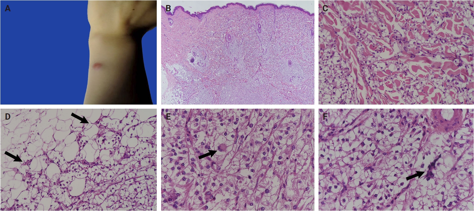
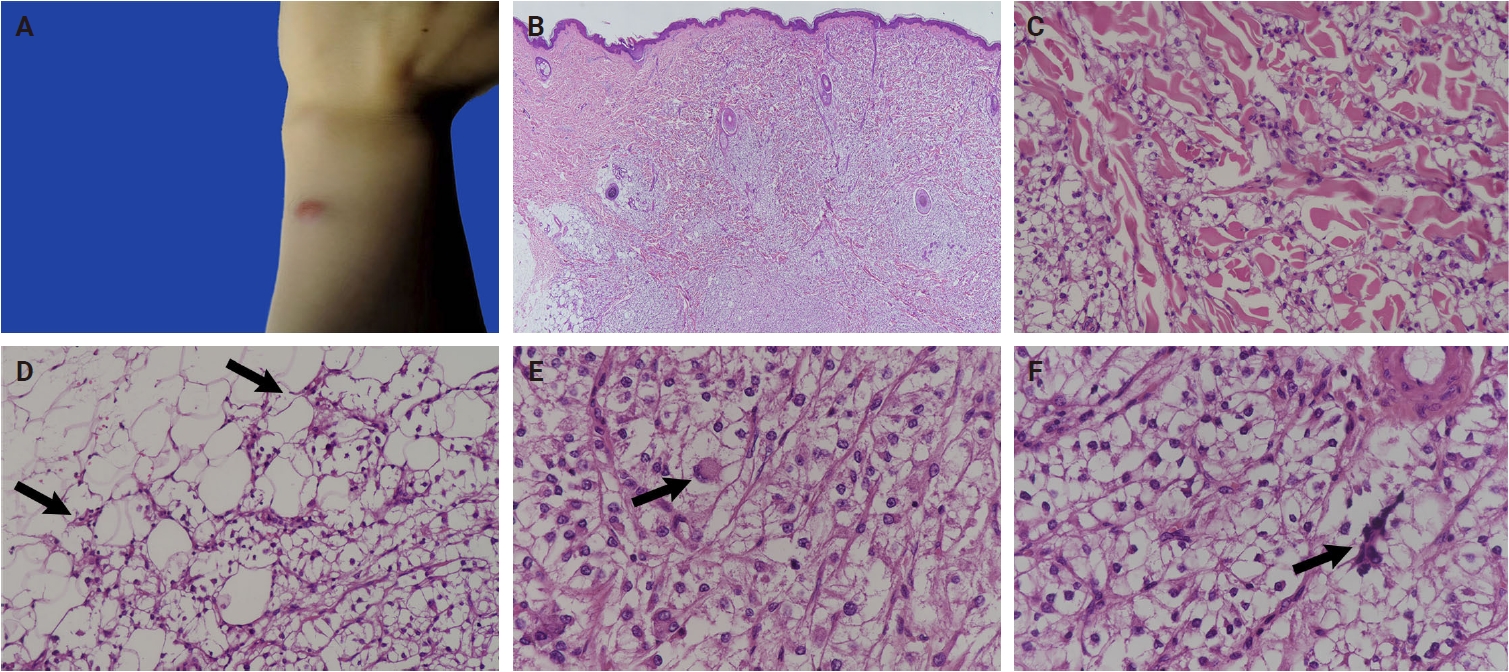
- A 19-year-old female college student from Harbin, originally residing in Heilongjiang Province, with no significant past medical history aside from anxiety, presented with a slowly enlarging cutaneous lesion on the medial aspect of her left anterior wrist. In December 2024, she noticed a small erythematous papule with slight surface elevation, initially resembling a mosquito-bite nodule. Over the following seven months, the lesion gradually progressed, exceeding 0.7 cm in diameter, darkening in color, and developing long hair growth, without associated tenderness or pain (Fig. 1A). By late August 2025, the lesion had evolved into a 1.1 cm exophytic red mass on the left upper limb. Initial clinical suspicion at the dermatology department of our hospital favored a hemangioma, which was further supported by dermatologic evaluation and ultrasonographic findings. The mass was completely excised and sent for histopathological examination. Following pathological diagnosis, the patient underwent a systematic evaluation, including computed tomography of the chest, abdomen, and pelvis, and ultrasonography of the abdomen and pelvis. All examinations were negative for metastatic disease. At 7 months of follow-up, the patient remained clinically stable with no change in condition.
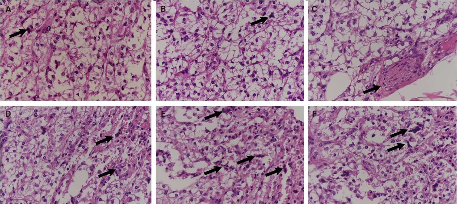
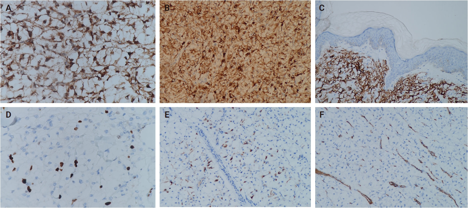
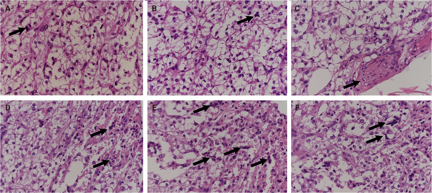
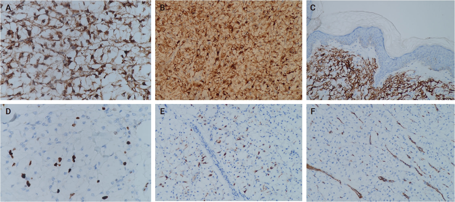
- The specimen measured 2.5×1.5×1 cm after removal of the overlying skin and the tumor itself measured 1.1×1.0×0.7 cm (Fig. 1B). The tumor was located in the dermis and exhibited invasive growth extending through the dermal layer into the subcutaneous adipose tissue (Fig. 1C, D). Histological examination revealed no evidence of tumor cells at the resection margin. The tumor was composed of nodules and nests of cells separated by numerous branching capillaries. The tumor cells exhibited abundant clear cell cytoplasm and nuclei of varying sizes that were morphologically similar to renal clear cell carcinoma. Some large epithelioid tumor cells displayed shrunken cytoplasm, similar to the spider-like cells in adult-type rhabdomyoma (Fig. 1E). Pleomorphic nuclei and multinucleated giant cells were observed at low magnification, with prominent nucleoli at 10× objective. Scattered multiple abnormal giant nuclear cells formed vascular lumen-like structures (Fig. 1F). Some areas of the tumor cells exhibited elevated cell density and hypercellularity, with a mitotic rate of 3 mitoses per 10 high-power fields (Fig. 2A, B). Perineurial involvement and vascular invasion were observed surrounding the tumor (Fig. 2C, D). In addition, the pleomorphism and significant nuclear atypia were noted (Fig. 2E, F). Immunohistochemical analysis revealed that tumor cells were positive for cathepsin K (Fig. 3A), HMB45 (Fig. 3B), CD10 (Fig. 3C), vimentin, and microphthalmia-associated transcription factor (MiTF). The proliferative index (Ki-67–positive cells) averaged 6%, reaching up to 15% in hotspot regions (Fig. 3D). Desmin was focally positive in this lesion (Fig. 3E). Tumor cells were negative for epithelial markers (cytokeratin [CK], epithelial membrane antigen [EMA], p63, paired box 8 [PAX8], and renal cell carcinoma [RCC]), neuroendocrine markers (synaptophysin and CD56), other melanin markers (Melan-A, S100, and SOX10), and myogenic markers (h-Caldesmon and smooth muscle actin [SMA]) (Fig. 3F) and transcription factor E3 (TFE3). SMA immunohistochemical staining highlighted numerous branching capillaries between the tumor cells. The patient was a young woman who underwent excision of the mass and subsequent clinical evaluation; no evidence of tumor involvement was identified in the viscera or other soft tissues. The diagnosis of primary malignant cutaneous PEComa was confirmed on the basis of histological and immunohistochemical features, particularly the expression of cathepsin K and melanocytic marker HMB45.
CASE REPORT
- PEComas are rare mesenchymal tumors derived from perivascular epithelioid cells, whose organizational composition is not well-defined, but show immunoreaction against melanocytic and smooth muscle markers [3]. The first case of primary malignant cutaneous PEComa was reported by Calder et al. in 2008 [4]. To date, fewer than 100 cases of primary cutaneous PEComas have been reported. After nearly 20 years since the initial description of primary malignant cutaneous PEComas, the present case represents the ninth such case reported in the literature [5-11].
- The present case showed epithelioid tumor cells with clear to eosinophilic cytoplasm. According to the classification criteria established by Folpe et al. [2], the cutaneous PEComa in the current case fulfills two criteria for malignancy: an invasive growth pattern and a mitotic activity exceeding 1 per 50 high-power fields. The tumor size is less than 5 cm and no obvious necrosis was observed. However, the presence of numerous high-grade atypical nuclei and atypical mitotic figures in focal areas, together with tumor cell infiltration around nerves and adjacent adipose tissue, combined with the two malignancy criteria outlined by Folpe et al. [2] leads us to classify this tumor as a primary malignant cutaneous PEComa. We have adapted and expanded the previously published tabulations by Elousrouti et al. [11], summarizing clinical, histopathological, and immunohistochemical characteristics of the disease (Tables 1, 2). Across the nine reported cases, a slight female predominance was observed (five females, four males), with a mean age of 52 years. The extremities were the most common sites, and the mean tumor size was 36.3 mm (range, 10 to 120 mm). Clinically, most cases presented as progressively enlarging exophytic erythematous tumors.
- In this case, CD10 immunostaining proved valuable in this context, as it distinctly highlighted tumor cell membranes and illustrated focal proximity of the lesion to the basal epidermal layer. To date, only one case of primary malignant cutaneous PEComa exhibiting pleomorphic sarcomatoid invasion of the epidermis, dermis, and subcutaneous soft tissue has been reported. The remaining seven cases of primary malignant cutaneous PEComas described no direct epidermal contact [12]. Among nine reported cases of primary malignant cutaneous PEComas, CD10 was expressed in all five tested cases. Similarly, in the reported primary benign cutaneous PEComas, CD10 was always diffusely positive, with partial expression in a few cases [13].
- The clear cell variant of PEComa and atypical fibroxanthoma (AFX) were the main differential diagnoses. AFX typically shows diffuse, strong positivity for CD10 and vimentin and may also show positivity for CD68, CD99, and SMA. However, they are negative for S100 and Melan-A. The clear cell subtype AFX is occasionally positive for HMB45, but this expression is restricted to giant cells [14]. It has also been reported that the clear cell variant of AFX tumor cells show diffuse positivity for MiTF, with significantly enhanced staining in multinucleated giant cells. In the present case, tumor cells were associated with small vascular networks, and no enhancement of MiTF staining was observed in giant cells [15]. The clear cell variant of AFX is primarily a diagnosis of exclusion and is commonly observed in older individuals over 60 years of age. This supports a diagnosis of PEComa rather than clear cell AFX in this young patient. In addition, dermatofibroma and xanthogranulomatous clear cell lesions of the skin (e.g., xanthoma, macula, and xanthogranuloma) can be excluded based on the absence of melanocytic marker expression despite CD10 positivity. They can be further distinguished by positivity for CD68 and CD163, or adipophilin positivity in some cases. Third, CD10 expression is also associated with metastatic clear cell RCC, which can be distinguished using additional immunohistochemical markers such as RCC, PAX8, carbonic anhydrase IX, and EMA. Clinical history and renal imaging further aid in this distinction. Fourth, cutaneous PEComas should be distinguished from clear cell sarcomas, clear cell melanomas, and balloon cell nevi. Positive cathepsin K immunostaining, along with negative S100 protein and SOX10, helps distinguish these three entities from PEComa. Clear cell sarcomas can also be distinguished by the presence of the t(12;22)(q13;q12) translocation [16]. Fifth, although PEComas exhibit epithelioid morphology, they do not express epithelial markers. Cutaneous PEComas can be distinguished from sebaceous carcinomas and clear cell hidradenomas using epithelial markers and HMB45. Sebaceous carcinomas and clear cell hidradenomas are positive CK immunostaining, whereas they are negative for HMB45 and Melan-A. Sixth, in contrast to dermal granulosa cell tumors, PEComas are negative for S100 and SOX10 (Table 2) [17]. Granular cell tumor, characterized by granular cytoplasm with focal clear cell changes, may morphologically resemble PEComas; however, they are distinguished by diffuse positivity for S100, nuclear expression of SOX10, positivity for inhibin, and frequent TFE3 reactivity. Seventh, epithelioid sarcoma shows positive CK immunostaining and loss of nuclear INI1 expression. Alveolar soft part sarcoma is typically positive for TFE3 and negative for HMB45/Melan-A. Although TFE3 expression can also occur in primary visceral or soft tissue PEComas, it is rarely observed in primary cutaneous PEComas [18]. To date, only one case of TFE3-positive primary cutaneous PEComa has been reported; however, this case exhibited histomorphologic features similar to those of pleomorphic sarcoma, differing from the typical epithelioid or mixed (epithelioid and spindle cell) morphology. All reported cases of TFE3-negative primary cutaneous PEComas have been described as containing epithelioid components.
- The molecular features of primary cutaneous PEComas also differ from those of systemic PEComas. Systemic PEComas predominantly arise from visceral organs and soft tissues. In systemic PEComas, loss-of-function mutations in tuberous sclerosis complex (TSC) 1 or TSC2 lead to activation of the mammalian target of rapamycin (mTOR) pathway and are associated with TFE3 rearrangements. These molecular mechanisms are largely not involved in most primary cutaneous PEComas. However, two reported cases showed genomic abnormalities, including baculovirus inhibitor of apoptosis protein duplication at the BIRC3 splice site 1622-27_1631del37 [13] and TP53 gene mutations or deletions [8]. One patient exhibited a homozygous/biallelic deletion of TSC2 along with deletion of NTHL1 [10], while another patient harbored a TSC1 mutation [8]. Previous studies suggest that primary cutaneous malignant PEComas are not associated with the TSC alterations and that hyperactivation of the mTOR pathway occurs via a distinct mechanism. Accordingly, in primary cutaneous malignant PEComa, overexpression of eukaryotic initiation factor 4E–binding protein 1 (an effector protein downstream of the mTOR pathway), supports the notion of mTOR overactivation that is independent of TSC1 or TSC2 mutations [19].
- Due to the rarity of primary cutaneous malignant PEComas, no consensus exists regarding their treatment or prognosis. In all reported cases of primary cutaneous PEComas, radical surgery with free margins has been the gold standard of treatment. Among the nine reported primary cases, complete surgical resection was the mainstay of treatment; three patients received radiotherapy, and one patient received nab-sirolimus, an mTOR inhibitor that appeared to be useful in cases with mTOR overactivation (Table 1). Follow-up data were available for eight of the nine patients, with follow-up periods ranging from 6 to 24 months (mean, 9 months), and no recurrent lesions or new metastatic lesions were observed in any of the reported primary cutaneous malignant PEComas.
- In conclusion, primary cutaneous PEComas are extremely rare and diagnostically challenging, and definitive diagnosis requires distinction between benign and malignant tumors based on the established pathological features. Given the rarity of malignant cases, next-generation sequencing analysis and long-term follow-up can be performed to improve understanding of this rare disease. Existing studies have not established a clear correlation between the specific number of mitotic figures and patient prognosis. Liegl et al. [20] reported that mitotic activity and occasional giant cells in cutaneous PEComas lack prognostic significance. Further research is therefore needed to elucidate the relationship between the biological behavior of PEComa and patient prognosis.
DISCUSSION
Ethics Statement
Informed consent was obtained from the patient. The study protocol was reviewed and approved by the Ethics Committee of Tianjin Union Medical Center (2026-B-009).
Availability of Data and Material
The datasets used during the current study are available from the corresponding author on reasonable request.
Code Availability
Not applicable.
Author Contributions
Conceptualization: HL, SZ. Data curation: SZ. Formal analysis: HL, DZ. Investigation: HL. Funding acquisition: SZ. Methodology: HL. Project administration: SZ. Resources: SZ. Software: HL, SZ. Supervision: SZ. Validation: HL, SZ. Visualization: HL, DZ. Writing—original draft: HL. Writing—review & editing: HL, SZ. Approval of final manuscript: all authors.
Conflicts of Interest
The authors declare that they have no potential conflicts of interest.
Funding Statement
This work was supported by grants from the Foundation of Tianjin Science and Technology Bureau (25JCZDJC01080), Foundation of Tianjin Key Medical Discipline Construction Project (TJYXZDXK-3-019C), Foundation sponsored by Tianjin Health Research Project (TJWJ2025ZK008). The funders had no roles in the design of the study, data collection, analysis, interpretation, or decision to write and publish the work. We also thank the patient and clinical staff at Tianjin Union Medical Center for their contributions.
| Case No. | Age (yr) | Sex | Location | Morphology | Contact with epidermis | Ulceration | Necrosis | Vascular invasion | Size (mm) | Treatment | Follow-up (mo) | Recurrence | Reference |
|---|---|---|---|---|---|---|---|---|---|---|---|---|---|
| 1 | 76 | M | Scalp | EpiCC | No | No | No | No | 16 | Excision | Not available | Not available | Calder et al. (2008) [4] |
| 2 | 44 | M | Left cheek | MES | No | No | No | No | 10 | Excision + radiotherapy | 24 | No | Greveling et al. (2013) [5] |
| 3 | 41 | F | Back of the left thigh | Epi; at least 1N+ | No | No | No | No | 35 | Excision | 20 | No | Haiges et al. (2018) [6] |
| 4 | 42 | F | Right shoulder | MES | No | No | No | No | 35 | Excision + radiotherapy | 10 | No | Cole et al. (2021) [7] |
| 5 | 43 | M | Distal left forearm | EpiCC | No | Yes | No | No | 10 | Excision | 6 | No | Cohen et al. (2022) [8] |
| 6 | 77 | F | Right shin | MES | No | Yes | Yes | No | 20 | Excision | 6 | No | Cornell et al. (2022) [9] |
| 7 | 34 | M | Right shoulder | Mimicking undifferentiated pleomorphic sarcoma | Yes | Yes | Yes | No | 120 | Excision | Not available | No | Neumann et al. (2022) [10] |
| 8 | 92 | F | Right thigh | EpiCC | No | Yes | No | No | 70 | Excision + radiotherapy + nab-sirolimus | 6 | No | Elousrouti et al. (2023) [11] |
| 9 | 19 | F | Distal left forearm | EpiCC | Yes | No | No | Yes | 11 | Excision | 6 | No | Current case (2026) |
| Case No. | HMB45 | MiTF | Melan-A | S100 | SOX10 | SMA | Desmin | h-Caldesmon | CD10 | Cathepsin K | TFE3 | Epithelial markers | Reference |
|---|---|---|---|---|---|---|---|---|---|---|---|---|---|
| 1 | Positive | Not performed | Positive | Negative | Not performed | Positive | Not performed | Not performed | Not performed | Not performed | Not performed | Negative | Calder et al. (2008) [4] |
| 2 | Positive | Positive | Positive | Negative | Not performed | Positive | Negative | Negative | Positive | Not performed | Not performed | Negative | Greveling et al. (2013) [5] |
| 3 | Positive | Negative | Negative | Negative | Negative | Negative | Not performed | Not performed | Positive | Not performed | Negative | Not performed | Haiges et al. (2018) [6] |
| 4 | Positive | Positive | Not performed | Not performed | Not performed | Positive | Negative | Positive | Positive | Not performed | Negative | Not performed | Cole et al. (2021) [7] |
| 5 | Positive | Not performed | Negative | Negative | Negative | Negative | Negative | Positive | Not performed | Not performed | Negative | Negative | Cohen et al. (2022) [8] |
| 6 | Negative | Positive | Negative | Not performed | Negative | Negative | Negative | Not performed | Not performed | Not performed | Not performed | Negative | Cornell et al. (2022) [9] |
| 7 | Positive | Negative | Negative | Negative | Negative | Negative | Negative | Positive | Not performed | Not performed | Positive | Not performed | Neumann et al. (2022) [10] |
| 8 | Negative | Not performed | Positive | Negative | Not performed | Positive | Negative | Negative | Positive | Not performed | Negative | Negative | Elousrouti et al. (2023) [11] |
| 9 | Positive | Positive | Negative | Negative | Negative | Negative | Positive | Negative | Positive | Positive | Negative | Negative | Current case (2026) |
- 1. Zambo I, Vesely K. WHO classification of tumours of soft tissue and bone 2013: the main changes compared to the 3rd edition. Cesk Patol 2014; 50: 64-70. PubMed
- 2. Folpe AL, Mentzel T, Lehr HA, Fisher C, Balzer BL, Weiss SW. Perivascular epithelioid cell neoplasms of soft tissue and gynecologic origin: a clinicopathologic study of 26 cases and review of the literature. Am J Surg Pathol 2005; 29: 1558-75. ArticlePubMed
- 3. Fisher C. Unusual myoid, perivascular, and postradiation lesions, with emphasis on atypical vascular lesion, postradiation cutaneous angiosarcoma, myoepithelial tumors, myopericytoma, and perivascular epithelioid cell tumor. Semin Diagn Pathol 2013; 30: 73-84. ArticlePubMed
- 4. Calder KB, Schlauder S, Morgan MB. Malignant perivascular epithelioid cell tumor ('PEComa'): a case report and literature review of cutaneous/subcutaneous presentations. J Cutan Pathol 2008; 35: 499-503. ArticlePubMed
- 5. Greveling K, Winnepenninckx VJ, Nagtzaam IF, et al. Malignant perivascular epithelioid cell tumor: a case report of a cutaneous tumor on the cheek of a male patient. J Am Acad Dermatol 2013; 69: e262-4. ArticlePubMed
- 6. Haiges D, Kurz P, Laaff H, Meiss F, Kutzner H, Technau-Hafsi K. Malignant PEComa. J Cutan Pathol 2018; 45: 84-9. ArticlePubMedPDF
- 7. Cole DW, Menge TD, Renati S, et al. Primary cutaneous malignant perivascular epithelioid cell tumor: case of a rare tumor with review of the literature. J Cutan Pathol 2021; 48: 1088-93. ArticlePubMedPDF
- 8. Cohen PR, Kato SM, Erickson CP, Calame A, Kurzrock R. Cutaneous perivascular epithelioid cell tumor (PEComa): case report and world literature review of clinical and molecular characteristics. Dermatol Online J 2022; 28. https://doi.org/10.5070/D328157058. ArticlePubMed
- 9. Cornell G, Jiang B, Ghaferi J. A rare case of primary cutaneous malignant perivascular epithelioid cell tumor and review of the literature. J Cutan Pathol 2023; 50: 301-5. ArticlePubMedPDF
- 10. Neumann NM, Cornelia Ding CK, Borgen A, et al. Primary cutaneous malignant perivascular epithelioid cell tumor mimicking undifferentiated pleomorphic sarcoma: a report of a rare entity. Am J Dermatopathol 2022; 44: 768-73. ArticlePubMed
- 11. Elousrouti LT, Mouaddine A, Fadlallah I, et al. Primary cutaneous malignant perivascular epithelioid cell tumor (PEComa): case report with review of the literature. Clin Pathol 2023; 16: 2632010X231178629.ArticlePubMedPMC
- 12. Zhang K, Yang X, Zheng M, Ning Y, Zhang S. Acetylated-PPARgamma expression is regulated by different P53 genotypes associated with the adipogenic differentiation of polyploid giant cancer cells with daughter cells. Cancer Biol Med 2023; 20: 56-76. Article
- 13. Cazzato G, Colagrande A, Lospalluti L, et al. Primitive cutaneous (P)erivascular (E)pithelioid (C)ell tumour (PEComa): a new case report of a rare cutaneous tumor. Genes (Basel) 2022; 13: 1153.ArticlePubMedPMC
- 14. Tardio JC, Pinedo F, Aramburu JA, et al. Clear cell atypical fibroxanthoma: clinicopathological study of 6 cases and review of the literature with special emphasis on the differential diagnosis. Am J Dermatopathol 2016; 38: 586-92. ArticlePubMed
- 15. Tallon B, Beer TW. MITF positivity in atypical fibroxanthoma: a diagnostic pitfall. Am J Dermatopathol 2014; 36: 888-91. ArticlePubMed
- 16. Ieremia E, Robson A. Cutaneous PEComa: a rare entity to consider in an unusual site. Am J Dermatopathol 2014; 36: e198-201. ArticlePubMed
- 17. Wong J, Mammino J, Seyffert J, Schmits K, Marks E, Rivlin D. Primary cutaneous perivascular epithelioid cell tumors: two cases and a review of the literature. Dermatol Online J 2021; 27: https://doi.org/10.5070/D3271055626. ArticlePubMed
- 18. Llamas-Velasco M, Mentzel T, Requena L, Palmedo G, Kasten R, Kutzner H. Cutaneous PEComa does not harbour TFE3 gene fusions: immunohistochemical and molecular study of 17 cases. Histopathology 2013; 63: 122-9. ArticlePubMed
- 19. Akumalla S, Madison R, Lin DI, et al. Characterization of clinical cases of malignant PEComa via comprehensive genomic profiling of DNA and RNA. Oncology 2020; 98: 905-12. ArticlePubMedPDF
- 20. Liegl B, Hornick JL, Fletcher CD. Primary cutaneous PEComa: distinctive clear cell lesions of skin. Am J Surg Pathol 2008; 32: 608-14. ArticlePubMed
REFERENCES
Figure & Data
References
Citations

 PubReader
 ePub Link
-
 Cite this Article
- Cite this Article
-
- Close
- Download Citation
- Close
 XML Download
- Figure
-
- Related articles
-
- Clinicopathological characteristics of digestive system angioleiomyomas: case report and literature review
- Cytological characteristics of Müllerian adenosarcoma of the uterine corpus: a case report and literature review
- Concurrent intestinal plasmablastic lymphoma and diffuse large B-cell lymphoma with a clonal relationship: a case report and literature review
- Intravascular NK/T-cell lymphoma: a case report and literature review
- Recurrent malignant solitary fibrous tumor of the scalp: a case report and literature review



Fig. 1.
Fig. 2.
Fig. 3.
| Case No. | Age (yr) | Sex | Location | Morphology | Contact with epidermis | Ulceration | Necrosis | Vascular invasion | Size (mm) | Treatment | Follow-up (mo) | Recurrence | Reference |
|---|---|---|---|---|---|---|---|---|---|---|---|---|---|
| 1 | 76 | M | Scalp | EpiCC | No | No | No | No | 16 | Excision | Not available | Not available | Calder et al. (2008) [4] |
| 2 | 44 | M | Left cheek | MES | No | No | No | No | 10 | Excision + radiotherapy | 24 | No | Greveling et al. (2013) [5] |
| 3 | 41 | F | Back of the left thigh | Epi; at least 1N+ | No | No | No | No | 35 | Excision | 20 | No | Haiges et al. (2018) [6] |
| 4 | 42 | F | Right shoulder | MES | No | No | No | No | 35 | Excision + radiotherapy | 10 | No | Cole et al. (2021) [7] |
| 5 | 43 | M | Distal left forearm | EpiCC | No | Yes | No | No | 10 | Excision | 6 | No | Cohen et al. (2022) [8] |
| 6 | 77 | F | Right shin | MES | No | Yes | Yes | No | 20 | Excision | 6 | No | Cornell et al. (2022) [9] |
| 7 | 34 | M | Right shoulder | Mimicking undifferentiated pleomorphic sarcoma | Yes | Yes | Yes | No | 120 | Excision | Not available | No | Neumann et al. (2022) [10] |
| 8 | 92 | F | Right thigh | EpiCC | No | Yes | No | No | 70 | Excision + radiotherapy + nab-sirolimus | 6 | No | Elousrouti et al. (2023) [11] |
| 9 | 19 | F | Distal left forearm | EpiCC | Yes | No | No | Yes | 11 | Excision | 6 | No | Current case (2026) |
| Case No. | HMB45 | MiTF | Melan-A | S100 | SOX10 | SMA | Desmin | h-Caldesmon | CD10 | Cathepsin K | TFE3 | Epithelial markers | Reference |
|---|---|---|---|---|---|---|---|---|---|---|---|---|---|
| 1 | Positive | Not performed | Positive | Negative | Not performed | Positive | Not performed | Not performed | Not performed | Not performed | Not performed | Negative | Calder et al. (2008) [4] |
| 2 | Positive | Positive | Positive | Negative | Not performed | Positive | Negative | Negative | Positive | Not performed | Not performed | Negative | Greveling et al. (2013) [5] |
| 3 | Positive | Negative | Negative | Negative | Negative | Negative | Not performed | Not performed | Positive | Not performed | Negative | Not performed | Haiges et al. (2018) [6] |
| 4 | Positive | Positive | Not performed | Not performed | Not performed | Positive | Negative | Positive | Positive | Not performed | Negative | Not performed | Cole et al. (2021) [7] |
| 5 | Positive | Not performed | Negative | Negative | Negative | Negative | Negative | Positive | Not performed | Not performed | Negative | Negative | Cohen et al. (2022) [8] |
| 6 | Negative | Positive | Negative | Not performed | Negative | Negative | Negative | Not performed | Not performed | Not performed | Not performed | Negative | Cornell et al. (2022) [9] |
| 7 | Positive | Negative | Negative | Negative | Negative | Negative | Negative | Positive | Not performed | Not performed | Positive | Not performed | Neumann et al. (2022) [10] |
| 8 | Negative | Not performed | Positive | Negative | Not performed | Positive | Negative | Negative | Positive | Not performed | Negative | Negative | Elousrouti et al. (2023) [11] |
| 9 | Positive | Positive | Negative | Negative | Negative | Negative | Positive | Negative | Positive | Positive | Negative | Negative | Current case (2026) |
Mitotic amount in all nine cases was greater than one per 50 high-power fields. PEComa, perivascular epithelioid cell tumors; EpiCC, epithelioid cells with clear cytoplasm; MES, mixed epithelioid and spindle cell; Epi, epithelioid cells.
PEComa, perivascular epithelioid cell tumors; HMB45, human melanoma black 45; MiTF, microphthalmia-associated transcription factor; SOX10, SRY-box transcription factor 10; SMA, smooth muscle actin; TFE3, transcription factor E3.


