 E-submission
Articles
- Page Path
- HOME > J Pathol Transl Med > Volume 56(1); 2022 > Article
-
Original Article
Clinicopathological differences in radiation-induced organizing hematomas of the brain based on type of radiation treatment and primary lesions -
Myung Sun Kim1
 , Se Hoon Kim1, Jong-Hee Chang2, Mina Park3, Yoon Jin Cha1
 -
Journal of Pathology and Translational Medicine 2022;56(1):16-21.
DOI: https://doi.org/10.4132/jptm.2021.08.30
Published online: October 15, 2021
1Department of Pathology, Yonsei University College of Medicine, Seoul, Korea
2Department of Neurosurgery, Yonsei University College of Medicine, Seoul, Korea
3Department of Radiology, Gangnam Severance Hospital, Yonsei University College of Medicine, Seoul, Korea
- Corresponding Author: Yoon Jin Cha, MD, PhD, Department of Pathology, Gangnam Severance Hospital, Yonsei University College of Medicine, 211 Eonju-ro, Gangnam-gu, Seoul 06273, Korea Tel: +82-2-2019-3540, Fax: +82-2-3463-2103, E-mail: yooncha@yuhs.ac
© 2022 The Korean Society of Pathologists/The Korean Society for Cytopathology
This is an Open Access article distributed under the terms of the Creative Commons Attribution Non-Commercial License (https://creativecommons.org/licenses/by-nc/4.0) which permits unrestricted non-commercial use, distribution, and reproduction in any medium, provided the original work is properly cited.


Figure & Data
References
Citations
- Impact of cranial irradiation on the clinical presentation of cerebral cavernous malformations
Neerav Kumar, Jeffrey Shi, Carlos Alcocer, Sara Luck, Andrew Garton, Maricruz Rivera, Mark M. Souweidane, Philip E. Stieg
Clinical Neurology and Neurosurgery.2026; 265: 109386. CrossRef - Radiation-Induced Cavernous Malformation in the Cerebellum: Clinical Features of Two Cases
Hyoung Soo Choi, Chae-Yong Kim, Byung Se Choi, Seung Hyuck Jeon, In Ah Kim, Joo-Young Kim, Kyu Sang Lee, Gheeyoung Choe
Brain Tumor Research and Treatment.2025; 13(2): 58. CrossRef - End-stage ADPKD with a low-frequency PKD1 mosaic variant accelerated by chemoradiotherapy
Hiroaki Hanafusa, Hiroshi Yamaguchi, Naoya Morisada, Ming Juan YE, Riki Matsumoto, Hiroaki Nagase, Kandai Nozu
Human Genome Variation.2024;[Epub] CrossRef - Recapitulating the Key Advances in the Diagnosis and Prognosis of High-Grade Gliomas: Second Half of 2021 Update
Guido Frosina
International Journal of Molecular Sciences.2023; 24(7): 6375. CrossRef - Earlier Age at Surgery for Brain Cavernous Angioma-Related Epilepsy May Achieve Complete Seizure Freedom without Aid of Anti-Seizure Medication
Ayataka Fujimoto, Hideo Enoki, Keisuke Hatano, Keishiro Sato, Tohru Okanishi
Brain Sciences.2022; 12(3): 403. CrossRef
 PubReader
 ePub Link
-
 Cite this Article
- Cite this Article
-
- Close
- Download Citation
- Close
- Figure
-
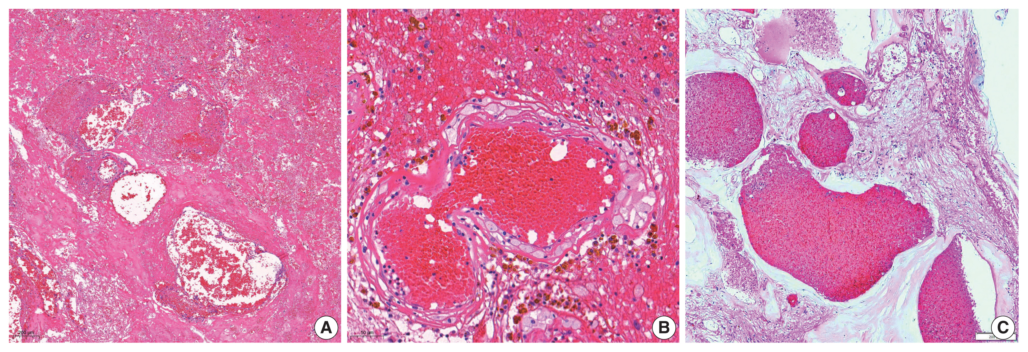
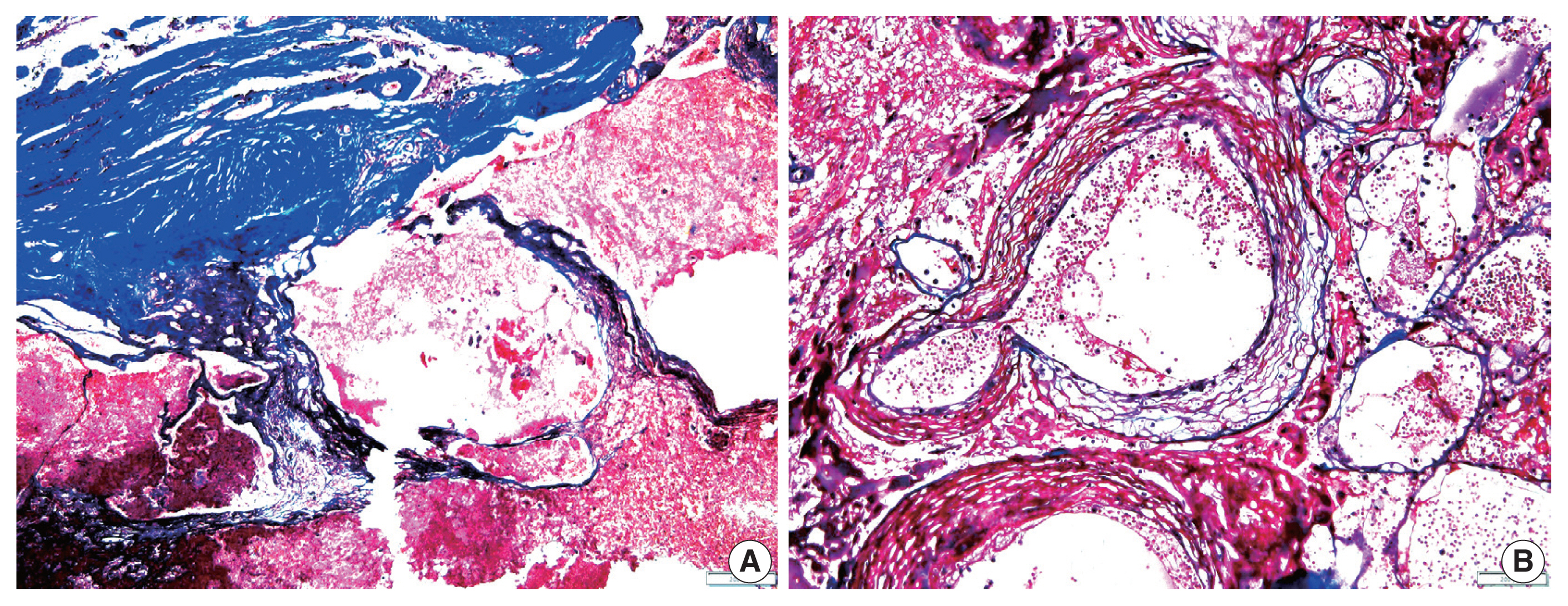


Fig. 1
Fig. 2
| Characteristic | No. (%) |
|---|---|
| Age at the time of RIOH detection (yr), mean ± SD | 46.57 ± 13.79 |
| Sex | |
| Male | 14 (37.8) |
| Female | 23 (62.2) |
| Original pathology | |
| AVM | 14 (37.8) |
| Brain tumor | 12 (32.4) |
| Metastasis | 4 (10.8) |
| Other tumors | 7 (19.0) |
| Type of treatment | |
| GKS | 24 (64.9) |
| RTx | 13 (35.1) |
| Tumor location | |
| Frontal lobe | 10 (27.0) |
| Parietal lobe | 5 (13.5) |
| Temporal lobe | 6 (16.2) |
| Occipital lobe | 4 (10.8) |
| Other (including sellar lesion) | 12 (32.5) |
| Variable | Radiation treatment | p-value | Original pathology | p-value | ||||
|---|---|---|---|---|---|---|---|---|
|
|
| |||||||
| GKS (n = 24) | RTx (n = 13) | AVM (n = 14) | Brain tumor (n = 12) | Metastasis (n = 4) | Others (n = 7) | |||
| Tumor size (cm) | ||||||||
| Primary tumor | 3.35 ± 1.22 | 3.04 ± 1.08 | .387 | 3.87 ± 1.26 | 2.85 ± 1.04 | 2.92 ± 0.60 | 2.85 ± 1.05 | .081 |
| RIOH | 2.01 ± 0.80 | 1.51 ± 0.55 | .055 | 2.06 ± 0.82 | 1.50 ± 0.68 | 2.20 ± 0.41 | 1.75 ± 0.75 | .459 |
| (Size differences) | 1.34 ± 1.33 | 1.53 ± 1.28 | .695 | 1.81 ± 1.49 | 1.35 ± 1.25 | 0.72 ± 0.45 | 1.10 ± 1.23 | .425 |
| Tumor wall thickness (μm) | 693.7 ± 565.7 | 406.9 ± 519.7 | .049 | 714.2 ± 610.9 | 560.0 ± 630.6 | 362.5 ± 47.8 | 538.5 ± 518.0 | .752 |
| Latency (yr) | 5.85 ± 4.06 | 11.15 ± 8.27 | .046 | 8.92 ± 7.01 | 7.55 ± 5.06 | 2.28 ± 3.45 | 8.68 ± 7.28 | .161 |
| Multiplicity | p-value | |||
|---|---|---|---|---|
|
| ||||
| Total | No | Yes | ||
| GKS | 24 | 21 (87.5) | 3 (12.5) | .586 |
| RTx | 13 | 11 (84.6) | 2 (15.4) | |
| AVM | 14 | 13 (92.8) | 1 (7.2) | .155 |
| Brain tumor | 12 | 10 (83.3) | 2 (16.7) | |
| Metastasis | 4 | 2 (50.0) | 2 (50.0) | |
| Others | 7 | 7 (100) | 0 | |
| Perilesional edema | p-value | Subacute stage hemorrhage (T1 high) | p-value | Hemosiderin deposit (T2 dark rim) | p-value | |||||
|---|---|---|---|---|---|---|---|---|---|---|
|
|
|
| ||||||||
| Total | Absent | Present | Absent | Present | Absent | Present | ||||
| GKS | 24 | 0 | 23 (95.8) |
.223 | 9 (37.5) | 7 (29.1) |
.059 | 2 (8.3) | 21 (87.5) |
.679 |
| RTx | 13 | 2 (15.3) | 11 (84.6) | 4 (30.7) | 9 (69.2) | 2 (15.3) | 11 (84.6) | |||
RIOH, radiation-induced organizing hematoma; SD, standard deviation; AVM, arteriovenous malformation; GKS, gamma knife surgery; RTx, radiation therapy.
Values are presented as mean±SD. GKS, gamma knife surgery; RTx, radiation therapy; AVM, arteriovenous malformation; RIOH, radiation-induced organizing hematoma; SD, standard deviation.
GKS, gamma knife surgery; RTx, radiation therapy; AVM, arteriovenous malformation.
GKS, gamma knife surgery; RTx, radiation therapy; RIOH; radiation-induced organizing hematoma; MRI, magnetic resonance imaging. One case was undetectable on MRI; Eight cases were excluded that did not include the T1 series in preoperative MRI.

