 E-submission
Articles
- Page Path
- HOME > J Pathol Transl Med > Volume 55(4); 2021 > Article
-
Original Article
Potential of AKT2 expression as a predictor of lymph-node metastasis in invasive breast carcinoma of no special type -
Primariadewi Rustamadji1,*
 , Elvan Wiyarta2,*, Kristina Anna Bethania1, Kusmardi Kusmardi3
 -
Journal of Pathology and Translational Medicine 2021;55(4):271-278.
DOI: https://doi.org/10.4132/jptm.2021.04.26
Published online: June 14, 2021
1Department of Anatomic Pathology, Faculty of Medicine, Universitas Indonesia, Jakarta, Indonesia
2Faculty of Medicine, Universitas Indonesia, Jakarta, Indonesia
3Department of Anatomic Pathology, Drug Development Research Cluster, Human Cancer Research Center, IMERI, Faculty of Medicine, Universitas Indonesia, Jakarta, Indonesia
- Corresponding Author: Elvan Wiyarta, MBBS, Faculty of Medicine, Universitas Indonesia, Salemba Raya No. 6. RW. 5, Kenari, Kec. Senen, Kota Jakarta Pusat, Jakarta 10430, Indonesia Tel: +62-81382222670, E-mail: elvan.wiyarta@ui.ac.id
*Primariadewi Rustamadji and Elvan Wiyarta contributed equally to this work.
© 2021 The Korean Society of Pathologists/The Korean Society for Cytopathology
This is an Open Access article distributed under the terms of the Creative Commons Attribution Non-Commercial License (http://creativecommons.org/licenses/by-nc/4.0) which permits unrestricted non-commercial use, distribution, and reproduction in any medium, provided the original work is properly cited.
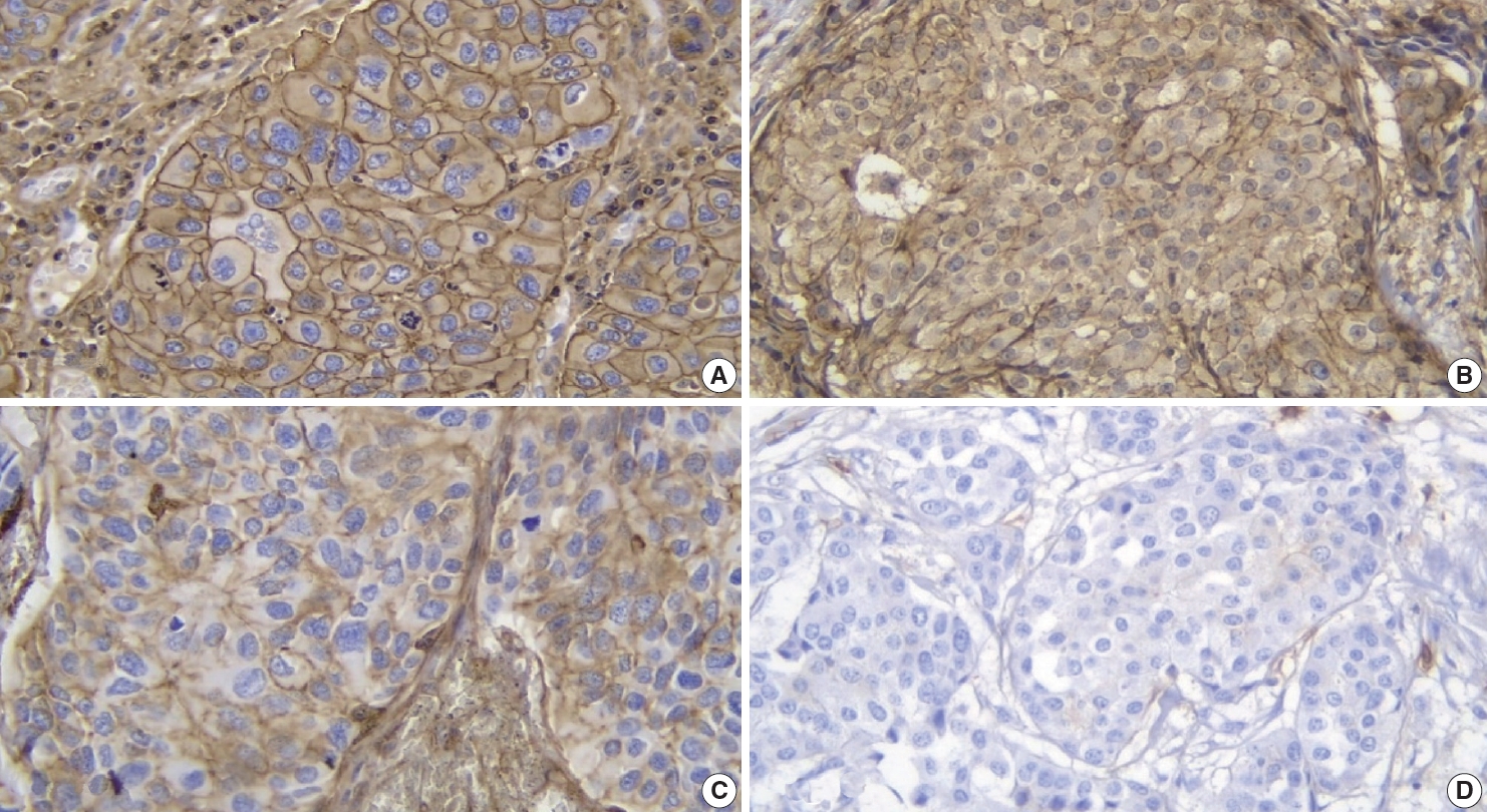
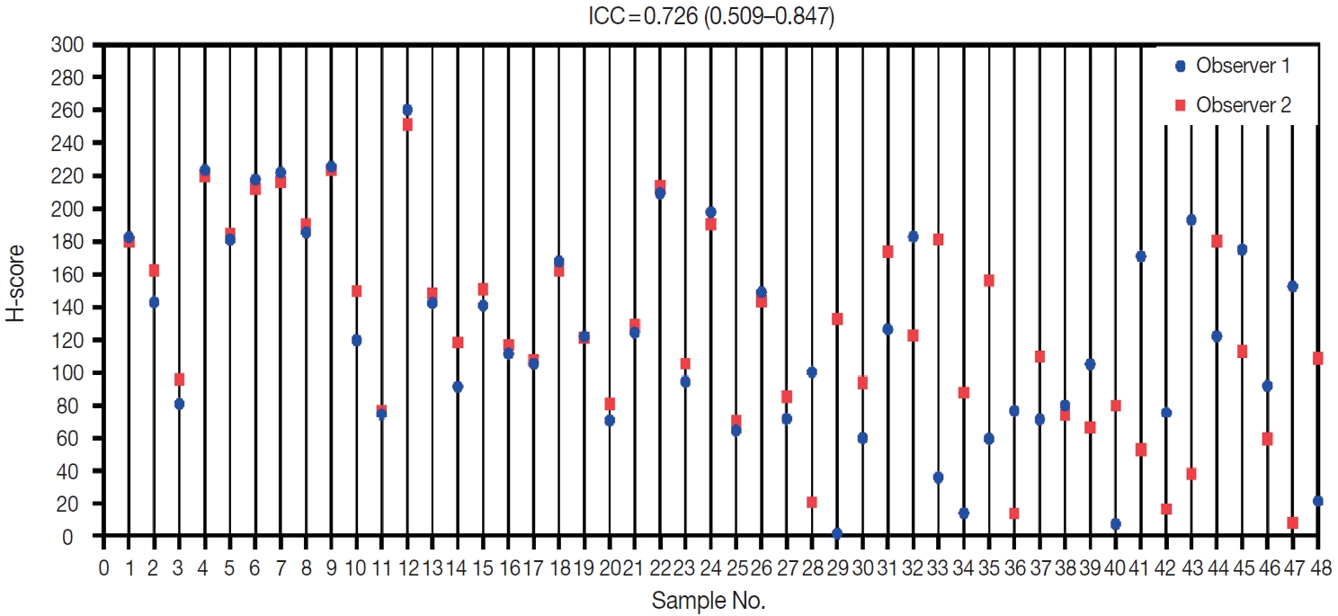
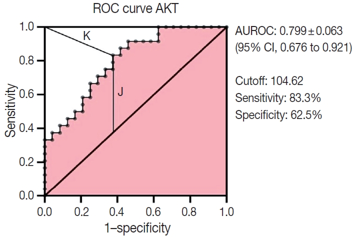
Figure & Data
References
Citations
- AKT2 Counteracts Hypoxia‐Induced Suppression of Homologous Recombination in Triple‐Negative Breast Cancer: A Targeted Approach to Sensitize Tumors to PARP Inhibition
Ying Liang, Deyu Kong, Yi Zhang, Zhijing Zhao, Li Lv, Zhe Yin, Ajaz Ahmad, Zongye Jiang
Journal of Biochemical and Molecular Toxicology.2026;[Epub] CrossRef - Association of Src with Nottingham Prognostic Index in Breast Cancer: Implications for Breast Cancer Prognostication
Primariadewi Rustamadji, Elvan Wiyarta
Journal of Nature and Science of Medicine.2024; 7(2): 90. CrossRef - CD4+ Tumor-infiltrating Lymphocytes in Neoadjuvant Chemotherapy-treated Invasive Breast Cancer of No Special Type
Primariadewi Rustamadji, Elvan Wiyarta, Meike Pramono, Sinta Chaira Maulanisa
Journal of Nature and Science of Medicine.2024; 7(3): 179. CrossRef - Potential of AKNA as a Predictive Biomarker for Ovarian Cancer and Its Relationship to Tumor Grading
P Rustamadji, E Wiyarta, M Miftahuzzakiyah, D Sukmawati, DA Suryandari, R Kodariah
Nigerian Journal of Clinical Practice.2024; 27(9): 1089. CrossRef - Exploring the Expression of Survivin on Neoadjuvant Chemotherapy in Invasive Breast Carcinoma
Primariadewi Rustamadji, Elvan Wiyarta, Ineke Anggreani
Open Access Macedonian Journal of Medical Sciences.2022; 10(B): 1440. CrossRef - Effect of Omega-3-Rich Fish Oil on TNF- Expression in Mice's Colonic Tissue Induced with Azoxymethane (AOM) and Dextran Sodium Sulphate (DSS)
Elvan Wiyarta, Kusmardi Kusmardi, Yurnadi Hanafi Midoen
Research Journal of Pharmacy and Technology.2022; : 3179. CrossRef - The potential of lunasin extract for the prevention of breast cancer progression by upregulating E-Cadherin and inhibiting ICAM-1
Kusmardi Kusmardi, Elvan Wiyarta, Numlil Khaira Rusdi, Andi Muh. Maulana, Ari Estuningtyas, Hadi Sunaryo
F1000Research.2021; 10: 902. CrossRef - CD44 Variant Exon 6 Isoform Expression as a Potential Predictor of Lymph Node Metastasis in Invasive Breast Carcinoma of No Special Type
Primariadewi Rustamadji, Elvan Wiyarta, Kristina A. Bethania, Rakesh Sathish Nair
International Journal of Breast Cancer.2021; 2021: 1. CrossRef - Correlation between CD 34 and CD 68 expression in placental malaria with maternal anemia
Primariadewi Rustamadji, Muhammad Takbir, Puspita Eka Wuyung, Kusmardi Kusmardi, Elvan Wiyarta
Tropical Parasitology.2021; 11(2): 92. CrossRef
 PubReader
 ePub Link
-
 Cite this Article
- Cite this Article
-
- Close
- Download Citation
- Close
- Figure
-


Fig. 1.
Fig. 2.
Fig. 3.
| Clinicopathological characteristic | No. (%) |
|---|---|
| Age (yr) | |
| ≥ 50 | 27 (56.3) |
| < 50 | 21 (43.8) |
| Mean ± SD | 50.9 ± 12.3 |
| Median (min–max) | 50 (29–75) |
| Tumor grade | |
| Grade I | 5 (10.4) |
| Grade II | 16 (33.3) |
| Grade III | 27 (56.3) |
| Tumor size (cm) | |
| < 2 | 2 (4.2) |
| 2–5 | 28 (58.3) |
| > 5 | 18 (37.5) |
| Lymphvovascular invasion | |
| Yes | 27 (56.3) |
| No | 21 (43.8) |
| Variable | Lymph-node metastasis, n (%) |
p-value | OR | 95% CI |
|||
|---|---|---|---|---|---|---|---|
| Yes | No | Total | Min | Max | |||
| Age (yr) |
> .990 | 1.19 | 0.38 | 3.71 | |||
| ≥ 50 | 14 (51.9) | 13 (48.1) | 27 | ||||
| < 50 | 10 (47.6) | 11 (52.4) | 21 | ||||
| Tumor grade |
.561 | 1.67 | 0.53 | 5.27 | |||
| High | 15 (55.6) | 12 (44.4) | 27 | ||||
| Low | 9 (42.9) | 12 (57.1) | 21 | ||||
| Tumor size (cm) |
.371 | 0.49 | 0.15 | 1.60 | |||
| > 5 | 7 (38.9) | 11 (61.1) | 18 | ||||
| ≤ 5 | 17 (56.7) | 13 (43.3) | 30 | ||||
| Lymphovascular invasion |
.020 | 5.00 | 1.45 | 17.27 | |||
| Yes | 18 (66.7) | 9 (33.3) | 27 | ||||
| No | 6 (28.6) | 15 (71.4) | 21 | ||||
| AKT2 expression |
.009 | 5.90 | 1.70 | 20.48 | |||
| High | 17 (70.8) | 7 (29.2) | 24 | ||||
| Low | 7 (29.2) | 17 (70.8) | 24 | ||||
| β | SE β | Wald’s χ2 | df | p-value | eβ (odds ratio) | |
|---|---|---|---|---|---|---|
| Predictor | ||||||
| AKT2 expression | 1.67 | 0.67 | 6.162 | 1 | .013 | 5.32 |
| Lymphovascular invasion | 1.49 | 0.68 | 4.789 | 1 | .028 | 4.46 |
| Constant | –1.69 | 0.63 | 7.100 | 1 | .008 | 0.19 |
| Test | ||||||
| Hosmer-Lemeshow | - | - | 0.002 | 2 | .999 | - |
| Goodness-of-Fit test | ||||||
| Overall model evaluation | - | - | 13.690 | 2 | .001 | - |
| AKT2 expression | Lymphovascular invasion | Probability of LNM (%) |
|---|---|---|
| High | Yes | 81.44 |
| High | No | 49.62 |
| Low | Yes | 45.18 |
| Low | No | 15.62 |
LNM, lymph-node metastasis; IBC-NST, invasive breast carcinoma of no special type. Bivariate analysis was performed using the chi-square test with continuity correlation.
LNM, lymph-node metastasis; IBC-NST, invasive breast carcinoma of no special type; SE, standard error; df, degree of freedom; e, euler number ≈ 2.718.
LNM, lymph-node metastasis; IBC-NST, invasive breast carcinoma of no special type.

