 E-submission
Articles
- Page Path
- HOME > J Pathol Transl Med > Volume 53(3); 2019 > Article
-
Case Study
A Rare Case of Adenosquamous Carcinoma Arising in the Background of IgG4-Related Lung Disease -
Sangjoon Choi
 , Sujin Park, Man Pyo Chung1, Tae Sung Kim2, Jong Ho Cho3, Joungho Han
 -
Journal of Pathology and Translational Medicine 2019;53(3):188-191.
DOI: https://doi.org/10.4132/jptm.2019.02.21
Published online: March 11, 2019
Department of Pathology and Translational Genomics, Samsung Medical Center, Sungkyunkwan University School of Medicine, Seoul, Korea
1Department of Internal Medicine, Samsung Medical Center, Sungkyunkwan University School of Medicine, Seoul, Korea
2Department of Radiology, Samsung Medical Center, Sungkyunkwan University School of Medicine, Seoul, Korea
3Thoracic and Cardiovascular Surgery, Samsung Medical Center, Sungkyunkwan University School of Medicine, Seoul, Korea
- Corresponding Author Joungho Han, MD Department and of Pathology and Translational Genomics, Samsung Medical Center, Sungkyunkwan University School of Medicine, 81 Irwon-ro, Gangnam-gu, Seoul 06351, Korea Tel: +82-2-3410-2800 Fax: +82-2-3410-0025 E-mail: hanjho@skku.edu
© 2019 The Korean Society of Pathologists/The Korean Society for Cytopathology
This is an Open Access article distributed under the terms of the Creative Commons Attribution Non-Commercial License (http://creativecommons.org/licenses/by-nc/4.0) which permits unrestricted non-commercial use, distribution, and reproduction in any medium, provided the original work is properly cited.
Abstract
- IgG4-related disease is a systemic inflammatory disease and is known as IgG4-related lung disease (IgG4-RLD) when it involves the respiratory system. Primary lung cancer arising from a background of IgG4-RLD is very rare. Herein, we report a case of adenosquamous carcinoma arising from the background of IgG4-RLD and presenting as an interstitial lung disease pattern. A 66-year-old man underwent lobectomy under the impression of primary lung cancer. Grossly, the mass was ill-defined and gray-tan colored, and the background lung was fibrotic. Microscopically, tumor cells showed both squamous and glandular differentiation. Dense lymphoplasmacytic infiltration with fibrosis and obliterative phlebitis were seen in the background lung. IgG4 immunohistochemical stain showed diffuse positivity in infiltrating plasma cells. Primary lung adenosquamous carcinoma has not been reported in a background of IgG4-RLD. Due to the rarity of IgG4-RLD, physicians must follow patients with IgG4-RLD over long periods of time to accurately predict the risk of lung cancer.
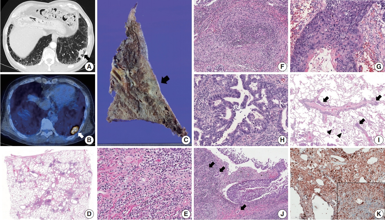
- A 66-year-old man who had a past medical history of idiopathic pulmonary fibrosis (IPF) and mass-forming IgG4-related autoimmune cholangitis was admitted to the hospital for a newly-identified consolidative lung mass discovered during follow-up. Chest computed tomography revealed a subpleural nodule in the left lower lobe of the lung in a background of reticular and honeycomb fibrosis (Fig. 1A). 18F-fluorodeoxyglucose uptake was detected in the subpleural nodule (Fig. 1B). The results of the pulmonary function tests were within normal range: forced vital capacity (FVC) 3.23 L (82% of the predicted value), forced expiratory volume in 1 second (FEV1) 2.35 L (80% of the predicted value), and FEV1/FVC 73%. Laboratory test showed an increased serum IgG4 level (232.4 mg/dL). The patient underwent lobectomy under the impression of lung cancer. Grossly, the tumor was ill-defined, gray-tan colored and measured 3.5 × 3.2 × 2.0 cm. The background lung was fibrotic and emphysematous (Fig. 1C). Microscopically, the background lung showed diffuse irregular interstitial fibrosis with dense lymphoplasmacytic infiltration and occasional obliterative phlebitis (Fig. 1D–F). Tumor cells showed both squamous and glandular differentiation. The squamous cell carcinoma component was composed of moderately to poorly differentiated tumor cells that contained keratin pearls (Fig. 1G). The glandular component was mainly acinar pattern with focal micropapillary pattern (Fig. 1H). Diffuse spread through air space of tumor cells was frequently found at the periphery of the mass (Fig. 1I). Multifocal lymphangitic spreading of tumor cells and metastatic lymph nodes were found (Fig. 1I). Dense fibrosis and lymphoplasmacytic infiltration were adjacent to the tumor cells (Fig. 1J). The final pathologic stage was pT2aN2M0 by the American Joint Committee on Cancer seventh staging system. Immunohistochemistry (IHC) staining revealed the squamous cell carcinoma component was focally positive for p63 (1:200, Biocare, Concord, CA, USA), and the glandular component was negative for TTF-1 (1:50, Dako, Glostrup, Denmark). Additional tests for anaplastic lymphoma kinase (ALK) IHC staining (1:40, NCL-ALK, clone 5A4, Novocastra, Newcastle upon Tyne, UK) and epidermal growth factor receptor gene mutation analysis using a PNA clamping kit (Panagene, Inc., Daejeon, Korea) were negative, and up to 10% of the tumor cells showed membrane positivity for programmed death-ligand 1 (RTU, 22C3, Dako). IgG4 (1:2,000, The Binding Site, Birmingham, UK) IHC stain showed diffuse positivity in infiltrating plasma cells (> 50 cells/high-power field), and the IgG4/IgG ratio was over 40% (Fig. 1K). Thus, the patient’s IPF was thought to be a manifestation of IgG4-RLD, and we concluded that primary adenosquamous carcinoma had developed in the background of IgG4-RLD. This study was approved by the Institutional Review Board of the Samsung Medical Center with a waiver of informed consent (IRB No. 2018-11-053) and performed in accordance with the principles of the Declaration of Helsinki.
CASE REPORT
- IgG4-RD was first reported as autoimmune pancreatitis in 2001 [7]. IgG4-RD is known to predominantly involve the pancreas, hepatobiliary tract, salivary glands, and lacrimal glands, and lung or pleural involvement can occur in up to 35% of patients [8]. The histologic patterns of IgG4-RLD are divided into three types: solid nodular type, bronchovascular type, and interstitial lung disease type [4].
- It is still debatable whether IgG4-RD is associated with malignancy. Yamamoto et al. [9] observed 106 IgG4-RD patients (primarily with Mikulicz’s disease), and the high standardized incidence rate (SIR) of 3.83 supported the association between IgG4-RD and increased incidence of total malignancies. In a different study, Hirano et al. [10] observed 113 patients with IgG4-RD (primarily with autoimmune pancreatitis), and the SIR was not significant (1.04). These different outcomes likely result from whether the studies considered cases that simultaneously found malignancies and IgG4-RD.
- However, none of these studies included patients with IgG4-RD that involved the pulmonary system. The association of IgG4-RLD with lung cancer has not been studied, and only three lung cancer cases in IgG4-RLD patients have been reported with their histopathologic findings [4-6].
- The patient in the present case had primary adenosquamous carcinoma, which has not been reported alongside IgG4-RD in the previous literature. The adenosquamous carcinoma was characterized by poorly differentiated squamous and glandular tumor cells with lymph node metastases, and the IgG4-RLD background presented as interstitial lung disease. Inoue et al. [5] and Tashiro et al. [6] reported a well differentiated lepidic pattern of adenocarcinoma accompanied by IgG4-RLD as a solid nodule or ground glass opacity pattern. There were no lymph node metastases in these two cases. Zen et al. [4] reported a moderately differentiated, mixed pattern (including acinar pattern) adenocarcinoma in a background of IgG4-RLD presenting as interstitial pneumonia. Lymph node metastases were found, and the pathologic stage was pT1N2M0. In the present case, similar to Zen’s report, moderately to poorly differentiated carcinoma occurred in a background of IgG4-RLD with an interstitial lung disease pattern. Numerous lymphovascular invasions and lymph node metastases were found, and the final pathologic stage was pT2aN2M0. Table 1 summarizes the clinicopathologic and radiological characteristics of the reported cases of concurrent IgG4-RLD and lung cancer.
- There have been no studies on whether IgG4-RLD increases the risk of malignancy. Although there have been a small number of cases, it is likely that lung cancer more frequently occurs in the solid nodular or interstitial lung disease type of IgG4-RLD rather than the bronchovascular type. Thus far, malignancy has not been reported in the bronchovascular type of IgG4-RLD. There is also a possibility that the differentiation or aggressiveness of the tumor may depend on the background type of IgG4-RLD, and the prognosis could be worse in patients with the interstitial lung disease background. Further studies with more cases are needed to elucidate the relationship between tumor aggressiveness and patterns of IgG4-RLD.
DISCUSSION
Author contributions
Conceptualization: SC.
Data curation: SC, JH.
Formal analysis: SC, SP.
Investigation: SC, SP.
Methodology: SC, SP, JH.
Project administration: SC, JH.
Resources: MPC, TSK, JHC.
Supervision: JH.
Validation: JH.
Writing—original draft: SC, JH.
Writing—review & editing: SC, SP, JH.
Conflicts of Interest
The authors declare that they have no potential conflicts of interest.
| Reference | Sex | Age (yr) | Location | Type of tumor | Pattern of ADC | Radiologic finding | Pattern of IgG4-RLD | TNM stage | Other manifestations | Serum IgG4 (mg/dL) |
|---|---|---|---|---|---|---|---|---|---|---|
| Present case | M | 66 | LLL | ASC | Acinar and focal micropapillary | Subpleural nodule in a background of reticular and honeycomb fibrosis | Interstitial | pT2aN2M0 | IHD | 232 |
| Zen et al. [4] | M | NA | RLL | ADC | Mixed, including acinar | Nodular lesion within the reticular shadow | Interstitial | pT1N2M0 | No | NA |
| Inoue et al. [5] | M | 78 | RUL | ADC | Lepidic | Ground-glass opacity with central collapse and pleural indentation | Nodular | pT1bN0M0 | Pancreas | 983 |
| Tashiro et al. [6] | M | 72 | RML | ADC | Lepidic | Spiculated nodule with pleural indentation | Nodular | pT1bN0M0 | No | 346 |
IgG4-RLD, IgG4-related lung disease; ADC, adenocarcinoma; M, male; LLL, left lower lobe; ASC, adenosquamous carcinoma; Interstitial, interstitial lung disease type; IHD, intrahepatic bile duct; RLL, right lower lobe; NA, not available; RUL, right upper lobe; Nodular, solid nodular type; RML, right middle lobe.
- 1. Deshpande V, Zen Y, Chan JK, et al. Consensus statement on the pathology of IgG4-related disease. Mod Pathol 2012; 25: 1181-92. PubMedPDF
- 2. Ahn JH, Hong SI, Cho DH, Chae EJ, Song JS, Song JW. A case of IgG4-related lung disease presenting as interstitial lung disease. Tuberc Respir Dis 2014; 77: 85-9. Article
- 3. Cho DH, An JH, Kang YM, Chae EJ, Song JS, Song JW. A case of IgG4-related lung disease mimicking non-specific interstitial pneumonia. Korean J Med 2015; 88: 308-12. ArticlePDF
- 4. Zen Y, Inoue D, Kitao A, et al. IgG4-related lung and pleural disease: a clinicopathologic study of 21 cases. Am J Surg Pathol 2009; 33: 1886-93. ArticlePubMed
- 5. Inoue T, Hayama M, Kobayashi S, et al. Lung cancer complicated with IgG4-related disease of the lung. Ann Thorac Cardiovasc Surg 2014; 20 Suppl: 474-7. ArticlePubMed
- 6. Tashiro H, Takahashi K, Nakamura T, Komiya K, Kimura S, Sueoka-Aragane N. Coexistence of lung cancer and immunoglobulin G4-related lung disease in a nodule: a case report. J Med Case Rep 2016; 10: 113.ArticlePubMedPMC
- 7. Hamano H, Kawa S, Horiuchi A, et al. High serum IgG4 concentrations in patients with sclerosing pancreatitis. N Engl J Med 2001; 344: 732-8. ArticlePubMed
- 8. Fei Y, Shi J, Lin W, et al. Intrathoracic involvements of immunoglobulin G4-related sclerosing disease. Medicine (Baltimore) 2015; 94: e2150.ArticlePubMedPMC
- 9. Yamamoto M, Takahashi H, Tabeya T, et al. Risk of malignancies in IgG4-related disease. Mod Rheumatol 2012; 22: 414-8. ArticlePubMed
- 10. Hirano K, Tada M, Sasahira N, et al. Incidence of malignancies in patients with IgG4-related disease. Intern Med 2014; 53: 171-6. ArticlePubMed
REFERENCES
Figure & Data
References
Citations
- Endometrioid Carcinomas of the Ovaries and Endometrium Involving Endocervical Polyps: Comprehensive Clinicopathological Analyses
Jihee Sohn, Yurimi Lee, Hyun-Soo Kim
Diagnostics.2022; 12(10): 2339. CrossRef - A Case of IgG4-related Disease Composed of a Paravertebral Tumor Alone with Multiple Lung Cancers
Mutsumi Ozasa, Toyomitsu Sawai, Yosuke Harada, Sumako Yoshioka, Nobuko Matsuo, Hiroshi Mukae
Haigan.2021; 61(3): 213. CrossRef - Serous Carcinoma of the Endometrium with Mesonephric-Like Differentiation Initially Misdiagnosed as Uterine Mesonephric-Like Adenocarcinoma: A Case Report with Emphasis on the Immunostaining and the Identification of Splice Site TP53 Mutation
Sangjoon Choi, Yoon Yang Jung, Hyun-Soo Kim
Diagnostics.2021; 11(4): 717. CrossRef - A Case of IgG4-related Thyroiditis Diagnosed by Total Thyroidectomy
Daiki Sakamoto, Masao Yagi, Hiroshi Iwai
Practica Oto-Rhino-Laryngologica.2021; 114(7): 547. CrossRef - Mesonephric-like Differentiation of Endometrial Endometrioid Carcinoma: Clinicopathological and Molecular Characteristics Distinct from Those of Uterine Mesonephric-like Adenocarcinoma
Sujin Park, Go Eun Bae, Jiyoung Kim, Hyun-Soo Kim
Diagnostics.2021; 11(8): 1450. CrossRef - Mesonephric-like Adenocarcinoma of the Uterine Corpus: Comprehensive Immunohistochemical Analyses Using Markers for Mesonephric, Endometrioid and Serous Tumors
Hyunjin Kim, Kiyong Na, Go Eun Bae, Hyun-Soo Kim
Diagnostics.2021; 11(11): 2042. CrossRef - Not Cancer After All: Two Rare Cases of IgG4-Related Lung Disease
Josué Pinto, Carla Damas, António Morais
Archivos de Bronconeumología.2020; 56(1): 53. CrossRef - Not Cancer After All: Two Rare Cases of IgG4-Related Lung Disease
Josuèc) Pinto, Carla Damas, António Morais
Archivos de Bronconeumología (English Edition).2020; 56(1): 52. CrossRef - Axillary lymphadenopathy with IgG4 positive plasma cell infiltration as differential diagnosis of metastatic lung adenocarcinoma
Yutaro Ito, Masanori Harada, Namio Kagoo, Tsutomu Kubota, Koshiro Ichijyo, Eisuke Mochizuki, Masahiro Uehara, Shun Matsuura, Masaru Tsukui, Naoki Koshimizu
Respiratory Medicine Case Reports.2020; 31: 101196. CrossRef
 PubReader
 ePub Link
-
 Cite this Article
- Cite this Article
-
- Close
- Download Citation
- Close
- Figure
-


Fig. 1.
| Reference | Sex | Age (yr) | Location | Type of tumor | Pattern of ADC | Radiologic finding | Pattern of IgG4-RLD | TNM stage | Other manifestations | Serum IgG4 (mg/dL) |
|---|---|---|---|---|---|---|---|---|---|---|
| Present case | M | 66 | LLL | ASC | Acinar and focal micropapillary | Subpleural nodule in a background of reticular and honeycomb fibrosis | Interstitial | pT2aN2M0 | IHD | 232 |
| Zen et al. [4] | M | NA | RLL | ADC | Mixed, including acinar | Nodular lesion within the reticular shadow | Interstitial | pT1N2M0 | No | NA |
| Inoue et al. [5] | M | 78 | RUL | ADC | Lepidic | Ground-glass opacity with central collapse and pleural indentation | Nodular | pT1bN0M0 | Pancreas | 983 |
| Tashiro et al. [6] | M | 72 | RML | ADC | Lepidic | Spiculated nodule with pleural indentation | Nodular | pT1bN0M0 | No | 346 |
IgG4-RLD, IgG4-related lung disease; ADC, adenocarcinoma; M, male; LLL, left lower lobe; ASC, adenosquamous carcinoma; Interstitial, interstitial lung disease type; IHD, intrahepatic bile duct; RLL, right lower lobe; NA, not available; RUL, right upper lobe; Nodular, solid nodular type; RML, right middle lobe.

