 E-submission
Articles
- Page Path
- HOME > J Pathol Transl Med > Volume 51(4); 2017 > Article
-
Case Study
A Rare Case of Intramural Müllerian Adenosarcoma Arising from Adenomyosis of the Uterus - Sun-Jae Lee, Ji Y. Park
-
Journal of Pathology and Translational Medicine 2017;51(4):433-440.
DOI: https://doi.org/10.4132/jptm.2017.06.11
Published online: June 29, 2017
Department of Pathology, Catholic University of Daegu School of Medicine, Daegu, Korea
- Corresponding Author Sun-Jae Lee, MD, PhD Department of Pathology, Catholic University of Daegu School of Medicine, 33 Duryugongwon-ro 17-gil, Nam-gu, Daegu 42472, Korea Tel: +82-53-650-4629 Fax: +82-53-650-4834 E-mail: pathosjlee@cu.ac.kr
© 2017 The Korean Society of Pathologists/The Korean Society for Cytopathology
This is an Open Access article distributed under the terms of the Creative Commons Attribution Non-Commercial License (http://creativecommons.org/licenses/by-nc/4.0) which permits unrestricted non-commercial use, distribution, and reproduction in any medium, provided the original work is properly cited.
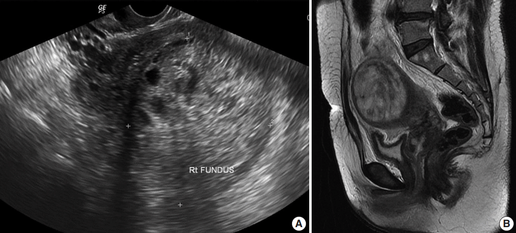

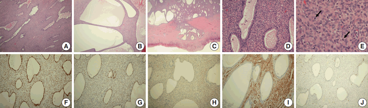
Figure & Data
References
Citations
- A Case of Intramural Adenosarcoma With Sarcomatous Overgrowth Associated With Adenomyosis and Endometriosis
Kyohei Kitamura, Sachiko Minamiguchi, Hiroaki Ito, Yosuke Yamada, Yuki Himoto, Koji Yamanoi, Ken Yamaguchi, Hironori Haga
Cureus.2025;[Epub] CrossRef - Whether surgical procedure can improve the prognosis of endometrial cancer arising in adenomyosis (EC-AIA)? A systematic review and meta-analysis
Yi Sun, Shitong Lin, Weijia Wu, Fangfang Nie, Yuchen Liu, Jing Wen, Xiaoran Cheng, Qianwen Liu, Yuanpei Wang, Fang Ren
International Journal of Surgery.2024; 110(5): 3072. CrossRef - Mullerian adenosarcoma accidentally detected and coexisting with cervical carcinoma in situ: a rare case report
Xuemei Qing, Min Xie, Hongying Guo, Liying Zhang, Jiatian Ye, Yong Zhang, Ying Ma
Frontiers in Oncology.2024;[Epub] CrossRef - Mucinous carcinoma originating from uterine adenomyosis: a case report
Satoshi Ohira, Ryota Tachibana, Sayaka Yasaki, Koji Tsunemi, Natsuki Uchiyama, Eri Ikeda, Kenji Sano
Journal of Medical Case Reports.2023;[Epub] CrossRef - Endometrial Cancer Arising in Adenomyosis (EC-AIA): A Systematic Review
Antonio Raffone, Diego Raimondo, Manuela Maletta, Antonio Travaglino, Federica Renzulli, Daniele Neola, Umberto De Laurentiis, Francesco De Laurentiis, Mohamed Mabrouk, Manuel Maria Ianieri, Renato Seracchioli, Paolo Casadio, Antonio Mollo
Cancers.2023; 15(4): 1142. CrossRef - Adenomyosis and Its Possible Malignancy: A Review of the Literature
Liviu Moraru, Melinda-Ildiko Mitranovici, Diana Maria Chiorean, Raluca Moraru, Laura Caravia, Andreea Taisia Tiron, Ovidiu Simion Cotoi
Diagnostics.2023; 13(11): 1883. CrossRef - Case Report: Uterine Adenosarcoma With Sarcomatous Overgrowth and Malignant Heterologous Elements
Yunuén I. García-Mendoza, Mario Murguia-Perez, Aldo I. Galván-Linares, Saulo Mendoza-Ramírez, Norma L. García-Salinas, Julio G. Moctezuma-Ramírez, Blanca O. Murillo-Ortiz, Luis Jonathan Bueno-Rosario, Marco A. Olvera-Olvera, Guillermo E. Corredor-Alonso
Frontiers in Medicine.2022;[Epub] CrossRef - Adenomyosis as a Risk Factor for Myometrial or Endometrial Neoplasms—Review
Maria Szubert, Edward Kozirog, Jacek Wilczynski
International Journal of Environmental Research and Public Health.2022; 19(4): 2294. CrossRef - Uterine adenosarcoma arising from a subserosal adenomyoma: A case report
Shazia Fakhar, Tehreem Zahid, Yamina Ishtiaq
Gynecologic Oncology Reports.2022; 40: 100957. CrossRef - Uterine Adenosarcoma Originating in Adenomyosis: Report of an Extremely Rare Phenomenon and Review of Published Literature
Karen L. Talia, Yael Naaman, W. Glenn McCluggage
International Journal of Gynecological Pathology.2021; 40(4): 342. CrossRef - Uterine adenosarcoma. Report of 5 cases and review of literature
I.V. Barinova, I.N. Voloshchuk, A.A. Fedorov, N.V. Puchkova, S.N. Buyanova, M.A. Chechneva, A.A. Popov, O.V. Kapitanova, N.I. Kondrikov
Arkhiv patologii.2021; 83(3): 25. CrossRef - New Aspects of Sarcomas of Uterine Corpus—A Brief Narrative Review
Stoyan Kostov, Yavor Kornovski, Vesela Ivanova, Deyan Dzhenkov, Dimitar Metodiev, Rafał Watrowski, Yonka Ivanova, Stanislav Slavchev, Dimitar Mitev, Angel Yordanov
Clinics and Practice.2021; 11(4): 878. CrossRef - Uterine Adenosarcoma with Sarcomatous Overgrowth: A Case Report of Aggressive Disease in a 16-Year-Old Girl and a Literature Review
Hanyuan Liu, Zhen Shen, Dabao Wu, Ying Zhou
Journal of Pediatric and Adolescent Gynecology.2018; 31(4): 426. CrossRef - Uterine Adenosarcoma
Uwe A. Ulrich, Dominik Denschlag
Oncology Research and Treatment.2018; 41(11): 693. CrossRef
 PubReader
 ePub Link
-
 Cite this Article
- Cite this Article
-
- Close
- Download Citation
- Close
- Figure
-

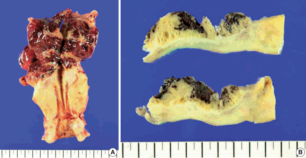


Fig. 1.
Fig. 2.
Fig. 3.
| Case No. (ref No.) | Clinical feature | Pathology | Treatment | Outcome | Remarks |
|---|---|---|---|---|---|
| 1 [15] | Age: 51 yr | Size: 4 cm | Unknown | Unknown | - |
| Gyn hx: unknown | Location: lateral wall of the uterine body | ||||
| Clinical sign: unknown | Micro: | ||||
| Glands with no epithelial cell atypia | |||||
| Sarcomatous component with cell pleomorphism and a high mitotic count | |||||
| Accompanied by adenomyosis | |||||
| Tumor marker: unknown | |||||
| 2 [13] | Age: 20 yr | Size: unknown | Hysterectomy | Two years after surgery, no evidence of recurrent disease | Stromal overgrowth |
| Gyn hx: null | Location: right anterolateral portion | ||||
| Clinical sign: a longstanding history of menorrhagia and vaginal bleeding | Micro: | ||||
| Florid adenomyosis with extensive myometrial invasion, expansile growth within the myometrium, and intravascular invasion in the myometrium | |||||
| Tumor marker: β-hCG 50–80 mIU/mL | |||||
| 3 [2] | Age: 46 yr | Size: unknown | Myomectomy | Unknown | - |
| Gyn hx: para 1 | Location: subserosal mass arising from the posterior surface of the uterus | Additional TAH, BSO, and bilateral pelvic lymphadenectomy | |||
| Clinical sign: vaginal bleeding | Micro: | ||||
| Adenomyoma with focal predominant endometrial stroma and periglandular cuffs | |||||
| Endometrial stromal cells in the periglandular cuffs showing mild and focal moderate cytological atypia with sparse mitotic figures, including an occasional atypical form | |||||
| Tumor marker: unknown | |||||
| 4 [5] | Age: 38 yr | Size: 1.5 cm | Exploratory laparotomy, TAH, LSO, and omentectomy | Disease-free 30 mo after treatment | Heterologous element (rhabdomyosarcoma) |
| Gyn hx: gravida 1, para 0 | Location: right cornual area | Adjuvant cisplatin, ifosfamide, and mesna | |||
| Clinical sign: chronic pelvic pain and dysmenorrhea | Micro: | 5,500 cGy to the abdominal wall | |||
| Irregular glands with benign epithelium surrounded by a hypercellular spindle cell stroma showing rare mitoses, mild nuclear hyperchromasia, and pleomorphism | |||||
| Tumor marker | |||||
| CEA and AFP: normal | |||||
| CA125: 45 U/mL | |||||
| 5 [10] | Age: 52 yr | Size: uncheckable (no distinct mass formation) | Radical hysterectomy with BSO and lymph node dissection and debulking of the pelvic mass | Unknown | Extrauterine pelvic mass (19 cm in diameter) diagnosed as adenosarcoma with rhabdomyosarcomatous differentiation and stromal overgrowth |
| Gyn hx: gravida 3, para 3 | Location: uterine fundus | ||||
| Peri-menopausal | Micro: | ||||
| Diffuse adenomyosis with focal stromal expansion, consisting of a hypercellular proliferation of moderately atypical spindle cells with mitotic activity around benign endometrial glands and infiltrating the anterior myometrium | |||||
| Clinical sign: none | |||||
| Tumor marker | |||||
| CA125: 258 U/mL | |||||
| 6 [14] | Age: 53 yr | Size: unknown | Unknown | Unknown | Developed breast carcinoma and received adjuvant chemotherapy including tamoxifen |
| Gyn hx: unknown | Location: unknown | ||||
| Clinical sign: unknown | Micro: | ||||
| Uterine adenosarcoma following an adenomyoma | |||||
| Tumor marker: unknown | |||||
| 7 | 7 Age: 40 yr | Size: 7.5 cm | Laparoscopically assisted TVH | No evidence of recurrence to date | This case |
| Gyn hx: gravida 2, para 2 | Location: uterine fundus | Additional BSO | |||
| Clinical sign: sudden-onset suprapubic pain and initial low back pain | Micro: | ||||
| Dilated glandular elements and abundant, hypercellular stromal elements | |||||
| Expansile growth within the myometrium with extensive myometrial invasion and focal infiltration with expansile margin into the subserosa | |||||
| Focal involvement of adenomyosis | |||||
| Tumor marker | |||||
| CA125: 5,000 U/mL | |||||
| CA19-9: 39 U/mL | |||||
| β-hCG, AFP: normal |
| Stage | Definition |
|---|---|
| I | Tumor limited to uterus |
| IA | Tumor limited to endometrium/endocervix with no myometrial invasion |
| IB | ≤ 50% myometrial invasion |
| IC | > 50% myometrial invasion |
| II | Tumor extension beyond the uterus, within the pelvis |
| IIA | Adnexal involvement |
| IIB | Involvement of other pelvic tissues |
| III | Tumor invasion of abdominal tissues (not just protruding into the abdomen) |
| IIIA | 1 site |
| IIIB | > 1 site |
| IIIC | Metastasis to pelvic and/or para-aortic lymph nodes |
| IV | |
| IVA | Tumor invasion of bladder and/or rectum |
| IVB | Distant metastasis |
Gyn Hx, gynecological history; hCG, human chorionic gonadotropin; Micro, microscopic findings; TAH, total abdominal hysterectomy; BSO, bilateral salpingooophorectomy; LSO, left salpingo-oophorectomy; CEA, carcinoembryonic antigen; AFP, α-fetoprotein; CA, carbohydrate antigen; TVH, total vaginal hysterectomy.
FIGO, International Federation of Gynecology and Obstetrics.

