 E-submission
Articles
- Page Path
- HOME > J Pathol Transl Med > Volume 49(6); 2015 > Article
-
Case Study
Mediastinal Glomus Tumor: A Case Report and Literature Review - Si-Hyong Jang, Hyun Deuk Cho, Ji-Hye Lee, Hyun Ju Lee, Hae Yoen Jung, Kyung-Ju Kim, Sung Sik Cho1, Mee-Hye Oh
-
Journal of Pathology and Translational Medicine 2015;49(6):520-524.
DOI: https://doi.org/10.4132/jptm.2015.07.02
Published online: August 4, 2015
Department of Pathology, Soonchunhyang University Cheonan Hospital, Soonchunhyang University College of Medicine, Cheonan, Korea
1Department of Radiology, Soonchunhyang University Cheonan Hospital, Soonchunhyang University College of Medicine, Cheonan, Korea
- Corresponding Author: Mee-Hye Oh, MD, PhD Department of Pathology,Soonchunhyang University Cheonan Hospital,31 Suncheonhyang 6-gil, Dongnam-gu,Cheonan 31151, Korea Tel: +82-41-570-3582 Fax: +82-41-570-3580 E-mail:mhoh0212@hanmail.net
© 2015 The Korean Society of Pathologists/The Korean Society for Cytopathology
This is an Open Access article distributed under the terms of the Creative Commons Attribution Non-Commercial License (http://creativecommons.org/licenses/by-nc/3.0/) which permits unrestricted non-commercial use, distribution, and reproduction in any medium, provided the original work is properly cited.
Abstract
- A glomus tumor in the mediastinum is very uncommon, and only five cases have been reported in the English literature. We recently encountered a 21-year-old woman with an asymptomatic mediastinal mass that measured 5.3 × 4.0 cm. Surgical excision was performed, and the tumor was finally diagnosed as mediastinal glomus tumor with an uncertain malignant potential. After reviewing this case and previous reports, we analyzed the clinicopathologic features associated with progression of such a tumor.
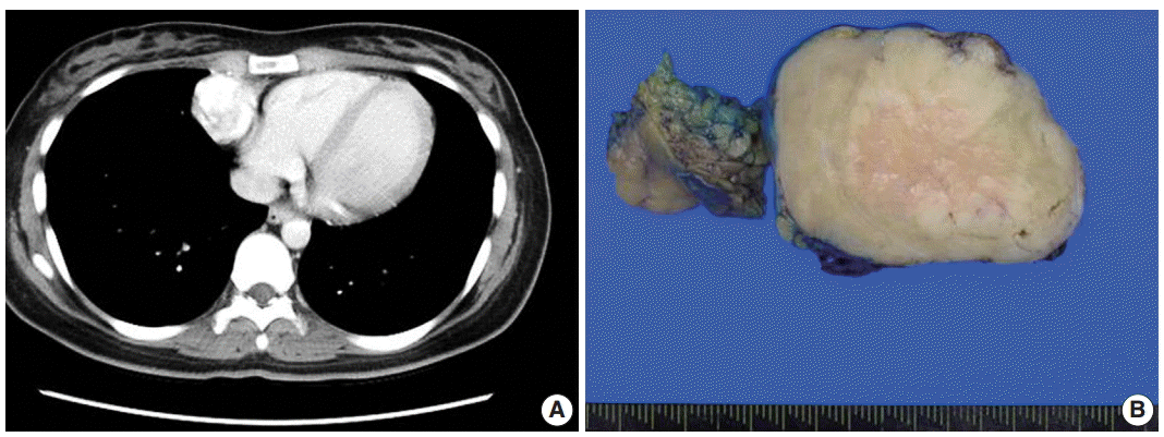
- A 21-year-old woman was referred to our hospital due to a mediastinal mass that was incidentally detected on a chest X-ray performed during health screening. The patient did not complain of any notable symptoms. Chest computed tomography scan revealed a densely-enhanced, round to oval-shaped mass in the right cardiophrenic angle of the anterior inferior mediastinum. Small tortuous vascular structures were noted near the tumor (Fig. 1A). The patient underwent thoracoscopic mediastinal mass excision.
- On gross examination, the specimen was a relatively well-demarcated solid mass with a rubbery consistency, measuring 5.3 ×4.0 cm. The cut surface of the tumor was tan yellow and had a nodular appearance without a necrotic or hemorrhagic area (Fig. 1B).
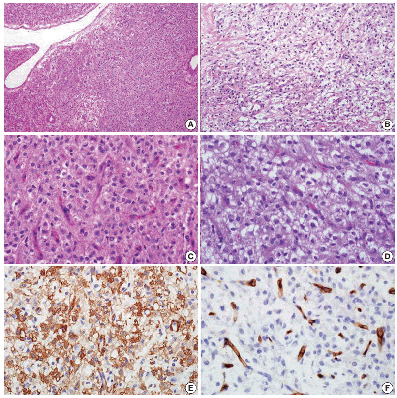
- Microscopically, the tumor was enveloped by a variable, thickened fibrous capsule with no evidence of infiltration into adjacent tissue. A solid growth pattern was evident, with prominent vascular structures composed of small- to medium-sized blood vessels. Medium-sized vessels occasionally appeared with staghorn features and myxoid changes in their walls (Fig. 2A). The tumor displayed both low and high cellular areas. The high cellular areas appeared in vague nodular configuration, and the low cellular area had edematous and myxoid change and was present between high cellular areas (Fig. 2A, B). High-powered examination revealed that the tumor was composed of round epithelioid cells, which had clear to eosinophilic cytoplasm with sharply defined borders. The centrally-located vesicular nuclei were round to polygonal, with a mild convoluted contour of nuclear membranes. One or two small inconspicuous nucleoli were noted. Nuclear pleomorphism was not identified (Fig. 2C, D). Mitotic activity was infrequent, observed in fewer than 1/50 high power fields. Atypical mitosis was not found. Lymphovascular or perineural invasion was not observed. Immunohistochemical staining for smooth muscle actin and vimentin showed diffuse and focal positivity, respectively. Tumor cells were not immunoreactive for CD34, calretinin, cytokeratin, desmin, or neuroendocrine markers, including synaptophysin and chromogranin (Fig. 2E, F). Based on the histology and immunohistochemical results, a pathologic diagnosis of glomus tumor with uncertain malignant potential was made. There was no clinicoradiologic evidence of recurrence or metastasis for seven months after surgery.
CASE REPORT
- Glomus tumors are unusual benign smooth muscle neoplasms that comprise fewer than 2% of all soft tissue tumors [10,11]. These tumors are preferentially located in the subungal area of extremities and have a characteristic clinical triad that includes pain, pinpoint tenderness and hypersensitivity to cold temperatures [12]. Clinically, this type of tumor is usually benign, and malignant cases have rarely been reported [13]. Additionally, mediastinal glomus tumors, first described in 1949, are extremely rare [2]; only five cases have been described in the English literature [1-3,8,9]. This is the sixth case of a glomus tumor in the mediastinum.
- The pathologic differential diagnoses of mediastinal glomus tumor include carcinoid tumor, hemangiopericytoma, epithelioid leiomyoma, primitive neuroectodermal tumor, and paraganglioma. Characteristic morphologic features that are helpful for distinguishing these tumors include uniform, round tumor cells with centrally-located nuclei, a well-defined cell membrane and immunohistochemial results that are positive for actin and equivocal or negative for CD34, neuroendocrine markers (chromogranin A, synaptophysin, neuron specific enolase) and CD99 [1].
- A previous article reported that mediastinal glomus tumors tend to have atypical histologic appearance with cytologic pleomorphism and infiltrate into surrounding tissue [1]. However, clinicopathologic features or distinct classification of glomus tumors in the mediastinum or visceral organs have not been established due to their infrequency. We comprehensively reviewed the clinicopathologic data of mediastinal glomus tumors; this data and the current case are presented in Table 1.
- Compared with previous cases, our patient was young and did not present with any symptoms. In addition, although previous mediastinal glomus tumors were frequently found in the posterior superior area, this case was noted in an unusual location, the anterior inferior mediastinum. To investigate the clinicopathologic characteristics in malignant mediastinal glomus tumors, we searched for histologic descriptions in former reports. A previous report indicated that, in a fatal mediastinal glomus tumor case [8], advanced age, extensive local invasion, and brisk mitotic activity were significant and correlated with poor prognosis in mediastinal glomus tumors.
- Glomus tumors with atypical features should be evaluated for variable clinicopathologic features, based on recently-defined classifications. These classifications include tumor location and size, nuclear atypia, and mitosis including atypical ones. The tumors can be categorized into four groups (Table 2) [4,7]. Since glomus tumors that arise in visceral organs, including the mediastinum, can be considered deeply-located tumors, they should be classified as malignant glomus tumor or glomus tumor of uncertain malignant potential. However, only one of six such reported patients died, and the remainder had no evidence of recurrence or metastasis during follow-up. Therefore, we collected and analyzed glomus tumor cases in visceral organs to define and outline characteristics for increased diagnostic accuracy, based on prognosis and adequate treatment.
- We described an extremely rare case of mediastinal glomus tumor with no subject symptoms and reviewed the clinicopathologic features of previously reported cases. Considering the unpredictable prognosis of mediastinal glomus tumor, we suggest the need for histopathologic assessment parameters through collection and observation of glomus tumors that originate in the visceral organs.
DISCUSSION
Acknowledgments
| Author | Location | Age (yr)/Sex | Symptom | Treatment | Clinical result | Follow-up period | Size (cm) | Local invasion | Nuclear grade | Atypical mitosis | Mitotic activity | Diagnosis |
|---|---|---|---|---|---|---|---|---|---|---|---|---|
| Brindley [2] | Posterior | 29/F | Chest pain | Resection | Free | 1 yr | 5 | No | ND | ND | ND | Glomus tumor |
| Choi et al. [8] | Superior | 78/F | Dysphagia, dyspnea, hoarseness | Radiation | Died | 2 wk | 4.5 | Extensive | Low | ND | More than 10/10 HPFs | Malignant glomus tumor |
| Gaertner et al. [1] | Superior | 46/F | Pleural effusion, dyspnea | Resection | Free | 3 yr | 7 | Partially | Low | No | Less than 2/10 HPFs | Glomus tumor, locally infiltrative |
| Bali et al. [3] | Posterior inferior | 26/F | Lower back pain | Resection | Free | 5 yr | 5 | No | High | ND | Scattered | Atypical glomus tumor |
| 4 mo | ||||||||||||
| Rychlik et al. [9] | Posterior | 59/M | Upper abdominal and chest pain | Resection | Free | 1 yr | 2 | No | Low | ND | ND | Glomus tumor |
| Present case | Anterior inferior | 21/F | No | Resection | Free | 7 mo | 5.3 | No | Low | No | Less than 1/50 HPFs | Glomus tumor of UMP |
- 1. Gaertner EM, Steinberg DM, Huber M, et al. Pulmonary and mediastinal glomus tumors: report of five cases including a pulmonary glomangiosarcoma: a clinicopathologic study with literature review. Am J Surg Pathol 2000; 24: 1105-14. PubMed
- 2. Brindley GV Jr. Glomus tumor of the mediastinum. J Thorac Surg 1949; 18: 417-20. ArticlePubMed
- 3. Bali GS, Hartman DJ, Haight JB, Gibson MK. A rare case of malignant glomus tumor of the esophagus. Case Rep Oncol Med 2013; 2013: 287078.ArticlePubMedPMCPDF
- 4. Folpe AL, Fanburg-Smith JC, Miettinen M, Weiss SW. Atypical and malignant glomus tumors: analysis of 52 cases, with a proposal for the reclassification of glomus tumors. Am J Surg Pathol 2001; 25: 1-12. PubMed
- 5. Tripodi SA, Rocca BJ, Mourmouras V, Barbanti G, Colecchia M, Ambrosio MR. Benign glomus tumor of the urinary bladder. Arch Pathol Lab Med 2013; 137: 1005-8. ArticlePubMedPDF
- 6. Lamba G, Rafiyath SM, Kaur H, et al. Malignant glomus tumor of kidney: the first reported case and review of literature. Hum Pathol 2011; 42: 1200-3. ArticlePubMed
- 7. Goldblum JR, Folpe AL, Weiss SW. Enzinger and Weiss’s soft tissue tumors. 6th ed. Philadelphia: Saunders/Elsevier, 2014; 749-62.
- 8. Choi YJ, Yang KH, Gang SJ, Kim BK, Kim SM. Malignant glomus tumor originating in the superior mediastinum: an immunohistochemical and ultrastructural study. J Korean Med Sci 1991; 6: 157-63. ArticlePubMedPMC
- 9. Rychlik IJ, O’Donnell ME, Davey P, Merard R, McGuigan J. Glomus tumor of the mediastinum. Asian Cardiovasc Thorac Ann 2014; 22: 95-7. ArticlePubMedPDF
- 10. Mravic M, LaChaud G, Nguyen A, Scott MA, Dry SM, James AW. Clinical and histopathological diagnosis of glomus tumor: an institutional experience of 138 cases. Int J Surg Pathol 2015; 23: 181-8. ArticlePubMedPMCPDF
- 11. Shugart RR, Soule EH, Johnson EW Jr. Glomus tumor. Surg Gynecol Obstet 1963; 117: 334-40. ArticlePubMed
- 12. Schiefer TK, Parker WL, Anakwenze OA, Amadio PC, Inwards CY, Spinner RJ. Extradigital glomus tumors: a 20-year experience. Mayo Clin Proc 2006; 81: 1337-44. ArticlePubMed
- 13. Hirose T, Hasegawa T, Seki K, et al. Atypical glomus tumor in the mediastinum: a case report with immunohistochemical and ultrastructural studies. Ultrastruct Pathol 1996; 20: 451-6. ArticlePubMed
REFERENCES
Figure & Data
References
Citations
- Primary glomus tumor of the thymus in a 66-year-old patient
Yibing Zang, Ruixing Zhao, Chengquan Ma, Dejun Gao
Journal of Cardiothoracic Surgery.2024;[Epub] CrossRef - Unexpected posterior mediastinal mass: A case report and literature review
Messaoudi Houssem, Bessrour Habib, Raghmoun Wafa, Dardour Syrine, Mansouri Nada, Hachicha Saber
International Journal of Surgery Case Reports.2024;[Epub] CrossRef - A case of mediastinal mesenchymal tumor with pericytic neoplasm feature that responded to radiation therapy
Miho Muramoto, Shintaro Kanda, Takashi Kobayashi, Hisashi Tamada, Ayumu Fukazawa, Keiichirou Koiwai, Tomonobu Koizumi
Thoracic Cancer.2023; 14(13): 1204. CrossRef - Report of a vagal paraganglioma at the cervicothoracic junction
Jun Yun, Danielle Kapustin, Aisosa Omorogbe, Samuel J. Rubin, Daniel G. Nicastri, Reade A. De Leacy, Azita Khorsandi, Mark L. Urken
Head & Neck.2023;[Epub] CrossRef - Total-Body Irradiation Is Associated With Increased Incidence of Mesenchymal Neoplasia in a Radiation Late Effects Cohort of Rhesus Macaques (Macaca mulatta)
W. Shane Sills, Janet A. Tooze, John D. Olson, David L. Caudell, Greg O. Dugan, Brendan J. Johnson, Nancy D. Kock, Rachel N. Andrews, George W. Schaaf, Richard A. Lang, J. Mark Cline
International Journal of Radiation Oncology*Biology*Physics.2022; 113(3): 661. CrossRef - Mesenchymal Tumors of the Mediastinum: An Update on Diagnostic Approach
Joon Hyuk Choi, Jae Y. Ro
Advances in Anatomic Pathology.2021; 28(5): 351. CrossRef - La glomangiomatose médiastinale postérieure : localisation exceptionnelle d’une tumeur rare. À propos d’un cas
A. Machboua, S. Hamraoui, S. Zarouki, I. Kamaoui, I. Alloubi
Revue des Maladies Respiratoires.2021; 38(8): 848. CrossRef - An unusual case of chest wall glomus tumor presenting with axillary pain: a case report and literature review
Leila Oryadi Zanjani, Bahman Shafiee Nia, Farzad Vosoughi, Elham Mirzaian, Leila Aghaghazvini, Aidin Arabzadeh
European Journal of Medical Research.2021;[Epub] CrossRef - Clinical Implications of 18F-FDG PET/CT in Malignant Glomus Tumors of the Esophagus
Romain-David Seban, Laurence Bozec, Laurence Champion
Clinical Nuclear Medicine.2020; 45(6): e301. CrossRef - Symplastic glomus tumor of the urinary bladder treated by robot-assisted partial cystectomy: a case report and literature review
Franco Palmisano, Franco Gadda, Matteo G. Spinelli, Marco Maggioni, Bernardo Rocco, Emanuele Montanari
Urologia Journal.2018; 85(3): 130. CrossRef - Thoracoscopic Surgery for Glomus Tumor: An Uncommon Mediastinal Neoplasm and Iatrogenic Tracheal Rupture
Zhongjie Fang, Dehua Ma, Baofu Chen, Huarong Luo
Case Reports in Surgery.2017; 2017: 1. CrossRef - Asymptomatic Glomus Tumor of the Mediastinum
Meletios Kanakis, Nikoletta Rapti, Maria Chorti, Achilleas Lioulias
Case Reports in Surgery.2015; 2015: 1. CrossRef
 PubReader
 ePub Link
-
 Cite this Article
- Cite this Article
-
- Close
- Download Citation
- Close
- Figure
-


- Related articles
-
- SDH-deficient renal cell carcinoma with intracytoplasmic mucinous material: a case report and literature review
- Intravascular NK/T-cell lymphoma: a case report and literature review
- Metastatic choroidal melanoma in the breast: a case report and review of the literature
- Recurrent malignant solitary fibrous tumor of the scalp: a case report and literature review


Fig. 1.
Fig. 2.
| Author | Location | Age (yr)/Sex | Symptom | Treatment | Clinical result | Follow-up period | Size (cm) | Local invasion | Nuclear grade | Atypical mitosis | Mitotic activity | Diagnosis |
|---|---|---|---|---|---|---|---|---|---|---|---|---|
| Brindley [2] | Posterior | 29/F | Chest pain | Resection | Free | 1 yr | 5 | No | ND | ND | ND | Glomus tumor |
| Choi et al. [8] | Superior | 78/F | Dysphagia, dyspnea, hoarseness | Radiation | Died | 2 wk | 4.5 | Extensive | Low | ND | More than 10/10 HPFs | Malignant glomus tumor |
| Gaertner et al. [1] | Superior | 46/F | Pleural effusion, dyspnea | Resection | Free | 3 yr | 7 | Partially | Low | No | Less than 2/10 HPFs | Glomus tumor, locally infiltrative |
| Bali et al. [3] | Posterior inferior | 26/F | Lower back pain | Resection | Free | 5 yr | 5 | No | High | ND | Scattered | Atypical glomus tumor |
| 4 mo | ||||||||||||
| Rychlik et al. [9] | Posterior | 59/M | Upper abdominal and chest pain | Resection | Free | 1 yr | 2 | No | Low | ND | ND | Glomus tumor |
| Present case | Anterior inferior | 21/F | No | Resection | Free | 7 mo | 5.3 | No | Low | No | Less than 1/50 HPFs | Glomus tumor of UMP |
| Group | Feature |
|---|---|
| Malignant glomus tumor | Severe atypia and increased mitotic activity (more than 5/50 HPFs) or presence of atypical mitosis |
| Glomus tumor of uncertain malignant potential | Superficial location and increased mitotic activity (more than 5/50 HPFs) or large size (more than 2 cm) and/or deep location |
| Symplastic glomus tumor | Lacks criteria for malignant glomus tumor and severe nuclear atypia |
| Glomangiomatosis | Lacks criteria for malignant glomus tumor or glomus tumor of uncertain malignant potential and diffuse growth resembling angiomatosis with prominent glomus component |
F, female; ND, not detected; HPF, high power fields; M, male; UMP, uncertain malignant potential.
HPF, high power fields.

