 E-submission
Articles
- Page Path
- HOME > J Pathol Transl Med > Volume 49(2); 2015 > Article
-
Case Study
Fallopian Metaplastic Papillary Tumour: An Atypical Transdifferentiation of the Tubal Epithelium? - Miguel Fdo. Salazar1,2, Isaías Estrada Moscoso1, Lorena Troncoso Vázquez1, Nubia Leticia López García2, Paola Andrea Escalante Abril2
-
Journal of Pathology and Translational Medicine 2015;49(2):148-155.
DOI: https://doi.org/10.4132/jptm.2014.10.15
Published online: March 12, 2015
1Anatomical Pathology Division, “Dr. Manuel Gea González” General Hospital, Mexico City, Mexico
2Pathology Unit, Mexico General Hospital, Mexico City, Mexico
- Corresponding Author: Miguel Fdo. Salazar, M.D. División de Anatomía Patológica, Hospital General “Dr. Manuel Gea González”, Calzada de Tlalpan 4800, Col. Sección XVI, Delegación Tlalpan, C.P. 14080, D.F. México Tel: +52-4000-3000 (ext. 3302) E-mail: k7nigricans@hotmail.com
• Received: August 3, 2014 • Revised: September 22, 2014 • Accepted: October 13, 2014
© 2015 The Korean Society of Pathologists/The Korean Society for Cytopathology
This is an Open Access article distributed under the terms of the Creative Commons Attribution Non-Commercial License (http://creativecommons.org/licenses/by-nc/3.0/) which permits unrestricted noncommercial use, distribution, and reproduction in any medium, provided the original work is properly cited.
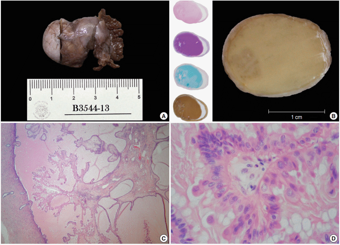

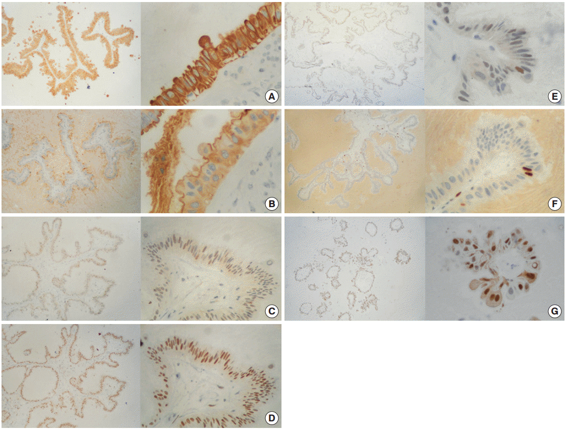
Figure & Data
References
Citations
Citations to this article as recorded by 
- Ungewöhnliche Proliferation des Eileiters
Angelina Vlaški, Vanessa Neukunft, Andrea Maria Gassel, Frederick Klauschen, Doris Mayr
Die Pathologie.2025; 46(1): 56. CrossRef - Fallopian tube papilloma
Shashank Mishra, Prerna Guleria
Indian Journal of Pathology and Microbiology.2021; 64(3): 608. CrossRef - Metaplastic papillary tumour of the fallopian tube, a rare entity, analysed by next‐generation sequencing
Sandra Sunitsch, Julia Reisinger, Luca Abete, Karl Kashofer, Peter Regitnig
Histopathology.2020; 76(6): 923. CrossRef
 PubReader
 Cite this Article

Fallopian Metaplastic Papillary Tumour: An Atypical Transdifferentiation of the Tubal Epithelium?


Fig. 1. Gross features/oxyphil metaplastic components. (A) Tubal ligation specimen. (B) Transverse cut surface. A crystallized syrup-like substance with a striking resemblance to a regional candy known as «acitrón» fills the lumen; there is also a left marginal blur with a snowflakelike appearance. The left column displays whole-mount sections stained with hematoxylin and eosin, periodic acid–Schiff, Alcian blue, and mucicarmine. (C) Panoramic photomicrograph showing a peninsular papillary framework in a lake of extracellular mucin. The stalk has a loose stroma and contains a small number of inflammatory cells (lymphocytes and neutrophils). (D) High magnification photomicrograph of the oncocytic epithelium. There is mild stratification and cell budding.
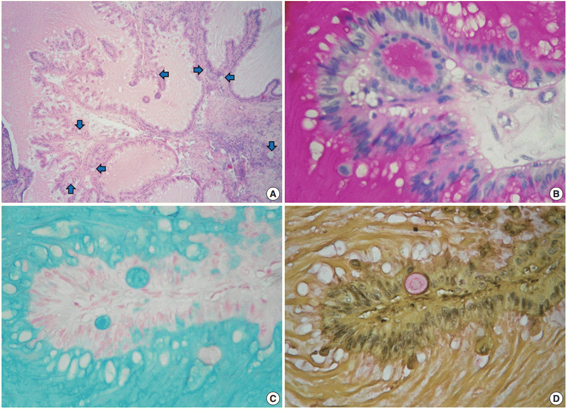
Fig. 2. Mucinous metaplastic components. (A) Some amphophilic mucin-filled cysts are noticeable beneath the epithelium (blue arrows). (B) Periodic acid−Schiff. (C) Alcian blue. (D) Mayer's mucicarmine.
Fig. 3. Immunohistochemistry panel. (A) Cytokeratin AE1/AE3 (+++/+++) in 100% of cells. (B) Epithelial membrane antigen (+++/+++) in 100% of cells. (C) Estrogen receptor (++/+++) in ~70% of cells. (D) Progesterone receptor (+++/+++) in ~95% of cells. (E) Androgen receptor (+/+++) in ~10% of cells. (F) Ki-67 (+++/+++) in ~3% of cells. (G) Cyclin-D1 (+++/+++) in ~90% of cells.
Fig. 1.
Fig. 2.
Fig. 3.
Fallopian Metaplastic Papillary Tumour: An Atypical Transdifferentiation of the Tubal Epithelium?
| Case No. | Year | Author (country) | Clinical data overview |
Histopathological finding | ||
|---|---|---|---|---|---|---|
| Landscape | Evdution and treatment | Follow-up | ||||
| 1 | 1978 | Starr et al. [2] (USA) | 26-Year-old woman (G2P1) | Normal delivery and immediate postpartum tubal ligation | Check-up every 3 months during a non-specified period | Interpretation as grade 1 primary adenocarcinoma of the Fallopian tube |
| Total abdominal hysterectomy and bilateral salpingo-oophorectomy performed due to report of malignancy | No anomalies reported | |||||
| 2-5 | 1980 | Saffos et al. [1] (USA) | Four women between 27 to 33 years of age (multiparous; non-specified number of pregnancies) | Normal delivery and tubal ligation in every case | No evidence of disease in three patients after different times of evaluation (1 year 6 months, 2 years and 6 years postoperatively) | Original description of the tumour: oncocytic and mucinous epithelia |
| Two patients had total abdominal hysterectomies with bilateral salpingo-oophorectomies because of uncertainty about the malignancy of the lesions | Mucinous component (intracytoplasmic vacuoles) reactive for mucicarmine stain | |||||
| One patient had used oral contraceptives several years previously | ||||||
| Only one mitosis noticed in one of the cases | ||||||
| One of the cases was lost during follow-up | ||||||
| 6 | 1988 | Keeney and Thrasher [3] (USA) | 27-Year-old Mexican woman (G3P1A1) | Premature rupture of foetal membranes with subsequent caesarean delivery | Without disease after 4 years of follow-up | No gross abnormalities |
| Description of a luminal acidophilic secretion mucicarmine-positive | ||||||
| Hypothyroidism requiring L-tiroxine therapy | ||||||
| Simultaneous tubal ligation | Recognition of only one tripolar mitosis | |||||
| First ultrastructural description by means of transmission electron microscopy | ||||||
| 7 | 1989 | Bartnik et al. [4] (USA) | 23-Year-old woman (G3P2) | Vaginal delivery and tubal ligation (Pomeroy technique) | Postoperative recovery and discharge | Gross description of a pale-yellow mucoid material (extracellular mucin within the tubal lumen) |
| Respiratory and urinary tract infections treated with ampicillin during the second and third trimesters | ||||||
| No mitotic activity | ||||||
| First description of the cell immunophenotype: CK (+) and EMA (+) | ||||||
| Minimal chronic salpingitis observed in both tubes | ||||||
| 8 | 1999 | Pang [5] (Taiwan) | 52-Year-old non-pregnant Taiwanese woman (G5P3A2) | Laparoscopic resection of both Fallopian tubes | Non-available information | Tumour localized in the right salpinx |
| Intracellular mucin reactive for PAS, alcian blue and mucicarmine stains | ||||||
| Consumption of progesterone-only oral contraceptives when she was 26 years old | ||||||
| Extracellular mucine present on the surface of some papillae | ||||||
| Complaint of lower abdominal fullness | ||||||
| Simultaneous finding of degenerated, partially calcified chorionic villi in both Fallopian tubes (bilateral ectopic pregnancies) | ||||||
| Bilateral hydrosalpinx by ultrasonography | ||||||
| 9 | 2003 | Solomon et al. [6] (USA) | 26-Year-old woman (G5P3A1) | Labor induction at 39 weeks using oxytocin | No complaints after her 6-week postpartum visit | Involvement of the left Fallopian tube by an exophytic, papillary lesion mucicarmine (+), EMA (+) and CK AE1/AE3 (+) |
| Last pregnancy conceived while using oral contraceptives | ||||||
| Vaginal delivery and bilateral tubal ligation via Pomeroy procedure on postpartum day 1 | ||||||
| Unexplained serum FP elevation during the 15th week of gestation | Associated chronic salpingitis and focal decidualization in the ipsilateral tube | |||||
| 10 | 2011 | DAdda et al. [7] (Italy) | 31-Year-old woman at her 40th week of gestation | Caesarean delivery due to podalic version | Non-available information | Microsatellital analysis of eight chromosomal regions involved in ovarian carcinogenesis: molecular profile probably similar to a subset of minimally altered low-grade borderline serous tumours |
Table 1. Metaplastic papillary tumor case list
CK, cytoketarin; EMA, epithelial membrane antigen; PAS, periodic acid–Schiff.

