 E-submission
Search
- Page Path
- HOME > Search
- Multicenter evaluation of the PASS score as a negative predictive tool and the impact of inter-observer variability in pheochromocytoma and paraganglioma risk stratification
- Sungyeon Jung, Hye-Ri Shin, Su-Jin Shin, Hee Young Na, Soon-Won Hong, So Yeon Park, Chan Kwon Jung, Kyeong Cheon Jung, Young Lyun Oh, Jae-Kyung Won
- J Pathol Transl Med. 2026;60(2):202-213. Published online February 23, 2026
- DOI: https://doi.org/10.4132/jptm.2025.11.05
- 2,150 View
- 127 Download
-
 Abstract
 PDF
PDF - Background
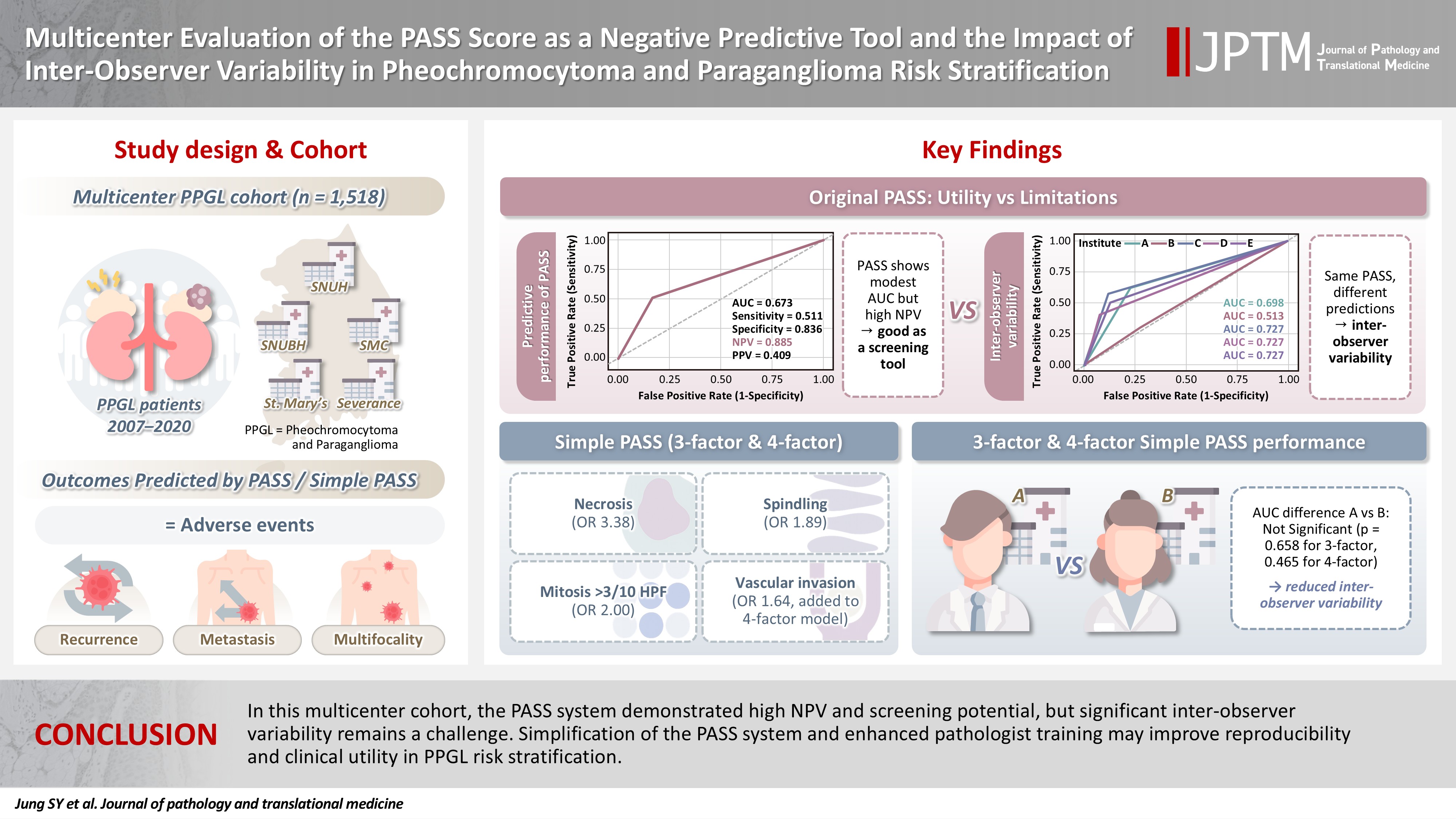
The Pheochromocytoma of the Adrenal Gland Scaled Score (PASS) is widely used for risk stratification in pheochromocytoma and paraganglioma (PPGL), but its clinical utility is limited by inter-observer variability of its parameters and inconsistent predictive performance. Methods: We conducted a multicenter retrospective study of 1,518 patients with PPGL from five tertiary referral centers in Korea. Prognostic utility of PASS system was assessed using logistic regression, Kaplan-Meier analysis, and receiver operating characteristic (ROC) curve analysis. Inter-observer variability was inferred by comparing area under the ROC curve (AUCs) across institutions. Simplified PASS systems were developed based on multivariable analysis of key histopathological parameters. Results: The PASS system was a significant predictor of adverse events and recurrence-free survival. Although the PASS system demonstrated only modest discriminative ability (AUC, 0.673), it showed a high negative predictive value (NPV, 0.885), supporting its usefulness as a screening tool for benign behavior. However, there was significant inter-institutional variability in PASS performance (AUC; range, 0.513 to 0.727; p < .05). The 3-factor Simple PASS, which incorporates necrosis, spindling, and mitotic figures, exhibited less inter-observer variation. The 4-factor Simple PASS, which adds vascular invasion to the 3-factor model, also showed reduced inter-observer variability and improved AUC and NPV compared to the original PASS system. Conclusions: In this multicenter cohort, the PASS system demonstrated high NPV and screening potential, but significant inter-observer variability remains a challenge. Simplification of the PASS system and enhanced pathologist training may improve reproducibility and clinical utility in PPGL risk stratification.
- Thyroid pathology, a clue to PTEN hamartoma tumor syndrome
- Yurimi Lee, Young Lyun Oh
- J Pathol Transl Med. 2023;57(3):178-183. Published online March 30, 2023
- DOI: https://doi.org/10.4132/jptm.2023.03.04
- 9,727 View
- 214 Download
- 10 Web of Science
- 9 Crossref
-
Abstract
PDF
- Phosphatase and tensin homolog (PTEN) hamartoma tumor syndrome (PHTS) is a hereditary disorder caused by germline inactivating mutations in the PTEN tumor suppressor gene. As a type of PHTS, Cowden syndrome is associated with abnormalities of the thyroid, breast, uterus, and gastrointestinal tract. A 52-year-old-woman visited the outpatient clinic of our endocrinology clinic with multiple thyroid nodules and Hashimoto's thyroiditis. Computed tomography imaging revealed a multinodular mass measuring up to 3.5 cm in the left thyroid lobe, causing laryngotracheal airway displacement. The total thyroidectomy specimen revealed multiple follicular adenomas and adenomatous nodules with lymphocytic thyroiditis and lipomatous metaplasia in the background. The patient was suspected of PTHS based on her thyroid pathology, family history, and numerous hamartomatous lesions of the breast, uterus, and skin. Her diagnosis was confirmed through molecular testing. This case demonstrates that pathologists must be well acquainted with thyroid pathology in PHTS.
-
Citations
Citations to this article as recorded by
- Risk of malignancy in PTEN-altered thyroid nodules detected on preoperative FNA molecular testing: a systematic review and meta-analysis
Patrizia Straccia, Vincenzo Fiorentino, Belen Padial Urtueta, Qianqian Zhang, Alessia Piermattei, Federica Cianfrini, Antonino Mule, Esther Diana Rossi
Human Pathology.2026; : 106104. CrossRef - Dual primary malignancies in Kashmir: A five-year analysis of temporal patterns, gender-specific presentations and treatment outcomes in a high gastrointestinal cancer risk population
Ubaid Jeelani, Mushood Ghulam Nabi, Asim Ahmad Dar, Gowher Ahmad Wagai, Aadil Najeed, Sheikh Owais Ahmad, Lande Sagar Janardhan, Md Mayeen Afsan Ahmad, Uzma Majeed
Journal of Family Medicine and Primary Care.2026; 15(2): 530. CrossRef - Recognizing Familial Thyroid Neoplasia: The Pathologist’s Role in Diagnosis and Management
Vania Nosé, Sule Canberk, Zubair Baloch
Advances in Anatomic Pathology.2026;[Epub] CrossRef - A clinical case of papillary thyroid cancer associated with a PTEN gene defect
R. A. Atanesyan, L. Ja. Klimov, T. M. Vdovina, G. A. Saneeva, E. I. Andreeva, I. A. Stremenkova, R. I. Arakelyan, I. K. Gasparyan
Rossiyskiy Vestnik Perinatologii i Pediatrii (Russian Bulletin of Perinatology and Pediatrics).2025; 69(6): 85. CrossRef - Pediatric cancer predisposition syndromes involving non-central nervous system solid pediatric tumors: a review on their manifestations with a focus on histopathology
B. Schurink, M. Reyes-Múgica, R. R. de Krijger
Virchows Archiv.2025; 486(1): 3. CrossRef - Dedifferentiated Leiomyosarcoma of the Uterine Corpus with Heterologous Component: Clinicopathological Analysis of Five Consecutive Cases from a Single Institution and Comprehensive Literature Review
Suyeon Kim, Hyunsik Bae, Hyun-Soo Kim
Diagnostics.2024; 14(2): 160. CrossRef - Case report: Rare oral manifestations in Cowden syndrome with PTEN mutation
Wei Yuan, Yanbin Liu, Haibin Sun, Ming Su, Lizheng Qin, Xin Huang
Frontiers in Oncology.2024;[Epub] CrossRef - Can thyroid histomorphology identify patients with PTEN hamartoma tumour syndrome?
Melad N Dababneh, Laura Rabinowitz, Gilman Plitt, Charis Eng, Christopher C Griffith
Histopathology.2024; 85(6): 929. CrossRef - A novel mutation in PTEN in anaplastic thyroid carcinoma: A case report
Yanli Zhao
Biomedical Reports.2024;[Epub] CrossRef
- Risk of malignancy in PTEN-altered thyroid nodules detected on preoperative FNA molecular testing: a systematic review and meta-analysis
- Papillary and medullary thyroid carcinomas coexisting in the same lobe, first suspected based on fine-needle aspiration cytology: a case report
- Hyun Hee Koh, Young Lyun Oh
- J Pathol Transl Med. 2022;56(5):301-308. Published online September 13, 2022
- DOI: https://doi.org/10.4132/jptm.2022.08.03
- 7,699 View
- 120 Download
- 6 Crossref
-
Abstract
PDF
- Because different types of thyroid malignancies have distinct embryological origins, coexisting tumors are rarely observed. We describe a coexisting papillary thyroid carcinoma (PTC) and medullary thyroid carcinoma (MTC) first suspected by fine-needle aspiration cytology (FNAC). A 57-year-old female presented with an irregular mass in the right thyroid lobe. The cytopathologic findings of fine-needle aspiration showed two components: a papillary-like arrangement consisting of cells with pale enlarged nuclei indicative of PTC and loose clusters comprised of oval cells with granular chromatin indicative of MTC. The diagnosis of a coexisting PTC and MTC was initially confirmed by calcitonin immunocytochemistry and later after total thyroidectomy. Although some surgical case reports of PTC and MTC coexisting in either the same or different lobes have been documented, a case suspected by FNAC before the surgery has rarely been reported. Because appropriate treatment and prognosis of PTC and MTC are different, cytopathologists should be aware of this rare entity.
-
Citations
Citations to this article as recorded by- Evaluation of Diagnostic Accuracy of Medullary Thyroid Carcinoma Using Fine‐Needle Aspiration Cytology—Based on a Single Tertiary Centre Experience
Si‐Yi Chen, Dong‐Mei Gu
Cytopathology.2026; 37(3): 255. CrossRef - Synchronous Presence of Papillary, Medullary, and Anaplastic Thyroid Tumors in a Single Patient: A Rare Case Report
Mohammed Al Essa, Reema Aldawish, Abdullah Alkhaldi, Ghaidaa Aljbli, Thamer Althunayan, Abdullah Alkarni, Abdullah Alsalamah
American Journal of Case Reports.2026;[Epub] CrossRef - Synchronous papillary and medullary thyroid carcinoma with distinct genetic mutations: A case report
Huanyu Jiang, Lijuan Zhou, Gang Zou, Haidong Zhang, Zhenkun Yu
Oral Oncology.2025; 161: 107191. CrossRef - Coexisting papillary and medullary thyroid carcinomas in a 60 year old male: a case report
Allahdad Khan, Anam Malik, Abdul Ahad Riaz, Muhammad Hussnain Sadiq, Muhammad Shahzaib Arshad, Alka Rani, Ibrahim Nagmeldin Hassan
Annals of Medicine & Surgery.2025; 87(10): 6740. CrossRef - Dedifferentiated Leiomyosarcoma of the Uterine Corpus with Heterologous Component: Clinicopathological Analysis of Five Consecutive Cases from a Single Institution and Comprehensive Literature Review
Suyeon Kim, Hyunsik Bae, Hyun-Soo Kim
Diagnostics.2024; 14(2): 160. CrossRef - Coexisting Medullary and Papillary Thyroid Carcinomas: A Case of Dual Neoplasia With a High Risk of Misdiagnosis
Santiago Sierra Castillo, Maria A Henao Rincón, David Aristizabal Colorado, David Alexander Vernaza Trujillo, Alin Abreu Lomba
Cureus.2024;[Epub] CrossRef
- Evaluation of Diagnostic Accuracy of Medullary Thyroid Carcinoma Using Fine‐Needle Aspiration Cytology—Based on a Single Tertiary Centre Experience
- Metastatic leiomyosarcoma of the thyroid gland: cytologic findings and differential diagnosis
- Jiyeon Lee, Yunjoo Cho, Kyue Hee Choi, Inwoo Hwang, Young Lyun Oh
- J Pathol Transl Med. 2021;55(5):360-365. Published online August 13, 2021
- DOI: https://doi.org/10.4132/jptm.2021.06.23
- 6,996 View
- 103 Download
- 8 Web of Science
- 6 Crossref
-
Abstract
PDF
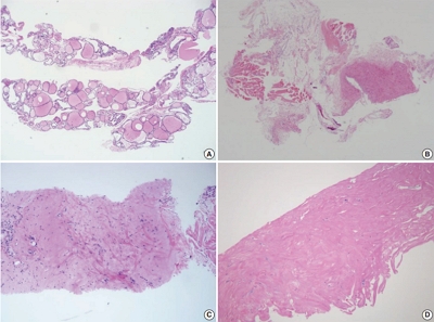
- Metastatic leiomyosarcoma to the thyroid is an extremely rare occurrence, and only 18 cases have been reported. Here, we report a case of a 37-year-old woman who presented with multiple masses on the scalp. Excisional biopsy was done and the mass revealed fascicles of smooth muscle fibers which showed positive staining for smooth muscle actin, thus confirming the diagnosis of leiomyosarcoma. The patient was also found to have a 0.9 cm mass within the left thyroid. Fine-needle aspiration was done and the cytological smear showed hypercellular spindle cell clusters with hyperchromatic and large nuclei. Normal thyroid follicular cells were found within or around tumor cells. In this report, we present the cytologic findings of metastatic leiomyosarcoma to the thyroid and offer differential diagnoses of the aspirated spindle cells.
-
Citations
Citations to this article as recorded by- Environmental Risk Factors and Thyroid Cancer: An Integrative Review and Future Directions
Muneer Nusir, Sajid Ullah Khan
International Journal of Computational Intelligence Systems.2026;[Epub] CrossRef - Cytological Features and Mimickers of Thyroid Gland Sarcomas: A Case-Based Study
Poorvi Mathur, Shipra Agarwal, Chanchal Rana
International Journal of Surgical Pathology.2025; 33(3): 711. CrossRef - A Rare Case of Metastatic Uterine Leiomyosarcoma to the Thyroid Gland
R. Sathish Kumar, H. Akshaykumar, C. Ramesan, J. Dipin
Indian Journal of Otolaryngology and Head & Neck Surgery.2024; 76(1): 1365. CrossRef - Neck Surgery for Non-Well Differentiated Thyroid Malignancies: Variations in Strategy According to Histopathology
Fernando López, Abir Al Ghuzlan, Mark Zafereo, Vincent Vander Poorten, K. Thomas Robbins, Marc Hamoir, Iain J. Nixon, Ralph P. Tufano, Gregory Randolph, Pia Pace-Asciak, Peter Angelos, Andrés Coca-Pelaz, Avi Khafif, Ohad Ronen, Juan Pablo Rodrigo, Álvaro
Cancers.2023; 15(4): 1255. CrossRef - Mesonephric-like Adenocarcinoma of the Ovary: Clinicopathological and Molecular Characteristics
Hyun Hee Koh, Eunhyang Park, Hyun-Soo Kim
Diagnostics.2022; 12(2): 326. CrossRef - Alveolar Soft Part Sarcoma of the Uterus: Clinicopathological and Molecular Characteristics
Yurimi Lee, Kiyong Na, Ha Young Woo, Hyun-Soo Kim
Diagnostics.2022; 12(5): 1102. CrossRef
- Environmental Risk Factors and Thyroid Cancer: An Integrative Review and Future Directions
- 2019 Practice guidelines for thyroid core needle biopsy: a report of the Clinical Practice Guidelines Development Committee of the Korean Thyroid Association
- Chan Kwon Jung, Jung Hwan Baek, Dong Gyu Na, Young Lyun Oh, Ka Hee Yi, Ho-Cheol Kang
- J Pathol Transl Med. 2020;54(1):64-86. Published online January 15, 2020
- DOI: https://doi.org/10.4132/jptm.2019.12.04
- 30,057 View
- 1,099 Download
- 57 Web of Science
- 63 Crossref
-
Abstract
PDF
- Ultrasound-guided core needle biopsy (CNB) has been increasingly used for the pre-operative diagnosis of thyroid nodules. Since the Korean Society of the Thyroid Radiology published the ‘Consensus Statement and Recommendations for Thyroid CNB’ in 2017 and the Korean Endocrine Pathology Thyroid CNB Study Group published ‘Pathology Reporting of Thyroid Core Needle Biopsy’ in 2015, advances have occurred rapidly not only in the management guidelines for thyroid nodules but also in the diagnostic terminology and classification schemes. The Clinical Practice Guidelines Development Committee of the Korean Thyroid Association (KTA) reviewed publications on thyroid CNB from 1995 to September 2019 and updated the recommendations and statements for the diagnosis and management of thyroid nodules using CNB. Recommendations for the resolution of clinical controversies regarding the use of CNB were based on expert opinion. These practical guidelines include recommendations and statements regarding indications for CNB, patient preparation, CNB technique, biopsy-related complications, biopsy specimen preparation and processing, and pathology interpretation and reporting of thyroid CNB.
-
Citations
Citations to this article as recorded by- Reevaluation of malignancy risk in nondiagnostic thyroid nodules with long‐term follow‐up via surgical resection or core needle biopsy: A retrospective study
Ji‐Seon Jeong, Young Jun Choi, Jeong Hyun Lee, Jung Hwan Baek, Yu‐Mi Lee, Tae‐Yon Sung, Dong Eun Song
Cancer Cytopathology.2026;[Epub] CrossRef - Clinical observation of thyroid metastatic lesion in a patient withrectal cancer
K. M. Blikyan, S. V. Lukyanov, A. B. Alnikin, N. S. Lukyanov, P. V. Konovalenko
Endocrine Surgery.2026; 19(3): 40. CrossRef - Current Evidence, Selective Indications, and the Role of Lymph-Node Assessment in Intraoperative Frozen Section in Thyroid Cancer Surgery: A Literature Review
Gregorio Scerrino, Marco Marciano', Bianca Vicari, Maria Aurora Bullaro, Renato Di Vuolo, Pierina Richiusa, Giuseppina Orlando, Vito Rodolico, Giuseppina Melfa
Journal of Clinical Medicine.2026; 15(4): 1611. CrossRef - Core needle biopsy as a first-line diagnostic tool for selected thyroid nodules: a real-world evaluation of diagnostic performance and safety
Xing Li, Yi Pan, Yanmei Ou, Xin Gao, Yue Gao, Luwei Liu, Yinze Li, Yong Xu, Wengui Xu
Frontiers in Oncology.2026;[Epub] CrossRef - Evaluation and Management of Thyroid Nodules: A Joint Consensus Statement From the British Thyroid Association (BTA), British Association of Endocrine and Thyroid Surgeons (BAETS) and Collaborating Bodies
Ram Moorthy, Saba P. Balasubramanian, Kate Farnell, Mairead Kelly, Gitta Madani, Mufaddal Moonim, Carla Moran, Julia Priestley, Michael Stechman, Emma Watts, Kristien Boelaert
Clinical Endocrinology.2026; 104(6): 682. CrossRef - Usefulness of core needle biopsy of thyroid for the diagnosis of IgG4 Hashimoto's thyroiditis
Chenxu Zhao, Yang Yu, Jumei Liu, Yang Zhang, Lei Chen, Guizhi Lu, Ying Gao
Journal of Translational Internal Medicine.2026; 14(2): 306. CrossRef - Comparison of ultrasound-guided biopsy techniques for level IV lymph nodes: semiautomatic vs. Menghini modified needles in a retrospective dual-center study
Gang Liu, Yixin Zhu, Guoru Wu, Guangyin Yu, Hao Luo, Lu Pang, Qiongxian Long, Lin Zhu, Yu Shi
BMC Medical Imaging.2026;[Epub] CrossRef - Clinical Value of Core Needle Biopsy as a Second-Line Approach After Non-Conclusive Fine-Needle Aspiration in Thyroid Nodules: A Paired Analysis
Vladan Markovic, Slobodanka Mitrovic, Tijana Maksic, Irfan Corovic, Marija Sekulic, Mladen Maksic, Vesna Grbovic
Diagnostics.2026; 16(7): 1104. CrossRef - Advantages of thyroid core needle biopsy: an emerging selective first-line biopsy modality
Jae Ho Shin, Yeseul Kim, Min Kyoung Lee, Jung Hwan Baek, So Lyung Jung
Ultrasonography.2026; 45(3): 205. CrossRef - Preoperative hydrodissection for predicting extrathyroidal extension in thyroid tumors
Yeseul Kim, Jae Ho Shin, Sung-Hye You, Bo Kyu Kim, Byungjun Kim, Kyeong Jin Kim
Minimally Invasive Therapy & Allied Technologies.2026; : 1. CrossRef - Clinicopathological profile of high-grade differentiated thyroid carcinoma in an Indonesian tertiary hospital
Novita, Agnes Stephanie Harahap, Maria Francisca Ham, Alfianto Widiono, Chan Kwon Jung
Journal of Pathology and Translational Medicine.2026; 60(3): 338. CrossRef - Comparison of core-needle biopsy and repeat fine-needle aspiration biopsy for thyroid nodules with initially inconclusive findings: a systematic review, diagnostic accuracy meta-analysis, and meta-regression
Hendra Zufry, Timotius Ivan Hariyanto
Journal of the American Society of Cytopathology.2025; 14(3): 159. CrossRef - Ultrasound-guided core-needle biopsy for diagnosis of thyroid cancer
D.D. Dolidze, S.D. Kovantsev, Z.A. Bagatelia, A.V. Bumbu, Yu.V. Barinov, G.M. Chechenin, N.V. Pichugina, D.G. Gogolashvili
Pirogov Russian Journal of Surgery.2025; (3): 87. CrossRef - Superior Diagnostic Yield of Core Needle Biopsy Over Fine Needle Aspiration in Diagnosing Follicular-Patterned Neoplasms: A Multicenter Study Focusing on Bethesda IV Results
Leehi Joo, Jung Hwan Baek, Jungbok Lee, Dong Eun Song, Sae Rom Chung, Young Jun Choi, Jeong Hyun Lee
Korean Journal of Radiology.2025; 26(6): 604. CrossRef - Diagnostic yield of fine needle aspiration with simultaneous core needle biopsy for thyroid nodules
Mohammad Ali Hasannia, Ramin Pourghorban, Hoda Asefi, Amir Aria, Elham Nazar, Hojat Ebrahiminik, Alireza Mohamadian
Journal of Pathology and Translational Medicine.2025; 59(3): 180. CrossRef - Lessons learned from the first 2 years of experience with thyroid core needle biopsy at an Indonesian national referral hospital
Agnes Stephanie Harahap, Maria Francisca Ham, Retno Asti Werdhani, Erwin Danil Julian, Rafi Ilmansyah, Chloe Indira Arfelita Mangunkusumso, Tri Juli Edi Tarigan
Journal of Pathology and Translational Medicine.2025; 59(3): 149. CrossRef - Preoperative Fine-Needle Aspiration in Goiter With Compressive Symptoms: A Systematic Review and Meta-analysis
Moeen Sbeit, Rania Faris, Ohad Ronen
Endocrine Practice.2025; 31(8): 1038. CrossRef - Risk Stratification of Thyroid Nodules Diagnosed as Follicular Neoplasm on Core Needle Biopsy
Byeong-Joo Noh, Won Jun Kim, Jin Yub Kim, Ha Young Kim, Jong Cheol Lee, Myoung Sook Shim, Yong Jin Song, Kwang Hyun Yoon, In-Hye Jung, Hyo Sang Lee, Wooyul Paik, Dong Gyu Na
Endocrinology and Metabolism.2025; 40(4): 610. CrossRef - Diagnostic performances of five US risk stratification systems for malignancy in focal [18F]FDG-PET/CT thyroid incidentalomas
Chae Young Shin, Hye Shin Ahn, Dong Gyu Na, Hyo Sang Lee, Eon-Woo Shin
European Radiology.2025; 35(12): 7701. CrossRef - Malignancy risk in AUS thyroid lesions: comparison between FNA and CNB with implications for NIFTP diagnosis
Yeseul Kim, Jae Ho Shin, You-Na Sung, Dawon Park, Harim Oh, Hyo Seon Ryu, Kyeong Jin Kim, Hyun Joo Kim, Sin Gon Kim, Hoon Yub Kim, Kwang Yoon Jung, Seung-Kuk Baek, Sangjeong Ahn
Frontiers in Endocrinology.2025;[Epub] CrossRef - Comparison of Diagnostic Yield Between Fine Needle Aspiration Cytology and Core Needle Biopsy in the Diagnosis of Thyroid Nodule
Yeongrok Lee, Myung Jin Ban, Do Hyeon Kim, Jin-Young Kim, Hyung Kwon Byeon, Jae Hong Park
Diagnostics.2025; 15(20): 2566. CrossRef - Repeatedly non-diagnostic thyroid nodules: the experience of two thyroid clinics
Filippo EGALINI, Mattia ROSSI, Chiara MELE, Yanina LIZET CASTILLO, Francesca MALETTA, Barbara PULIGHEDDU, Ezio GHIGO, Ruth ROSSETTO GIACCHERINO, Loredana PAGANO, Mauro PAPOTTI
Minerva Endocrinology.2025;[Epub] CrossRef - A comparative analysis of core needle biopsy and repeat fine needle aspiration in patients with inconclusive initial cytology of thyroid nodules
Xuejiao Su, Can Yue, Wanting Yang, Buyun Ma
Frontiers in Endocrinology.2024;[Epub] CrossRef - A Narrative Review of the 2023 Korean Thyroid Association Management Guideline for Patients with Thyroid Nodules
Eun Kyung Lee, Young Joo Park, Chan Kwon Jung, Dong Gyu Na
Endocrinology and Metabolism.2024; 39(1): 61. CrossRef - Doing more with less: integrating small biopsies in cytology practice
Anjali Saqi, Michiya Nishino, Mauro Saieg, Amy Ly, Abberly Lott Limbach
Journal of the American Society of Cytopathology.2024; 13(4): 233. CrossRef - 2023 Update of the Korean Thyroid Association Guidelines for the Management of Thyroid Nodules
Eun Kyung Lee, Young Joo Park
Clinical Thyroidology®.2024; 36(4): 153. CrossRef - Korean Thyroid Association Guidelines on the Management of Differentiated Thyroid Cancers; Part I. Initial Management of Differentiated Thyroid Cancers - Chapter 2. Surgical Management of Thyroid Cancer 2024
Yoon Young Cho, Cho Rok Lee, Ho-Cheol Kang, Bon Seok Koo, Hyungju Kwon, Sun Wook Kim, Won Woong Kim, Jung-Han Kim, Dong Gyu Na, Young Joo Park, Kyorim Back, Young Shin Song, Seung Hoon Woo, Ho-Ryun Won, Chang Hwan Ryu, Jee Hee Yoon, Min Kyoung Lee, Eun Ky
International Journal of Thyroidology.2024; 17(1): 30. CrossRef - Korean Thyroid Association Management Guidelines for Patients with Thyroid Nodules 2024
Young Joo Park, Eun Kyung Lee, Young Shin Song, Su Hwan Kang, Bon Seok Koo, Sun Wook Kim, Dong Gyu Na, Seung-Kuk Baek, So Won Oh, Min Kyoung Lee, Sang-Woo Lee, Young Ah Lee, Yong Sang Lee, Ji Ye Lee, Dong-Jun Lim, Leehi Joo, Yuh-Seog Jung, Chan Kwon Jung,
International Journal of Thyroidology.2024; 17(1): 208. CrossRef - Korean Thyroid Association Guidelines on the Management of Differentiated Thyroid Cancers; Overview and Summary 2024
Young Joo Park, Eun Kyung Lee, Young Shin Song, Bon Seok Koo, Hyungju Kwon, Keunyoung Kim, Mijin Kim, Bo Hyun Kim, Won Gu Kim, Won Bae Kim, Won Woong Kim, Jung-Han Kim, Hee Kyung Kim, Hee Young Na, Shin Je Moon, Jung-Eun Moon, Sohyun Park, Jun-Ook Park, J
International Journal of Thyroidology.2024; 17(1): 1. CrossRef - Educational exchange in thyroid core needle biopsy diagnosis: enhancing pathological interpretation through guideline integration and peer learning
Agnes Stephanie Harahap, Chan Kwon Jung
Journal of Pathology and Translational Medicine.2024; 58(5): 205. CrossRef - Current role of interventional radiology in thyroid nodules
Onur Taydas, Erbil Arik, Omer Faruk Sevinc, Ahmet Burak Kara, Mustafa Ozdemir, Hasret Cengiz, Zulfu Bayhan, Mehmet Halil Ozturk
Frontiers in Endocrinology.2024;[Epub] CrossRef - Neck Schwannoma Masking as Thyroid Tumour: Into the Deep of Diagnostics and Anatomy
Serghei Covantsev, Anna Bumbu, Anna Sukhotko, Evghenii Zakurdaev, Ivan Kuts, Andrey Evsikov
Diagnostics.2024; 14(20): 2332. CrossRef - Thermal ablation for Bethesda III and IV thyroid nodules: current diagnosis and management
Wen-Hui Chan, Pi-Ling Chiang, An-Ni Lin, Yen-Hsiang Chang, Wei-Che Lin
Ultrasonography.2024; 43(6): 395. CrossRef - A new LNC89/LNC60-Col11a2 axis revealed by whole-transcriptome analysis may be associated with goiters related to excess iodine nutrition
Guanying Nie, Shuang Li, Wei Zhang, Fangang Meng, Zixuan Ru, Jiahui Li, Dianjun Sun, Ming Li
Frontiers in Endocrinology.2024;[Epub] CrossRef - A simplified four-tier classification for thyroid core needle biopsy
M. Paja, J. L. Del Cura, R. Zabala, I. Korta, Mª T. Gutiérrez, A. Expósito, A. Ugalde
Journal of Endocrinological Investigation.2024; 48(4): 895. CrossRef - Risk of thyroid cancer in a lung cancer screening population of the National Lung Screening Trial according to the presence of incidental thyroid nodules detected on low-dose chest CT
Hyobin Seo, Kwang Nam Jin, Ji Sang Park, Koung Mi Kang, Eun Kyung Lee, Ji Ye Lee, Roh-Eul Yoo, Young Joo Park, Ji-hoon Kim
Ultrasonography.2023; 42(2): 275. CrossRef - Preoperative Risk Stratification of Follicular-patterned Thyroid Lesions on Core Needle Biopsy by Histologic Subtyping and RAS Variant-specific Immunohistochemistry
Meejeong Kim, Sora Jeon, Chan Kwon Jung
Endocrine Pathology.2023; 34(2): 247. CrossRef - Differential regional importance mapping for thyroid nodule malignancy prediction with potential to improve needle aspiration biopsy sampling reliability
Liping Wang, Yuan Wang, Wenliang Lu, Dong Xu, Jincao Yao, Lijing Wang, Lei Xu
Frontiers in Oncology.2023;[Epub] CrossRef - Preoperative evaluation of thyroid nodules – Diagnosis and management strategies
Tapoi Dana Antonia, Lambrescu Ioana Maria, Gheorghisan-Galateanu Ancuta-Augustina
Pathology - Research and Practice.2023; 246: 154516. CrossRef - 2023 Korean Thyroid Association Management Guidelines for Patients with Thyroid Nodules
Young Joo Park, Eun Kyung Lee, Young Shin Song, Soo Hwan Kang, Bon Seok Koo, Sun Wook Kim, Dong Gyu Na, Seung-Kuk Baek, So Won Oh, Min Kyoung Lee, Sang-Woo Lee, Young Ah Lee, Yong Sang Lee, Ji Ye Lee, Dong-Jun Lim, Leehi Joo, Yuh-Seog Jung, Chan Kwon Jung
International Journal of Thyroidology.2023; 16(1): 1. CrossRef - Fast Track Management of Primary Thyroid Lymphoma in the Very Elderly Patient
Pierre Yves Marcy, Frederic Bauduer, Juliette Thariat, Olivier Gisserot, Edouard Ghanassia, Bruno Chetaille, Laurys Boudin, Jean Baptiste Morvan
Current Oncology.2023; 30(6): 5816. CrossRef - Reevaluating diagnostic categories and associated malignancy risks in thyroid core needle biopsy
Chan Kwon Jung
Journal of Pathology and Translational Medicine.2023; 57(4): 208. CrossRef - Diagnostic performance of shear wave elastography in thyroid nodules with indeterminate cytology: A systematic review and meta-analysis
Yuxuan Qiu, Zhichao Xing, Qianru Yang, Yan Luo, Buyun Ma
Heliyon.2023; 9(10): e20654. CrossRef - Comparison of the diagnostic value of fine needle aspiration and ultrasound in thyroid pathology
P. S. Glushkov, R. Kh. Azimov, N. L. Aleshenko, E. A. Maruchak, Y. P. Sych, G. N. Minkova, K. A. Shemyatovsky, V. A. Gorsky
Endocrine Surgery.2023; 17(3): 43. CrossRef - Comparison of Core Needle Biopsy and Repeat Fine-Needle Aspiration in Avoiding Diagnostic Surgery for Thyroid Nodules Initially Diagnosed as Atypia/Follicular Lesion of Undetermined Significance
Leehi Joo, Dong Gyu Na, Ji-hoon Kim, Hyobin Seo
Korean Journal of Radiology.2022; 23(2): 280. CrossRef - Diagnostic efficacy, performance and safety of side-cut core needle biopsy for thyroid nodules: comparison of automated and semi-automated biopsy needles
Ji Yeon Park, Seong Yoon Yi, Soo Heui Baek, Yu Hyun Lee, Heon-Ju Kwon, Hee Jin Park
Endocrine.2022; 76(2): 341. CrossRef - Thyroid Cancer Diagnostics Related to Occupational and Environmental Risk Factors: An Integrated Risk Assessment Approach
Gabriela Maria Berinde, Andreea Iulia Socaciu, Mihai Adrian Socaciu, Andreea Cozma, Armand Gabriel Rajnoveanu, Gabriel Emil Petre, Doina Piciu
Diagnostics.2022; 12(2): 318. CrossRef - Approach to Bethesda system category III thyroid nodules according to US-risk stratification
Jieun Kim, Jung Hee Shin, Young Lyun Oh, Soo Yeon Hahn, Ko Woon Park
Endocrine Journal.2022; 69(1): 67. CrossRef - Clinicopathological and Molecular Features of Secondary Cancer (Metastasis) to the Thyroid and Advances in Management
Marie Nguyen, George He, Alfred King-Yin Lam
International Journal of Molecular Sciences.2022; 23(6): 3242. CrossRef - Diagnostic Performance of Thyroid Core Needle Biopsy Using the Revised Reporting System: Comparison with Fine Needle Aspiration Cytology
Kwangsoon Kim, Ja Seong Bae, Jeong Soo Kim, So Lyung Jung, Chan Kwon Jung
Endocrinology and Metabolism.2022; 37(1): 159. CrossRef - Core Needle Biopsy Can Early and Precisely Identify Large Thyroid Masses
Antonio Matrone, Luigi De Napoli, Liborio Torregrossa, Aleksandr Aghababyan, Piermarco Papini, Carlo Enrico Ambrosini, Rosa Cervelli, Clara Ugolini, Fulvio Basolo, Eleonora Molinaro, Rossella Elisei, Gabriele Materazzi
Frontiers in Oncology.2022;[Epub] CrossRef - Primary thyroid leiomyosarcoma with transvenous extension to the right atrium: a case report
Juraj Dubrava, Peter Martanovic, Marina Pavlovicova, Pavel Babal, Akhil Narang, Maria Mattioli, Nidhish Tiwari, Zhiyu Liu, Mariame Chakir
European Heart Journal - Case Reports.2022;[Epub] CrossRef - Radiofrequency ablation for management of thyroid nodules in quarantine zone of COVID-19 pandemic setting in Indonesia
Kristanto Yuli Yarso, Sumadi Lukman Anwar
Annals of Medicine and Surgery.2022; 81: 104132. CrossRef - A Matched-Pair Analysis of Nuclear Morphologic Features Between Core Needle Biopsy and Surgical Specimen in Thyroid Tumors Using a Deep Learning Model
Faridul Haq, Andrey Bychkov, Chan Kwon Jung
Endocrine Pathology.2022; 33(4): 472. CrossRef - Diagnostic performance of core needle biopsy as a first‐line diagnostic tool for thyroid nodules according to ultrasound patterns: Comparison with fine needle aspiration using propensity score matching analysis
Hye Shin Ahn, Inyoung Youn, Dong Gyu Na, Soo Jin Kim, Mi Yeon Lee
Clinical Endocrinology.2021; 94(3): 494. CrossRef - Hydrodissection: A Novel Approach for Safe Core Needle Biopsy of Small High-Risk Subcapsular Thyroid Nodules
Hojat Ebrahiminik, Hossein Chegeni, Javad Jalili, Rambod Salouti, Hadi Rokni, Afshin Mohammadi, Ali Mosaddegh Khah, Seyed Mohammad Tavangar, Zahra Ebrahiminik
CardioVascular and Interventional Radiology.2021; 44(10): 1651. CrossRef - Application of biomarkers in the diagnosis of uncertain samples of core needle biopsy of thyroid nodules
Yan Xiong, Xin Li, Li Liang, Dong Li, Limin Yan, Xueying Li, Jiting Di, Ting Li
Virchows Archiv.2021; 479(5): 961. CrossRef - VE1 immunohistochemistry is an adjunct tool for detection of BRAFV600E mutation: Validation in thyroid cancer patients
Faiza A. Rashid, Sobia Tabassum, Mosin S. Khan, Hifzur R. Ansari, Muhammad Asif, Ahmareen K. Sheikh, Syed Sameer Aga
Journal of Clinical Laboratory Analysis.2021;[Epub] CrossRef - The Diagnostic Value of the American College of Radiology Thyroid Imaging Reporting and Data System Classification and Shear-Wave Elastography for the Differentiation of Thyroid Nodules
Gül Bora Makal, Aydın Aslan
Ultrasound in Medicine & Biology.2021; 47(5): 1227. CrossRef - Comparison of the diagnostic performance of the modified Korean Thyroid Imaging Reporting and Data System for thyroid malignancy with three international guidelines
Eun Ju Ha, Jung Hee Shin, Dong Gyu Na, So Lyung Jung, Young Hen Lee, Wooyul Paik, Min Ji Hong, Yeo Koon Kim, Chang Yoon Lee
Ultrasonography.2021; 40(4): 594. CrossRef - VE1 Immunohistochemistry Improves the Limit of Genotyping for Detecting BRAFV600E Mutation in Papillary Thyroid Cancer
Sonam Choden, Somboon Keelawat, Chan Kwon Jung, Andrey Bychkov
Cancers.2020; 12(3): 596. CrossRef - The 2019 core-needle biopsy practice guidelines
So Yeong Jeong, Jung Hwan Baek
Ultrasonography.2020; 39(3): 311. CrossRef - Re: The 2019 core-needle biopsy practice guidelines
Ji-hoon Kim
Ultrasonography.2020; 39(3): 313. CrossRef
- Reevaluation of malignancy risk in nondiagnostic thyroid nodules with long‐term follow‐up via surgical resection or core needle biopsy: A retrospective study
- Do Helper T Cell Subtypes in Lymphocytic Thyroiditis Play a Role in the Antitumor Effect?
- Seok Woo Yang, Seong-Ho Kang, Kyung Rae Kim, In Hong Choi, Hang Seok Chang, Young Lyun Oh, Soon Won Hong
- J Pathol Transl Med. 2016;50(5):377-384. Published online September 15, 2016
- DOI: https://doi.org/10.4132/jptm.2016.07.25
- 11,058 View
- 108 Download
- 3 Web of Science
- 4 Crossref
-
Abstract
PDF
- Background
Papillary thyroid carcinoma (PTC) is frequently accompanied by lymphocytic thyroiditis (LT). Some reports claim that Hashimoto’s thyroiditis (the clinical form of LT) enhances the likelihood of PTC; however, others suggest that LT has antitumor activity. This study was aimed to find out the relationship between the patterns of helper T cell (Th) cytokines in thyroid tissue of PTC with or without LT and the clinicopathological manifestation of PTC.
Methods
Fresh surgical samples of PTC with (13 cases) or without (10 cases) LT were used. The prognostic parameters (tumor size, extra-thyroidal extension of PTC, and lymph node metastasis) were analyzed. The mRNA levels of two subtypes of Th cytokines, Th1 (tumor necrosis factor α [TNF-α], interferon γ [IFN-γ ], and interleukin [IL] 2) and Th2 (IL-4 and IL-10), were analyzed. Because most PTC cases were microcarcinomas and recent cases without clinical follow-up, negative or faint p27 immunoreactivity was used as a surrogate marker for lymph node metastasis.
Results
PTC with LT cases showed significantly higher expression of TNF-α (p = .043), IFN-γ (p < .010), IL-4 (p = .015) than those without LT cases. Although the data were not statistically significant, all analyzed cytokines (except for IL-4) were highly expressed in the cases with higher expression of p27 surrogate marker.
Conclusions
These results indicate that mixed Th1 (TNF-α, IFN-γ , and IL-2) and Th2 (IL-10) immunity might play a role in the antitumor effect in terms of lymph node metastasis. -
Citations
Citations to this article as recorded by- Papillary thyroid carcinoma with Hashimoto’s thyroiditis: impact and correlation
Shengpeng Yao, Hong Zhang
Frontiers in Endocrinology.2025;[Epub] CrossRef - Obesity and Thyroid Cancer Risk: An Update
Fabiana Franchini, Giuseppe Palatucci, Annamaria Colao, Paola Ungaro, Paolo Emidio Macchia, Immacolata Cristina Nettore
International Journal of Environmental Research and Public Health.2022; 19(3): 1116. CrossRef - Association between Hashimoto thyroiditis and clinical outcomes of papillary thyroid carcinoma: A meta-analysis
Qizhi Tang, Weiyu Pan, Liangyue Peng, Francis Moore
PLOS ONE.2022; 17(6): e0269995. CrossRef - The Heat Shock Protein Story—From Taking mTORC1,2 and Heat Shock Protein Inhibitors as Therapeutic Measures for Treating Cancers to Development of Cancer Vaccines
Peter Chin Wan Fung, Regina Kit Chee Kong
Journal of Cancer Therapy.2017; 08(11): 962. CrossRef
- Papillary thyroid carcinoma with Hashimoto’s thyroiditis: impact and correlation
- Comparison of Three
BRAF Mutation Tests in Formalin-Fixed Paraffin Embedded Clinical Samples - Soomin Ahn, Jeeyun Lee, Ji-Youn Sung, So Young Kang, Sang Yun Ha, Kee-Taek Jang, Yoon-La Choi, Jung-Sun Kim, Young Lyun Oh, Kyoung-Mee Kim
- Korean J Pathol. 2013;47(4):348-354. Published online August 26, 2013
- DOI: https://doi.org/10.4132/KoreanJPathol.2013.47.4.348
- 10,815 View
- 60 Download
- 9 Crossref
-
Abstract
PDF
Background Recently,
BRAF inhibitors showed dramatic treatment outcomes inBRAF V600 mutant melanoma. Therefore, the accuracy ofBRAF mutation test is critical.Methods BRAF mutations were tested by dual-priming oligonucleotide-polymerase chain reaction (DPO-PCR), direct sequencing and subsequently retested with a real-time PCR assay, cobas 4800 V600 mutation test. In total, 64 tumors including 34 malignant melanomas and 16 papillary thyroid carcinomas were analyzed. DNA was extracted from formalin-fixed paraffin embedded tissue samples and the results of cobas test were directly compared with those of DPO-PCR and direct sequencing.Results BRAF mutations were found in 23 of 64 (35.9%) tumors. There was 9.4% discordance among 3 methods. Out of 6 discordant cases, 4 cases were melanomas; 3 cases wereBRAF V600E detected only by cobas test, but were not detected by DPO-PCR and direct sequencing. One melanoma patient withBRAF mutation detected only by cobas test has been on vemurafenib treatment for 6 months and showed a dramatic response to vemurafenib. DPO-PCR failed to detect V600K mutation in one case identified by both direct sequencing and cobas test.Conclusions In direct comparison of the currently available DPO-PCR, direct sequencing and real-time cobas test for
BRAF mutation, real-time PCR assay is the most sensitive method.-
Citations
Citations to this article as recorded by- Preoperative BRAFV600E mutation detection in thyroid carcinoma by immunocytochemistry
Kristine Zøylner Swan, Stine Horskær Madsen, Steen Joop Bonnema, Viveque Egsgaard Nielsen, Marie Louise Jespersen
APMIS.2022; 130(11): 627. CrossRef - Strategy to reduce unnecessary surgeries in thyroid nodules with cytology of Bethesda category III (AUS/FLUS): a retrospective analysis of 667 patients diagnosed by surgery
Yong Joon Suh, Yeon Ju Choi
Endocrine.2020; 69(3): 578. CrossRef - A new primer construction technique that effectively increases amplification of rare mutant templates in samples
Jr-Kai Huang, Ling Fan, Tao-Yeuan Wang, Pao-Shu Wu
BMC Biotechnology.2019;[Epub] CrossRef - BRAF and NRAS mutations and antitumor immunity in Korean malignant melanomas and their prognostic relevance: Gene set enrichment analysis and CIBERSORT analysis
Kyueng-Whan Min, Ji-Young Choe, Mi Jung Kwon, Hye Kyung Lee, Ho Suk Kang, Eun Sook Nam, Seong Jin Cho, Hye-Rim Park, Soo Kee Min, Jinwon Seo, Yun Joong Kim, Nan Young Kim, Ho Young Kim
Pathology - Research and Practice.2019; 215(12): 152671. CrossRef - The association between dermoscopic features and BRAF mutational status in cutaneous melanoma: Significance of the blue-white veil
Miquel Armengot-Carbó, Eduardo Nagore, Zaida García-Casado, Rafael Botella-Estrada
Journal of the American Academy of Dermatology.2018; 78(5): 920. CrossRef - Comparison of Five Different Assays for the Detection of BRAF Mutations in Formalin-Fixed Paraffin Embedded Tissues of Patients with Metastatic Melanoma
Claire Franczak, Julia Salleron, Cindy Dubois, Pierre Filhine-Trésarrieu, Agnès Leroux, Jean-Louis Merlin, Alexandre Harlé
Molecular Diagnosis & Therapy.2017; 21(2): 209. CrossRef - Validation of an NGS mutation detection panel for melanoma
Anne Reiman, Hugh Kikuchi, Daniela Scocchia, Peter Smith, Yee Wah Tsang, David Snead, Ian A Cree
BMC Cancer.2017;[Epub] CrossRef - Transformation to Small Cell Lung Cancer of Pulmonary Adenocarcinoma: Clinicopathologic Analysis of Six Cases
Soomin Ahn, Soo Hyun Hwang, Joungho Han, Yoon-La Choi, Se-Hoon Lee, Jin Seok Ahn, Keunchil Park, Myung-Ju Ahn, Woong-Yang Park
Journal of Pathology and Translational Medicine.2016; 50(4): 258. CrossRef - Immunohistochemistry with the anti-BRAF V600E (VE1) antibody: impact of pre-analytical conditions and concordance with DNA sequencing in colorectal and papillary thyroid carcinoma
Katerina Dvorak, Birte Aggeler, John Palting, Penny McKelvie, Andrew Ruszkiewicz, Paul Waring
Pathology.2014; 46(6): 509. CrossRef
- Preoperative BRAFV600E mutation detection in thyroid carcinoma by immunocytochemistry
- Prognostic Significance of Methylation Profiles in Urothelial Carcinomas of the Bladder.
- Hee Jung Park, Eui Jin Lee, Sang Yun Ha, Ghee Young Kwon, Young Lyun Oh, Kyoung Mee Kim, Dae Shick Kim, Seongil Seo, Hyun Moo Lee, Han Yong Choi
- Korean J Pathol. 2010;44(6):623-630.
- DOI: https://doi.org/10.4132/KoreanJPathol.2010.44.6.623
- 4,600 View
- 22 Download
- 1 Crossref
-
Abstract
PDF
- BACKGROUND
Study on epigenetics of urothelial carcinomas has expanded and allowed better understanding of their correlation with clinicopathologic features. The aim of this study was to determine reliable predictive epigenetic markers for patients with urothelial carcinoma of urinary bladder.
METHODS
In 64 urothelial carcinomas of the urinary bladder, methylationspecific polymerase chain reaction with RAS association domain family 1A (RASSF1A), adenomatous polyposis coli (APC), death-associated protein-kinase (DAPK), runt-related transcription factor 3 (RUNX3), p14, p16 and MGMT was performed and correlated the results with p53 mutations, DNA ploidy, clinicopathologic parameters and recurrences.
RESULTS
Hypermethyation of RASSF1A, APC, DAPK, RUNX3, p14, p16 and MGMT promoters was observed in 35 (54.7%), 29 (45.3%), 18 (28.1%), 18 (28.1%), 9 (14.1%), 2 (3.1%), and 6 (9.4%) cases, respectively. Hypermethylation of RUNX3 and APC was significantly associated with high histologic grades and aneuploidy. Methylation of DAPK was significantly associated with muscle invasion. Methylation of DAPK and RUNX3 genes was significantly associated with recurrence. In survival analyses, methylation of RUNX3 gene and methylation-high (methylation at two or more loci) phenotype was significantly associated with poor recurrence-free survival.
CONCLUSIONS
Methylation of RUNX3 gene and methylation-high phenotype are significant indicator of recurrence. -
Citations
Citations to this article as recorded by- DAPK Promoter Methylation and Bladder Cancer Risk: A Systematic Review and Meta-Analysis
Lihe Dai, Chong Ma, Zhensheng Zhang, Shuxiong Zeng, Anwei Liu, Shijie Tang, Qian Ren, Yinghao Sun, Chuanliang Xu, Shengtao Zhou
PLOS ONE.2016; 11(12): e0167228. CrossRef
- DAPK Promoter Methylation and Bladder Cancer Risk: A Systematic Review and Meta-Analysis
- The Frequency of BRAF Mutation in Very Small Papillary Thyroid Carcinomas.
- Taeeun Kim, Ji Hyun Roh, Hee Jung Park, Jee Eun Kwon, So Young Kang, Yoon La Choi, Young Lyun Oh
- Korean J Pathol. 2010;44(3):308-314.
- DOI: https://doi.org/10.4132/KoreanJPathol.2010.44.3.308
- 5,828 View
- 21 Download
- 3 Crossref
-
Abstract
PDF
- BACKGROUND
Papillary thyroid carcinoma (PTC) is the most common malignant tumor of the thyroid and BRAF (V600E) is the most frequent genetic alteration in PTCs. The aim of this study was to investigate the frequency of BRAF mutation, especially in very small PTCs.
METHODS
We analyzed the presence of the BRAF mutation in PTCs in subgroups defined by tumor size (0.5 cm intervals).
RESULTS
Of 140 patients, 85 (60.7%) showed a BRAF mutation. The frequency of BRAF mutation in the subgroup was: 45/70 (64.3%) in tumors less than 0.5 cm in size, 18/28 (64.3%) in 0.6-1 cm tumors, 10/22 (45.5%) in 1.1-1.5 cm tumors, and 12/20 (60.0%) in 1.6-2 cm tumors. There was no statistically significant association between BRAF mutation and tumor size (p = 0.44). Similarly, BRAF mutation was not statistically related to age, sex, stage, perithyroidal extension or lymph node metastasis. On multivariate logistic regression analysis, tumor sizes larger than 0.5 cm were associated with lymph node metastasis (odds ratio, 3.79; 95% confidence interval, 1.81 to 7.91; p < 0.01).
CONCLUSIONS
The BRAF mutation is not related to tumor size even in very small PTCs. The similar frequency of BRAF mutation in very small PTCs suggests that the BRAF mutation is a very early event in the tumorigenesis of PTCs. -
Citations
Citations to this article as recorded by- BRAF mutation detection in indeterminate thyroid cytology specimens
N. Paul Ohori, Rashi Singhal, Marina N. Nikiforova, Linwah Yip, Karen E. Schoedel, Christopher Coyne, Kelly L. McCoy, Shane O. LeBeau, Steven P. Hodak, Sally E. Carty, Yuri E. Nikiforov
Cancer Cytopathology.2013; 121(4): 197. CrossRef - BRAFV600E mutation does not serve as a prognostic factor in Korean patients with papillary thyroid carcinoma
Dongbin Ahn, June Sik Park, Jin Ho Sohn, Jae Hyug Kim, Sun-Kyun Park, An Na Seo, Ji Young Park
Auris Nasus Larynx.2012; 39(2): 198. CrossRef - Mutational Patterns and Novel Mutations of the BRAF Gene in a Large Cohort of Korean Patients with Papillary Thyroid Carcinoma
Chan-Kwon Jung, So-Young Im, Yeo-Ju Kang, Hyoungnam Lee, Eun-Sun Jung, Chang-Suk Kang, Ja-Seong Bae, Yeong-Jin Choi
Thyroid.2012; 22(8): 791. CrossRef
- BRAF mutation detection in indeterminate thyroid cytology specimens
- Comparison of Liqui-PREP(TM) and Conventional Preparations in Thyroid Fine Needle Aspiration.
- Eun Su Park, Eun Yoon Cho, In Gu Do, Soon Jae Kim, Jung Hee Shin, Boo Kyung Han, Young Lyun Oh
- Korean J Pathol. 2009;43(6):550-556.
- DOI: https://doi.org/10.4132/KoreanJPathol.2009.43.6.550
- 5,999 View
- 29 Download
- 3 Crossref
-
Abstract
PDF
- BACKGROUND
Liqui-PREP(TM) (LP) is a new liquid-based cytologic preparation that produces a thin layer of cells.
METHODS
Thyroid aspirates were obtained from 189 patients and divided to prepare pairs of conventional preparation (CP) and LP slides. The CP slides were routinely diagnosed by attending staffs and classified into the six categories. LP slides were independently evaluated by three cytopathologists and classified in an identical manner. Agreements between CP and LP diagnoses were investigated and interobserver variability of thyroid aspiration cytology results obtained using the LP method was determined using kappa values. RESULTS: CP and LP slides from 155 patients (83%) were identically classified by all of three cytopathologists. Concurrences between CP and LP diagnoses for the three cytopathologists were 89% (kappa=0.78), 92% (kappa=0.87), and 85% (kappa=0.70), respectively. Interobserver agreement among the three cytopathologists for LP slides ranged from substantial to almost perfect (kappa=0.84, 0.74 and 0.84). However, a lack of interobserver agreement was found for LP slides of the undetermined category as determined by original CP-based diagnoses. Moreover, cytomorphological alterations in the benign category appeared more worrisome for LP slides.
CONCLUSIONS
An awareness of the novel cytomorphologic changes induced by the LP method is needed to avoid misinterpretations. -
Citations
Citations to this article as recorded by- Liquid base cytology in evaluation of thyroid nodules
Elahe Keyhani, Sasan A Sharghi, Rana Amini, Sina A Sharghi, Masoud Karimlou, Fatemeh A Moghaddam, Bagher Larijani
Journal of Diabetes & Metabolic Disorders.2014;[Epub] CrossRef - Diagnostic value of liquid‐based (Liqui‐PREP) preparations and interobserver reproducibility in fine needle aspiration cytology of the nodular thyroid lesions
U. S. Tetikkurt, F. Oz Puyan, F. Oz, N. Erdogan, S. Ceylan, A. Yakupoglu
Diagnostic Cytopathology.2012; 40(5): 388. CrossRef - Application of Bethesda System for Reporting Thyroid Aspiration Cytology
Kyungji Lee, Chan-Kwon Jung, Kyo-Young Lee, Ja-Seong Bae, Dong-Jun Lim, So-Lyung Jung
The Korean Journal of Pathology.2010; 44(5): 521. CrossRef
- Liquid base cytology in evaluation of thyroid nodules
- Cytologic Features of Cancers Metastatic to the Lung and Diagnostic Usefulness of Immunohistochemistry: Distinction Between Primary and Secondary Lung Tumors.
- Young Lyun Oh
- J Pathol Transl Med. 2008;19(1):16-26.
- DOI: https://doi.org/10.3338/kjc.2008.19.1.16
- 2,749 View
- 22 Download
-
Abstract
PDF
- The lungs are one of the most common visceral sites for metastatic disease. The identification of a metastasis from a second primary lung tumor is clinically important for patients with pulmonary metastases of an extrathoracic origin. Although the cytologic features of metastatic tumors involving the lung have been extensively described, making the cytologic diagnosis is usually not easy in the absence of clinical information. However, the immunohistochemical staining for many tumor markers and the different expressions of cytokeratin 7 and 20 are very useful in the diagnosis. This review presents the cytomorphological spectrum of metastatic tumors along with the immunohistochemical findings.
- Alveolar Soft Part Sarcoma of the Lung Diagnosed by Fine Needle Aspiration Cytology: A Case Report .
- Dae Su Kim, Young Lyun Oh, Young Hyeh Ko
- J Pathol Transl Med. 1998;9(2):187-192.
- 1,968 View
- 12 Download
-
Abstract
PDF
- Alveolar soft part sarcoma(ASPS) is a rare malignant neoplasm with a distinct clinicopathologic entity of which fine needle aspiration(FNA) cytologic findings have been described in only a few reports. Although patients usually present with an isolated soft-tissue mass in the extremity, metastasis can occur in about 13 % of total cases and the most frequent metastatic site is the lung. We have recently experienced a FNA cytologic case of ASPS in the lung. A 23-year-old female patient was admitted to this hospital due to 2-month-history of cough. She had been good in health before the visit. Chest computed tomography revealed multiple, variable sized, bilateral pulmonary nodules. Physical examination and other staging work up revealed no other lesions except for pulmonary nodules. A percutaneous transthoracic FNA was performed from the pulmonary nodules. The smear was cellular and most cells were arranged singly. In addition, a few clusters lined by thin-walled vasculature with a pseudoalveolar pattern were present. Some of the tumor cells were large and polygonal to oval with abundant granular or vacuolated cytoplasm. Most cells were naked nuclei showing finely granular chromatin pattern with prominent central nucleoli.
- Diagnostic Usefulness and Limitation of Fine Needle Aspiration Cytology of Lymph Node: Analysis of 176 Cases Confirmed by Biopsy .
- Hee Sung Kim, Dae Soo Kim, Young Lyun Oh, Young Hyeh Ko, Howe J Ree
- J Pathol Transl Med. 1999;10(1):35-42.
- 3,715 View
- 53 Download
-
Abstract
PDF
- The accuracy of fine needle aspiration cytology(FNAC) of the lymph node was investigated through a review of 176 FNAC cases and the corresponding biopsies. We chose 157 FNAC cases after the exclusion of 19 inadequate ones. Sensitivity of malignancy was 94.0%, specificity 100%, false negativity 6.0%, and false positivity 0.0%. The overall diagnostic accuracy was 96.8%. Sensitivity of metastatic carcinoma was 98.0% and that of malignant lymphoma was 87.9%. False negative cases included one metastatic carcinoma and four malignant lymphomas. The aspirates of metastatic carcinoma with false negativity exhibited a diffuse smear of keratin debris without viable cells, which led to the difficulty in differentiation from benign epithelial cyst. The cases of malignant lymphoma with false negative diagnosis were two Hodgkin diseases, one Lennert's lymphoma, and one peripheral T cell lymphoma in the histologic sections. On the analysis of 39 cases of tuberculosis, 17 cases(43.6%) were diagnosed as tuberculosis, 4(10.3%) as granulomatous lymphadenitis, 3(7.7%) as necrotizing lymphadenitis, and 15(38.5%) as reactive hyperplasia or pyogenic inflammation. Sensitivity of tuberculosis was 53.9%. In conclusion, lymph node FNAC is an excellent non-invasive diagnostic tool for the diagnosis of metastatic carcinoma. The diagnostic accuracy of malignant lymphoma could be improved with flow cytometry or polymerase chain reaction for antigen receptor genes. For the FNAC diagnosis of tuberculosis, AFB stain, culture, and PCR would be helpful as adjuvant techniques.
- A Clinicopathological Study of Posttransplant Liver Biopsy.
- Na Rae Kim, Dae Su Kim, Young Lyun Oh, Mi Kyung Kim, Young Hyeh Ko
- Korean J Pathol. 1999;33(3):169-178.
- 2,162 View
- 15 Download
-
Abstract
PDF
- Liver biopsies are used routinely in the assessment of graft dysfunction following liver transplantation and generally considered to be the most reliable method for the diagnosis of posttransplant complications with overlapping clinical and laboratory findings. To investigate posttransplant complications causing graft dysfunction and usefulness of liver biopsy, we analysed clinicopathologic features of 65 posttransplant liver biopsies, 2 autopsies and an explanted liver, taken from 20 patients. The frequencies of posttransplant complications were acute cellular rejection in 9 patients (45%), postoperative infection in 11 patients (55%), of which cytomegalovirus (CMV) infection and systemic invasive aspergillosis with candidiasis occured in 10 patients (50%) and 1 patient (5%), respectively. Remainders were hepatic arterial thrombosis in two (10%), primary graft dysfunction due to fatty donor liver in one (5%), and posttransplant lymphoproliferative disorder (PTLD) in two (10%). There were no chronic rejection or recurrent disease. Postoperative mortality was 25%. Histologic grade by Banff schema was well correlated with clinical parameters associated with unfavorable short term prognosis. CMV infection was associated with acute cellular rejection in 6 out of 10 patients (60%). Immunohistochemical staining for CMV was more sensitive method than CMV in situ hybridization or histologic detection of viral inclusion on tissue section. It was unique that one case of PTLD developed under the circumstances of the lowest dosage of immunosuppression and took grave outcome. Based on these results, we concluded that clinicopathologic correlation with integration of all the clinical and laboratory findings is necessary in the interpretation of accurate and early diagnosis of posttransplant liver biopsies. The interrelationship between chronic rejection and CMV infection as well as pathogenetic factors of PTLD remains to be clarified through further ongoing observation.
- Fine Needle Aspiration Cytology for Secretory Carcinoma of the Breast in a Female Adult: A Case Report.
- Na Rae Kim, Young Hyeh Ko, Young Lyun Oh
- J Pathol Transl Med. 2000;11(1):25-30.
- 2,318 View
- 32 Download
-
Abstract
PDF
- Secretory carcinoma of the breast is a rare tumor of the ductal origin with a more favorable prognosis than the conventional ductal carcinoma. To the best of our knowledge, there are a few reports on fine needle aspiration cytology (FNAC) of secretory carcinoma in the English literature and one in the Korean literature. Recently, we experienced a case of secretory carcinoma of the breast performed by FNAC. The cytologic smears revealed several clusters and sheets of cohesive neoplastic cells in eosinophilic secretory background. Individually scattered cells were rarely found. Intracytoplasmic vacuolization and occasional signet ring cells with lacy cytoplasm were detected. To make the diagnosis and differentiation of this rare tumor, an identification of the secretory background and microcystic spaces filled with bluish mucin and occasional nuclear atypism of tumor cells is crucial.
- Fine Needle Aspiration Cytology of Solitary Fibrous Tumor of the Pleura: Report of a case misdiagnosed as denocarcinoma of lung.
- Yoon La Choi, Young Lyun Oh, Mee Sook Lee, Jung Ho Han, Geung Hwan Ahn
- J Pathol Transl Med. 2001;12(2):111-115.
- 2,537 View
- 24 Download
-
Abstract
PDF
- Solitary fibrous tumor of the pleura is rare but should be included in the differential diagnosis of a peripheral pulmonary nodule. Cytologic features of solitary fibrous tumor of the pleura is not familar to the pathologist and may be misdiagnosed as malignancy. We report fine needle aspiration cytologic(FNAC) findings of a case of solitary fibrous tumor misdiagnosed as adenocarcinoma in a 48-year-old woman. The FNAC displayed a mixture of bland-looking spindle cells and clusters of epithelioid cells, which have hyperchromatic nuclei with prominent nucleoli. The helpful finding to distinguish it from other circumscribed benign and malignant lesions is the presence of fibromyxoid matrix admixed with blood vessels and thin collagen fibers. Familiarity with these features is essential to avoid misdiagnosis and overtreatment.
- Histopathological Features of Endoscopic Biopsies in Ischemic Colitis.
- Young Lyun Oh, Cheol Keun Park
- Korean J Pathol. 1999;33(7):490-496.
- 4,234 View
- 118 Download
-
Abstract
PDF
- Ischemic colitis still remains largely underdiagnosed despite the fact that it is one of the most common disorders of the large bowel. The aims of the present study were to evaluate the variable histologic findings of ischemic colitis and to find out helpful histopathological features in diagnosis. Retrospective review of the clinical symptoms, underlying diseases, endoscopic findings of 23 patients, and the histologic features of 37 biopsies was done. We analyzed the significant pathologic features in the histologically diagnosed ischemic colitis group and compared the biopsy time between the histologically diagnosed ischemic colitis group and the non-diagnosed group. Comparison of the endoscopic biopsy time between the group that showed significant histologic features and the group that showed no significant histologic features was also done. The age of the patients ranged from 27 to 87 years. Most patients had abdominal pain, hematemesis, and melena. Endoscopic differential diagnoses included ischemic colitis, ulcerative colitis, infectious colitis, tuberculous colitis, Crohn's disease, and pseudomembranous colitis. Histologic features and diagnoses were also variable. The coagulative necrosis of mucosa and the epithelial desquamation were frequently detected in the group pathologically diagnosed as ischemic colitis. The most pathognomonic finding was coagulative necrosis of the mucosa that was almost always detected within seven days after the onset of clinical symptoms. Recognition of variable patterns of ischemic colitis in a biopsy specimen will direct the clinician to evaluate the vascular system. Early endoscopic biopsy is essential for the precise diagnosis of ischemic colitis.
- Adenomyoma of Ampulla of Vater or the Common Bile Duct: A Report of Three Cases.
- Kee Taek Jang, Jin Seok Heo, Seoung Ho Choi, Dong Il Choi, Jae Hoon Lim, Young Lyun Oh, Geung Hwan Ahn
- Korean J Pathol. 2005;39(1):59-62.
- 2,475 View
- 33 Download
-
Abstract
PDF
- Adenomyoma is a rare non-neoplastic lesion of the biliary tract. Here we report on three cases of adenomyoma; one located in the ampulla of Vater and two located in the common bile duct. Although preoperative endoscopic and radiological evaluations could not determine whether lesions were benign or malignant, intra-operative frozen section histologic examinations aided the differential diagnosis. Microscopic features of a lobular gland architecture with basally located nuclei and the absence of desmoplastic stromal reaction were found to be characteristic in frozen and paraffin sections.
- Detection Rate of Helicobacter Pylori in Gastric Adenocarcinoma and Effect of Helicobacter Pylori Infection on Proliferative Activity of Gastric Epithelium.
- Young Lyun Oh, Geung Hwan Ahn
- Korean J Pathol. 1999;33(8):581-588.
- 2,360 View
- 14 Download
-
Abstract
PDF
- Helicobacter pylori infection has been shown to be associated with gastric carcinoma. However, despite the frequent detection of seropositivity for H. pylori and histologic detection in biopsy specimen, histologic detection rate of H. pylori in surgical specimens has been low. In this study, we investigated the prevalence of H. pylori infection in gastrectomy specimens bearing gastric adenocarcinoma and compared it with both endoscopic biopsy and serologic results. H. pylori infection was identified by Giemsa stain in the mucosa stripped from the tumor, body, and antrum in 61 gastrectomy specimens. We evaluated the effect of H. pylori infection on gastric mucosal cell proliferation by using monoclonal antibody for Ki-67. H. pylori detection rate using Giemsa stain was higher in gastrectomy specimens (67.3%) compared to that (48.1%) of biopsy specimens (p=0.006). The detection rate was higher in body than that of antrum or tumor site in the same patients (p=0.001). The H. pylori seropositivity was 60.5% and relatively nonspecific. The mean value of Ki-67 labeling index in the H. pylori-positive group was higher than that in the H. pylori-negative group (p<0.05). The increase in gastric epithelial cell proliferation was not influenced by the location of the tumor or the site of the specimen. The results suggest that the actual prevalence of H. pylori infection in patients with gastric carcinoma is considerably higher than that evaluated on endoscopic biopsy specimens. In addition, the increased cell proliferation in the H. pylori-positive group suggests some evidence that H. pylori may be involved in gastric carcinogenesis.
- Flow Cytometric DNA Analysis of Hepatocellular Carcinoma.
- Young Lyun Oh, Yong Il Kim
- Korean J Pathol. 1993;27(6):581-589.
- 2,149 View
- 15 Download
-
Abstract
PDF
- A flow cytometric analysis of the nuclear DNA content of solid tumors using paraffin-embedded tissues has become available since 1983, and its ploidy pattern has been designated as an important prognostic parameter in many human tumors. Hepatocellular carcinoma(HCC) is one of the most common malignant tumors among Koreans, but little information is consolidated about the significance of ploidy pattern. We measured the nuclear DNA content of 62 surgically resected HCCs and 45 non-neoplastic tissues from the surrounding parenchyma by flow cytometry. Aneuploid was detected in 18 cases(29.0%) in HCCs and 2 cases(4.4%) in nonneoplastic hepatic parenchyma(p<0.005). Correlations between the DNA ploidy pattern and various clinicopathologic findings of HCCs were analized. The mean tumor size was significantly different(p<0.05) between the aneuploid group(8.8 cm) and the diploid group(6.1 cm). Mean age of the aneuploid group was younger(47 year) than the diploid group(51 years), but the difference was not statistically significant(p=0.052). The DNA pattern did not show any meaningful correlation with the gross and microscopic features of HCC except for the presence of capsule. These results suggest that DNA ploidy correlates with growth rate of the tumor and it may be a possibly useful prognostic factor in HCCs.
- Expression of p27kip1, Cyclin D1 and p53 Protein in Ductal Carcinoma In Situ of the Breast.
- Young Lyun Oh, Sang Yong Song, Jong Sun Choi, Young Hyeh Ko, Hwoe J Ree, Geung Hwan Ahn
- Korean J Pathol. 1999;33(9):709-716.
- 2,232 View
- 19 Download
-
Abstract
PDF
- p27(kip1) protein, a cyclin-dependent kinase inhibitor, has been reported to be a powerful negative prognostic marker in patients with breast carcinoma. However, to this day, studies on p27(kip1) protein expression in ductal carcinoma in situ (DCIS) have been extremely limited. We studied the immunohistochemical expression of p27(kip1) protein in 49 cases of the DCIS and compared the findings to the clinicopathologic parameters, cyclin D1, p53 and estrogen receptor (ER). Positive nuclear staining of p27(kip1) protein was identified in 23 (46.9%) cases. The p27(kip1) protein expression correlated positively with the cyclin D1 immunopositivity (p<0.005) and ER expression (p<0.005). No significant associations were seen in the p27(kip1) protein expression and clinicopathologic parameters. The overexpression of cyclin D1 (59.2% of the cases) correlated positively with ER expression (p<0.001). The p53 protein expression was identified in 30.6% and seemed to be correlated inversely with ER expression (p=0.06). The DCISs with high grade nuclei were more likely to be p53-positive (p<0.05). Our data suggest that the expression of p27(kip1) protein as well as cyclin D1 and p53 protein may be influenced by the ER status in DCIS. The significantly positive correlation of p27(kip1) protein and cyclin D1 expression (p<0.005) supports the theory that the balance of the two opposing signals is important in determining the cell proliferation in breast cancers. Therefore, a comprehensive understanding of loop reaction of p27(kip1)-cyclin D1-ER may be necessary for the treatment of DCIS.
- Correlation between p53 Immunohistochemical Expression, DNA Ploidy and Ki-67 Expression in Gastric Carcinoma.
- Young Lyun Oh, Joung Ho Han, Young Hyeh Ko, Cheol Keun Park, Hwoe J Ree
- Korean J Pathol. 1997;31(12):1264-1271.
- 2,523 View
- 18 Download
-
Abstract
PDF
- We examined the p53 protein overexpression and evaluated its correlation with pathobiological variables, including: (1) patient age, sex, tumor size, histological type and grade, invasion depth, vascular invasion, perineural invasion and lymph node status; (2) the Ki-67 labeling index in 100 gastric carcinomas; and (3) the DNA ploidy pattern, S phase fraction (SPF), and the proliferation index (PI) in 84 cases using flow cytometry. The positive rate of p53 staining was 48% and the p53 immunoreactivity was independent of variable clinicopathologic factors. No correlation was made between the Ki-67 labeling index with p53 immunostaining and DNA ploidy parameters. Aneuploidy rate was slightly higher in the p53 positive group (55.6%) than the p53 negative group (44.4%)(p=0.097). The mean values of SPF and PI were significantly higher in the p53 protein positive group. Aneuploidy was more often observed in the intestinal type (p=0.038), advanced gastric carcinoma (p=0.015) and lymph node positive group(p=0.039). The above results suggest that although the p53 protein overexpression has no significant correlation with pathological factors and the Ki-67 labeling index, it may play an important role in tumor cell proliferation. Since p53 protein overexpression was slightly higher in the aneuploidy group showing significant correlation with poor prognostic parameters, it is thought that re-evaluation of the p53 mutation by molecular biological study is needed.
- Quality Assuarance on Fine Needle Aspiration Cytology of Malignant Salivary Gland Neoplasms.
- Young Hyeh Ko, Young Lyun Oh
- J Pathol Transl Med. 2004;15(1):40-44.
- 2,235 View
- 11 Download
-
Abstract
PDF
- To evaluate the quality of fine needle aspiration cytology diagnosis on malignant salivary gland neoplasms, cytologic findings were correlated with histologic diagnosis of 56 surgically removed malignant salivary gland tumors. Seven cases (12.5%) were insufficient, 23 cases (41.1%) were diagnosed as malignant, 17 (30.4%) cases were accurately diagnosed by histologic subtype, and 9 cases (16%) were diagnosed as benign. Five out of 9 false negative cases were misdiagnosed as pleomorphic adenomas. Except the cases with insufficient specimen, overall sensitivity was 81.6%, and the sensitivity varied according to the histologic subtype; 91% in salivary duct carcinoma, 100% in carcinoma ex pleomorphic adenoma, 50% in mucoepidermoid carcinoma, 63% in adenoid cystic carcinoma, and 50% in acinic cell carcinoma. The diagnostic accuracy differed among cytopathologists irrespective of periods after acquisition of board of pathologists. These results confirm that salivary gland neoplasm can be easily misdiagnosed in fine needle aspiration cytology and a great caution should be given in diagnosing the benign appearing salivary aspirates to avoid under-diagnosis of malignant neoplasm with low grade cytologic atypia.
- Fine Needle Aspiration Cytology of High Grade Neoplasm and Spindle Cell Lesion of Salivary Gland.
- Young Lyun Oh
- J Pathol Transl Med. 2005;16(2):75-87.
- 2,139 View
- 21 Download
-
Abstract
PDF
- Fine needle aspiration cytology (FNAC) is a very useful tool in the preoperative diagnosis of lesions of the salivary gland. Surgical therapy of high-grade malignancies (salivary duct carcinoma, mucoepidermoid carcinoma, squamous cellcarcinoma, carcinoma ex pleomorphic adenoma, small cell carcinoma, and sebaceous carcinoma) is different from that of benign lesions or low-grade malignancies. Therefore, the recognition of high-grade malignancies is important in salivary gland FNAC. Although recognition of high-grade malignancies of the salivary gland by FNAC is not difficult, precise classification of these malignancies is often impossible. Additionally, because of its rarity, FNAC of spindle cells and mesenchymal lesions of the salivary glands is a tool that is not familiar to many cytopathologists. The characteristic cytomorphologic features of these lesions are reviewed here with a discussion of specific diagnostic problems.
- Congenital Cystic Adenomatoid Malformation of the Lung: Clinicopathologic analysis of 22 cases.
- Young Lyun Oh, Yeon Lim Suh, Je G Chi
- Korean J Pathol. 1994;28(3):219-227.
- 2,298 View
- 10 Download
-
Abstract
- Congenital cystic adenomatoid malformation of the lung(CCAML) is a rare developmental anomaly characterized by an "adenomatoid" hyperplasia of terminal respiratory structures with formation of the cysts of varying sizes. CCAML is separated into three major types based on the gross and microscopic findings. We have analyzed 22 cases of CCAML, those consisted of 6 autopsy cases and 16 surgical specimens. Out of 22 cases, 5 cases were composed of large cysts(type I) and 9 cases had multiple small cysts(type II). Remaining one case revealed features of solid type(type III), and 7 cases were mixed form. There were 16 boys and 6 girls. All cases were below the age of 14 years. There was no clear-cut age difference between different types of CCAML. However, inflammation, fibrosis and pseudostratification of epithelium were often found in older age. All fetal autopsy cases of CCAML had hydrops fetalis and were associated with maternal hydramnios. One case of type III showed definite mucinogenic cells in the cysts unexpectedly, and one case of the mixed form(typeI+II+III) was found in a fetus of 22 weeks of gestational age. Above findings contradicted the classical description of the CCAML, and suggested that arbitrary classification into three types may not be the best way in understanding this condition.
- Cyclin D1 Expression in 101 Cases of Breast Carcinoma.
- Duck Hwan Kim, Eun Sook Nam, Hyung Sik Shin, Jin Woo Ryu, Jai Hyang Go, Young Lyun Oh, Sang Yong Song, Dae Shick Kim, Min Chul Lee
- Korean J Pathol. 1998;32(4):266-272.
- 2,554 View
- 18 Download
-
Abstract
PDF
- Cyclin D1, a cell cycle regulator essential for G1 phase progression, is a candidate proto-oncogene implicated in pathogenesis of several human carcinomas including breast carcinoma. We studied the cyclin D1 expression in 101 cases of primary breast carcinoma tissues. The overexpression of cyclin D1 was immunohistochemically demonstrated in 34 (37.8%) of 90 cases of invasive breast carcinoma. Positive cyclin D1 staining was seen in 32 of 79 invasive ductal carcinomas, and 2 of 3 mucinous carcinomas. All 5 medullary carcinomas, 2 invasive lobular carcinomas, and 1 metaplastic carcinoma were negative. Cyclin D1 overexpression was observed in 9 of 11 ductal carcinoma in situ (DCIS). Normal epithelial components, either ductal or lobular, were not immunoreactive for cyclin D1. No significant correlations were observed between cyclin D1 immunoreactivity and other parameters including tumor size, clinical stage, nuclear or histologic grades, lymphatic or angioinvasion, lymph node metastasis, and immunohistochemical status of progesterone receptor, p53 and c-erbB-2. The overexpression of cyclin D1 was positively correlated with estrogen receptor status (p=0.025). Based on our results, the cyclin D1 protein aberration may play a role in tumorigenesis of breast carcinoma, but does not seem to have prognostic value in invasive breast carcinoma without hormonal treatment.
- The Cytology of a Cellular Variant of Cerebellar Hemangioblastoma in Squash Preparation : Pitfalls in Diagnosis.
- Young Lyun Oh, Yeon Lim Suh
- J Pathol Transl Med. 2006;17(2):148-152.
- 2,478 View
- 70 Download
-
Abstract
PDF
- Due to its nuclear pleomorphism, knowledge regarding the cytological findings of cerebellar hemangioblastoma can lead to misdiagnosis when using squash specimens, which in other circumstances serves as a useful adjunct in the diagnosis of brain tumors on frozen section. We recently experienced the cytological findings of a cellular variant of cerebellar hemangioblastoma in a 51-year-old man. Squash specimens revealed scattered single tumor cells, with pleomorphic nuclei and cytoplasmic vacuoles, on a hemorrhagic background. The cellular clusters were composed of spindle-shaped endothelial cellsin addition to densely clustered stromal cells. Intranuclear inclusions were frequently seen. The nuclear pleomorphism, bubbly cytoplasmic vacuoles and presence of intranuclear inclusions, seen in the squash specimen, may increase the difficulty of frozen section diagnosis of cerebellar hemangioblastoma. Awareness of the cytologicalfindings of hemangioblastoma is needed to avoid the pitfalls in the intraoperative diagnosis of cerebellar hemangioblastomas.
- Analysis of Fine Needle Aspiration Cytology and Ultrasonography of Metastatic Tumors to the Thyroid.
- Eun Yoon Cho, Young Lyun Oh
- J Pathol Transl Med. 2007;18(2):133-142.
- 2,251 View
- 15 Download
-
Abstract
PDF
- Cytologic diagnosis of the metastatic tumors to the thyroid is important in the management of the patients. There have been rare reports analyzing fine-needle aspiration (FNA) cytology of metastatic tumors to the thyroid. This study examines comprehensive cytologic findings of metastatic tumors to the thyroid with radiologic findings. The FNA cytology slides obtained from 12 cases with metastatic tumors of the thyroid; lung cancer (n=5), tongue and tonsil cancer (n=3), esophageal cancer (n=2), and breast cancer (n=2) were reviewed. Radiological study showed single mass with heterogeneous texture or multiple masses without calcification. Metastatic tumor was easily considered in a differential diagnosis of FNA cytology because they had peculiar cytological features which were not seen in primary thyroid tumor. The smear background varied from predominantly necrotic, bloody, and inflammatory to colloid. The aspirates exhibited a mixture of benign follicular cells and malignant cells in 6 cases. The characteristic cytoplasmic features of the tumor cells, such as keratin, mucin and melanin, were found in 9 cases. Although some cases mimic primary thyroid neoplasm, a careful examination of the cytological characteristics may help cytopathologists to recognize a metastatic tumor in the thyroid by FNA, and may help the clinicians to establish a proper treatment plan.
- Gastric Metaplasia in Duodenum.
- Young Lyun Oh, Mi Kyung Kim, Woo Ho Kim, Yong Il Kim
- Korean J Pathol. 1992;26(3):242-246.
- 5,373 View
- 135 Download
-
Abstract
PDF
- The partial replacement of the human duodenal mucosa by epithelial cells containing gastric-type mucus(gastric metaplasia) is not an uncommon finding, and an emphasis on its etiological role in duodenal ulcerogenesis has been proposed. It is unclear, Furthermore, all the previous studies were done with endoscopic biopsy specimens. We reviewed a total of 118 surgically resected stomachs with attached duodenal stumps(24 cases of gastric ulcer, 15 duodenal ulcer and 79 advanced gastric cancer). The gastric-type mucous cells were homogeneously stained red with PAS in contrast to the intestinal cells which gave a strong PAS stainability only along the the brush border. The gastric metaplasia was seen near the tips or on the sides of the villi and occasionally in the crypts. It was observed in 8 cases(53%) in duodenal ulcer, 12 cases(50%) in gastric ulcer and 29 cases(37%) in gastric cancer. There were no significant statistical differences in incidence among the groups. Nevertheless, diffuse form of gastric metaplasia was more prevalent in patients with duodenal ulcer(p<0.05).
- Fine Needle Aspiration Cytology of Anaplastic Large Cell Lymphoma: A case mimicking malignant fibrous histiocytoma.
- Jung Won Lee, Young Lyun Oh, Young Hyeh Ko
- J Pathol Transl Med. 1998;9(1):99-104.
- 2,381 View
- 21 Download
-
Abstract
PDF
- Anaplastic large cell lymphoma(ALCL) is an uncommon type of non-Hodgkin's lymphoma(NHL) populated with anaplastic, often bizarre cells that express CD30 (Ki-1) antigen. The unusual histologic and cytologic features may cause confusion with other neoplasms, such as poorly differentiated carcinoma, melanoma, Hodgkin's disease, or true histiocytic lymphoma. Although the cytologic features of ALCL have been well described, there are few reports about cytologic findings of the sarcomatoid variant of ALCL. We experienced a case of fine needle aspiration(FNA) cytologic findings of ALCL which mimicks malignant fibrous histiocytoma. FNA cytology of chest wall mass in a 62-year-old female with a history of peripheral T-cell lymphoma(Lennert lymphoma) revealed a heterogeneous population of single cells and poorly cohesive cells with large, pleomorphic nuclei and spindle cells gathering around vascular structures within an inflammatory background. Additional features of the neoplastic cells were eccentric, multilobated nuclei with occasional "wreath-like" configuration; abundant cytoplasm with vacuolization; and prominent nucleoli. The cytologic features suggested sarcoma, especially malignant fibrous histiocytoma. The diagnosis was made retrospectively with an aid of immunocytochemical staining.
- Fine Needle Aspiration Cytology of Metastatic Melanoma in the Breast: A Case Report.
- Young Lyun Oh, Young Hyeh Ko
- J Pathol Transl Med. 1998;9(1):111-116.
- 2,516 View
- 30 Download
-
Abstract
PDF
- Although the characteristic cytologic features of melanoma have been well described, the diagnosis of metastatic melanoma by fine needle aspiration cytology (FNAC) may be difficult in the case of amelanotic melanoma and in the absence of awareness of clinical history. Furthermore, when the breast is the site of initial presentation, it could simulate a primary breast carcinoma leading to misdiagnosis. The recognition of metastatic malignant melanoma in FNAC material is essential to avoid an unnecessary mastectomy and to ensure appropriate chemotherapy. We experienced a case of metastatic melanoma of breast which presented as solitary breast mass in a 56-year-old woman. She had a history of surgical excision of right foot for melanoma one year ago. The cytologic smears were composed of noncohesive epithelioid cells with round or eccentric nuclei, bi-or multi-nucleation, prominent nucleoli, fine chromatin, and intranuclear inclusions. The cytoplasm of tumor cells had scanty melanin pigment but were diffusely positive for S-100 protein.
- Does the Colorectal Cancer Among Koreans Share the Same Pathological Features by Geographical Distribution: A Nationwide Survey of Surgically Resected 1,676 Cancers from 1,602 Patients.
- Mee Soo Chang, Jin Hee Sohn, Dae Young Kang, Gyeong Hoon Kang, Myung Sook Kim, Woo Ho Kim, Jong Hee Nam, Woo Sung Moon, Sun Hoo Park, Cheol Jeun Park, Ro hyun Sung, Young Lyun Oh, Eun Sook Chang, Hee Kyung Chang, Mee Yon Cho, Kyung Ja Cho, Yong Il Kim
- Korean J Pathol. 2001;35(1):14-19.
- 2,544 View
- 15 Download
-
Abstract
PDF
- BACKGROUND
This nationwide survey was undertaken to characterize the general pathological features of colorectal cancer in Korea, and especially to elucidate the geographical characteristics by means of their anatomical distribution.
METHODS
We analysed 1,676 colorectal cancers (from 1,602 patients) surgically resected in 1998 at 15 institutions from nine geographical sites in Korea.
RESULTS
The topographic incidence of colorectal cancer in seven out of the total nine geographical sites, was the highest in the rectum (32-54%); and those from Wonju and Cheongju were in the sigmoid colon (28% for both). The right colon cancer incidence was 42% in Wonju and 36% in Cheongju, while it was 17-22% in the other areas. The cecal cancer incidences in Wonju and in Taegu were 7% and 8%, respectively, but 0-4% in the other areas. As for histology, moderately differentiated adenocarcinoma was the most frequent (46-84%), except for in Wonju and Chonju, where the most predominant type was well differentiated (63% and 52%, respectively).
CONCLUSION
The incidence of right colon cancer was higher in Wonju and Cheongju, than in the other geographical sites. The cecal predilection was prominent in Taegu and Wonju. The Elucidation of geographical differences in degree of differentiation for tubular adenocarcinoma seems to require further cumulative study with strict guidelines.
 First
First Prev
Prev


