 E-submission
Search
- Page Path
- HOME > Search
- Primary Merkel cell carcinoma of the salivary gland: a clinicopathologic study of four cases with a review of literature
- Gyuheon Choi, Joon Seon Song, Hee Jin Lee, Gi Hwan Kim, Young Ho Jung, Yoon Se Lee, Kyung-Ja Cho
- J Pathol Transl Med. 2025;59(3):171-179. Published online April 30, 2025
- DOI: https://doi.org/10.4132/jptm.2025.03.25
- 5,136 View
- 164 Download
- 1 Crossref
-
 Abstract
 PDF
PDF - Background
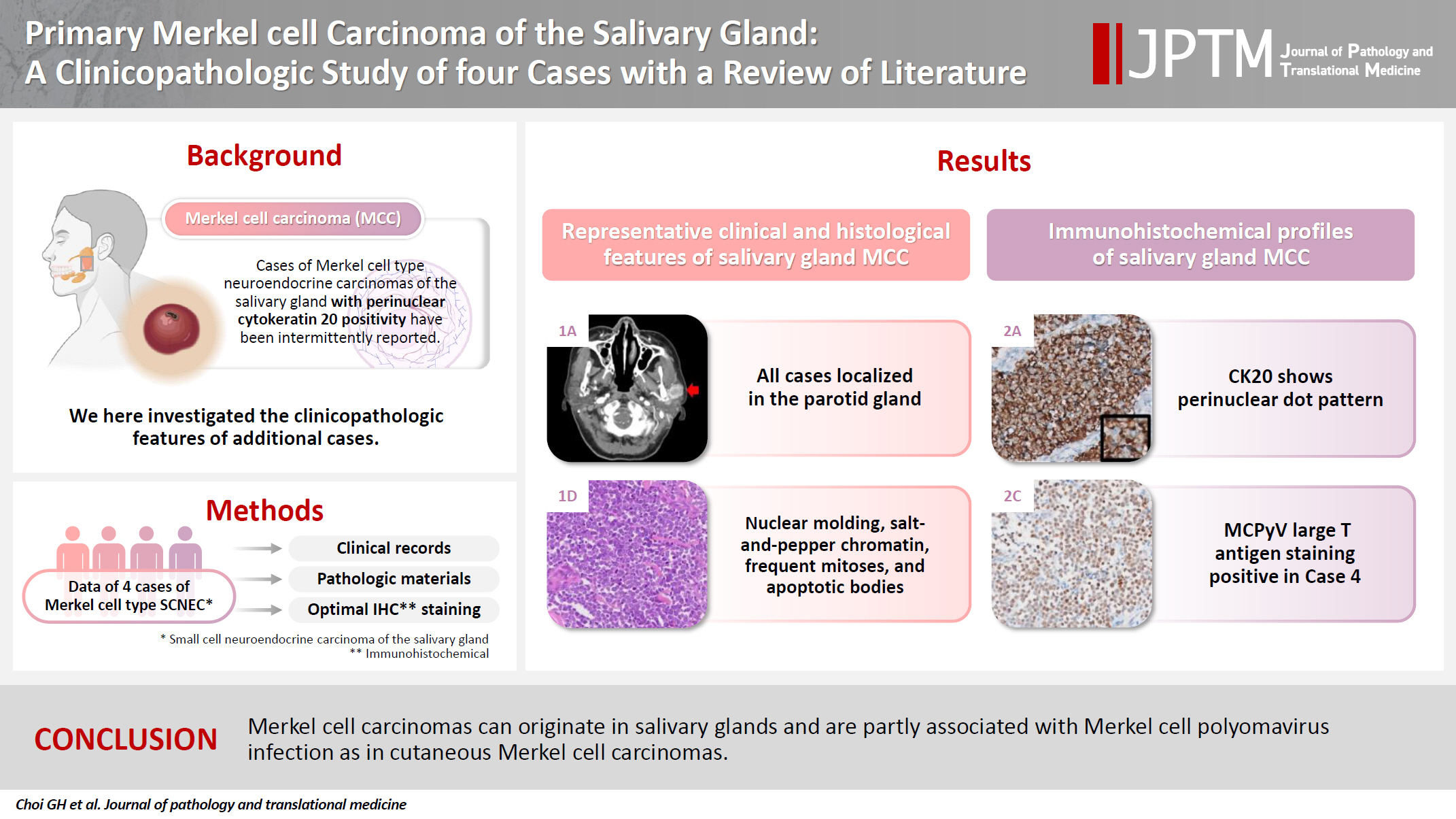
Primary Merkel cell carcinoma of the salivary gland is currently not listed in the World Health Organization classification. However, cases of Merkel cell type neuroendocrine carcinomas of the salivary gland with perinuclear cytokeratin 20 positivity have been intermittently reported. We here investigated the clinicopathologic features of additional cases.
Methods
Data of four cases of Merkel cell type small cell neuroendocrine carcinoma of the salivary gland were retrieved. To confirm the tumors’ primary nature, clinical records and pathologic materials were reviewed. Optimal immunohistochemical staining was performed to support the diagnosis.
Results
All tumors were located in the parotid gland. Possibilities of metastasis were excluded in all cases through a meticulous clinicopathological review. Tumor histology was consistent with the diagnosis of small cell neuroendocrine carcinoma. Tumors’ immunohistochemical phenotypes were consistent with Merkel cell carcinoma, including Merkel cell polyomavirus large T antigen positivity in two of the four cases.
Conclusions
Merkel cell carcinomas can originate in salivary glands and are partly associated with Merkel cell polyomavirus infection as in cutaneous Merkel cell carcinomas. -
Citations
Citations to this article as recorded by
- Parotid intranodal metastasis of Merkel cell carcinoma: a rare case report
Tong Gao, Dengshun Wang, Hongwei Yu, Yu’e Wang, Haibin Lu
BMC Oral Health.2025;[Epub] CrossRef
- Parotid intranodal metastasis of Merkel cell carcinoma: a rare case report
- Urinary Decoy Cell Grading and Its Clinical Implications
- Myoung Ju Koh, Beom Jin Lim, Songmi Noh, Yon Hee Kim, Hyeon Joo Jeong
- Korean J Pathol. 2012;46(3):233-236. Published online June 22, 2012
- DOI: https://doi.org/10.4132/KoreanJPathol.2012.46.3.233
- 12,667 View
- 95 Download
- 7 Crossref
-
Abstract
PDF
Background Examination of urine for decoy cells (DCs) is a useful screening test for polyomavirus (PV) activation. We explored the significance of the amount of DCs in persistent shedding, PV nephropathy and acute rejection.
Methods A case-controlled study was performed in 88 renal allograft patients who had DCs detected at least once in four or more urine samples.
Results Fifty one patients were classified into the high-grade shedding group (HG) and 37 patients into the low-grade shedding group (LG) according to DC shedding (≥10 or <10 DCs/10 high power field [HPF]). DC shedding of more than three consecutive months was significantly more prevalent in the HG as compared with their LG counterparts (p<0.0001). Urinary DCs were present for more than one year in 29.4% of the HG and 8.1% of the LG. Real-time polymerase chain reaction for PV was higher in both urine (51.4% vs. 11.1%) and plasma (9.1% vs. 0%) of the HG than the LG. The prevalence of PV nephropathy was higher in the HG than the LG (p=0.019). However, there was no significant difference in the prevalence of acute rejection.
Conclusions Shedding of ≥10 DCs/10 HPF is associated with sustained shedding, polymerase chain reaction positivity and PV nephropathy, but not a predictor of acute rejection.
-
Citations
Citations to this article as recorded by- Urinary VP1 Flow Cytometry as a Complementary Approach for BK Polyomavirus Monitoring: A Proof-Of-Concept Study
Haris Omic, David Vecsei, Michael Eder, Karim Abd El-Ghany, Wolfgang Winnicki, Alice Schmidt, Sebastian Kapps, Daniela Gerges, Robert Strassl, Ludwig Wagner, Farsad Eskandary
Transplant International.2026;[Epub] CrossRef - Polyomavirus nephropathy: diagnosis, histologic features, and differentiation from acute rejection
Cynthia C. Nast
Clinical Transplantation and Research.2024; 38(2): 71. CrossRef - Challenges and opportunities in research on BK virus infection after renal transplantation
Yukun Tang, Zipei Wang, Dunfeng Du
International Immunopharmacology.2024; 141: 112793. CrossRef - BK Virus-Associated Nephropathy after Renal Transplantation
Yasuhito Funahashi
Pathogens.2021; 10(2): 150. CrossRef - Diagnostic utility of urine cytology in detection of decoy cells in renal transplant patients: Report of five cases and review of literature
Santosh Tummidi, Kanchan Kothari, Mona Agnihotri, Leena Naik, Amey Rojekar
Diagnostic Cytopathology.2020; 48(3): 222. CrossRef - Association of Pretransplant BK Polyomavirus Antibody Status with BK Polyomavirus Infection After Kidney Transplantation: A Prospective Cohort Pilot Study of 47 Transplant Recipients
Yu Hisadome, Hiroshi Noguchi, Yuki Nakafusa, Kukiko Sakihama, Takanori Mei, Keizo Kaku, Yasuhiro Okabe, Kosuke Masutani, Yuki Ohara, Kazuyuki Ikeda, Yoshinao Oda, Masafumi Nakamura
Transplantation Proceedings.2020; 52(6): 1762. CrossRef - Association Between the Polyomaviruses Titers and Decoy Cell Positivity Rates After Renal Transplantation
Y. Funahashi, M. Kato, T. Fujita, S. Ishida, A. Mori, M. Gotoh
Transplantation Proceedings.2016; 48(3): 921. CrossRef
- Urinary VP1 Flow Cytometry as a Complementary Approach for BK Polyomavirus Monitoring: A Proof-Of-Concept Study
- Detection of JC Virus T-Ag in Early Gastric Cancer.
- Eun Jeong Jang, Jung Sik Jang, Jae Hoon Kim, Han Ik Bae, In Soo Suh
- Korean J Pathol. 2010;44(5):456-461.
- DOI: https://doi.org/10.4132/KoreanJPathol.2010.44.5.456
- 4,558 View
- 41 Download
- 1 Crossref
-
Abstract
PDF
- BACKGROUND
JC virus (JCV) is a polyomavirus that commonly infects humans and can cause progressive multifocal leukoencephalopathy in immunocompromised patients. Recently, many reports have documented detection of JCV in gastrointestinal tract cancers. We investigated the presence of JCV in gastric adenocarcinoma, adenoma, and non-neoplastic gastric mucosa.
METHODS
We selected paraffin-embedded tissue from endoscopic mucosal resections performed from January 2007 to September 2008. DNA was extracted from the paraffin-embedded specimens of 30 adenocarcinomas, 20 adenomas of the stomach, and 20 non-neoplastic gastric mucosa. Polymerase chain reaction amplifications were performed using gene-specific primers to detect the JCV gene sequences, and immunohistochemical staining was performed to detect the T-antigen (T-Ag) protein.
RESULTS
The T-Ag sequence was detected in nine of 30 gastric cancers (30%), two of 20 adenomas (10%), and eight of 20 non-neoplastic gastric mucosa specimens (40%). T-Ag protein expression was found in five of 30 gastric cancers (16.7%) and one of 20 non-neoplastic gastric mucosa specimens (5%), whereas no expression was observed in any of the adenomas.
CONCLUSIONS
Although we could not detect a correlation between JCV and gastric cancer, we demonstrated the presence of JCV T-Ag expression in human gastric cancers. These findings suggest a possible role for JCV in gastric carcinogenesis. -
Citations
Citations to this article as recorded by- Associations Between Gastric Cancer Risk and Virus Infection Other Than Epstein-Barr Virus: A Systematic Review and Meta-analysis Based on Epidemiological Studies
Hui Wang, Xiao-Long Chen, Kai Liu, Dan Bai, Wei-Han Zhang, Xin-Zu Chen, Jian-Kun Hu
Clinical and Translational Gastroenterology.2020; 11(7): e00201. CrossRef
- Associations Between Gastric Cancer Risk and Virus Infection Other Than Epstein-Barr Virus: A Systematic Review and Meta-analysis Based on Epidemiological Studies
- Detection of SV40 Large T Antigen in Malignant Lymphomas.
- Young A Kim, MeeSoo Chang, Jinho Paik, Sun Och Yoon, Yoon Kyung Jeon, Chul Woo Kim, Ji Eun Kim
- Korean J Pathol. 2009;43(4):312-316.
- DOI: https://doi.org/10.4132/KoreanJPathol.2009.43.4.312
- 5,435 View
- 71 Download
- 1 Crossref
-
Abstract
PDF
- BACKGROUND
The association of simian virus 40 (SV40) with certain types of human cancers, including malignant lymphomas, has been a topic of interest for some time. Although the virus is distributed worldwide, its incidences vary according to the specific types of tumors, and the epidemiological areas. The aim of this study was to investigate the frequency of SV40 in malignant lymphomas among Korean patients. METHODS: One hundred seventy three cases of malignant lymphomas were evaluated by immunohistochemical staining for SV40 large T antigen (TAg), using an extremely sensitive, tyramide based, catalyzed signal amplification method. RESULTS: From 158 non-Hodgkin's lymphomas, including 115 diffuse large B-cell lymphomas, and 15 Hodgkin's lymphomas, none of the cases were positive for SV40 TAg. CONCLUSIONS: SV40 does not appear to be related to the pathogenesis of malignant lymphomas among Koreans. -
Citations
Citations to this article as recorded by- No Detection of Simian Virus 40 in Malignant Mesothelioma in Korea
Minseob Eom, Jamshid Abdul-Ghafar, Sun-Mi Park, Joung Ho Han, Soon Won Hong, Kun Young Kwon, Eun Suk Ko, Lucia Kim, Wan Seop Kim, Seung Yeon Ha, Kyo Young Lee, Chang Hun Lee, Hye Kyoung Yoon, Yoo Duk Choi, Myoung Ja Chung, Soon-Hee Jung
Korean Journal of Pathology.2013; 47(2): 124. CrossRef
- No Detection of Simian Virus 40 in Malignant Mesothelioma in Korea
- Polyomavirus Renal Infection Confirmed by Electron Microscopy in a Patient with Acquired Immunodeficiency Syndrome: An Autopsy Case Report.
- Na Rae Kim, Byoung Kwon Kim, Je G Chi
- Korean J Pathol. 2001;35(2):168-171.
- 1,998 View
- 56 Download
-
Abstract
PDF
- Polyomavirus infection commonly occurs in childhood and adolescence, remaining in a latent status and reactivated in an immunocompromised status. We report herein an autopsy case of HIV-positive 41-year-old male, who succumbed to disseminated Kaposi sarcoma and cytomegalovirus infection involving the gastrointestinal tract, lung and brain. The involved kidney showed minimal inflammatory infiltrates and tubular injury: the nuclei of tubular epithelial cells were markedly enlarged with central clearing and peripheral chromatin margination or bore basophilic nuclear inclusions. Inclusion-bearing tubular epithelial cells were negative for the viral immunostains including herpes simplex virus, Epstein-Barr virus and adenovirus. Electron microscopy disclosed 42 nm intranuclear viral particles compatible with the BK polyomavirus. The viral particles were icosahedral in paracrystalline array and nonenveloped.
- Cytologic Findings of Polyomavirus Infection in the Urine: A Case Report.
- Mi Seon Kwon, Young Shin Kim, Kyo Young Lee, Yeong Jin Choi, Chang Suk Kang, Sang In Shim
- J Pathol Transl Med. 1996;7(2):192-196.
- 2,773 View
- 28 Download
-
Abstract
PDF
- The principal significance of the urothelial changes caused by polyomavirus activation is in an erroneous diagnosis of urothelial cancer; however, the clue to their benign nature is the smooth structureless nuclear configuration and the relative paucity of affected cells. Though virologic studies and electron microscopy are usually needed to firmly establish the diagnosis, cytology is the most readily available and rapid means of establishing a presumptive diagnosis of human polyomavirus infection. A urine specimen of a 24-year-old man with hemorrhagic cystitis beginning two months after bone marrow transplantation for acute myeloblastic leukemia(M2) was submitted for cytologic evaluation. Cytologic findings revealed a few inclusion-bearing epithelial cells intermingled with erythrocytes, neutrophils, lymphocytes, and macrophages. Most of the inclusion-bearing -cells had large, round to ovoid nuclei almost completely filed with homogeneous dark, basophilic inclusion. The chromatin was clumped along the periphery and the cytoplasm was mostly degenerated. The other cells exhibited irregular inclusions attached to the nuclear membrane surrounded by an indistinct halo. These findings were consistent with polyomavirus infection.
 First
First Prev
Prev


