 E-submission
Search
- Page Path
- HOME > Search
Case Study
- Drug-induced phospholipidosis of the kidney suspected to be caused by atomoxetine
- Sung-Eun Choi, Kee Hyuck Kim, Minsun Jung, Jeong Hae Kie
- J Pathol Transl Med. 2026;60(1):124-128. Published online January 14, 2026
- DOI: https://doi.org/10.4132/jptm.2025.12.10
- 1,825 View
- 146 Download
- 1 Crossref
-
 Abstract
 PDF
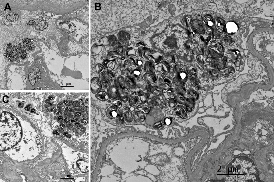
PDF - Drug-induced phospholipidosis (DIP) is characterized by intracellular accumulation of phospholipids with lamellar body formation secondary to drug-altered lipid metabolism, which can trigger inflammation and histopathological changes. Fabry disease and DIP both exhibit zebra bodies on electron microscopy, complicating differential diagnosis. A 17-year-old male with microscopic hematuria and proteinuria had received atomoxetine (40 mg) for 11 months to treat attention-deficit hyperactivity disorder. Light microscopy showed one glomerulus with perihilar sclerosis and periglomerular fibrosis. Kidney biopsy revealed zebra bodies in podocytes, initially suggesting Fabry disease. However, α-galactosidase A enzyme activity was normal on tandem mass spectrometry. Next-generation sequencing of GLA identified only three benign variants. This represents the first reported case of atomoxetine-induced DIP. When zebra bodies are observed, clinicians should consider DIP caused by cationic amphiphilic drugs alongside Fabry disease. Atomoxetine meets the structural criteria for inducing DIP, and awareness of this potential complication is essential.
-
Citations
Citations to this article as recorded by
- Atomoxetine
Reactions Weekly.2026; 2095(1): 19. CrossRef
- Atomoxetine
Original Articles
- Podocyte Expression of Osteopontin and FSP-1/S100A4 in Human Crescentic Glomerulonephritis.
- Ghil Suk Yoon, Tae Sook Kim
- Korean J Pathol. 2011;45(3):237-246.
- DOI: https://doi.org/10.4132/KoreanJPathol.2011.45.3.237
- 4,013 View
- 35 Download
-
Abstract
PDF
- BACKGROUND
Osteopontin (OPN) is a cytokine associated with a cell-matrix via integrins. Fibroblast specific protein-1 (FSP-1), known as S100A4, has been implicated in cell migration by non-muscle myosin. We investigated whether the role of OPN and FSP-1/S100A4 expression in their contribution to the podocyte phenotype change to form podocyte bridge and cellular crescent.
METHODS
Glomerular expression of OPN and FSP-1/S100A4 in renal biopsies of 16 patients with crescentic glomerulonephritis (CrGN) and 13 normal renal biopsies were studied by immunohistochemistry.
RESULTS
The expression of OPN and FSP-1/S100A4 was increased in the podocytes of glomeruli, with and without crescents, in patients with CrGN. Neither OPN nor FSP-1/S100A4 was expressed in glomeruli from the normal controls (p<0.01). A significant positive correlation was found between the expression of OPN in glomerular tufts and cellular crescents, and the expression of OPN and FSP-1/S100A4 in glomerular tufts (p<0.05).
CONCLUSIONS
The results suggest that OPN plays a role in early podocyte attachment to Bowman's capsule, and FSP-1/S100A4 potentiate podocyte contribution to cellular crescent formation by inducing cellular migration and growth.
- Significance of Osteopontin Expression in the Progression of Human Focal Segmental Glomerulosclerosis.
- Ghil Suk Yoon, Tae Sook Kim
- Korean J Pathol. 2010;44(5):462-468.
- DOI: https://doi.org/10.4132/KoreanJPathol.2010.44.5.462
- 4,300 View
- 29 Download
- 1 Crossref
-
Abstract
PDF
- BACKGROUND
Osteopontin (OPN) is a cytokine related to cell-matrix adhesion and cell survival and is expressed in the distal convoluted tubules in normal adult kidneys. Only one in vitro study has investigated the role of OPN in mechanically stretched podocytes and their actin cytoskeleton rearrangement.
METHODS
Glomerular OPN expression was investigated in biopsies from patients with human idiopathic focal segmental glomerulosclerosis (FSGS) (n = 25) and in normal renal biopsies (n = 16) by immunohistochemistry.
RESULTS
OPN was expressed in the podocytes from patients with FSGS. OPN expression increased in podocytes from both non-sclerotic hypertrophic and sclerotic glomerular tufts in patients with FSGS compared to the podocytes in normal controls.
CONCLUSIONS
The results suggest that OPN plays a role in the early adaptive response of podocytes to the increased mechanical load caused by glomerular hypertrophy preceding FSGS. OPN was involved in cell-matrix adhesion and influenced the detachment delay of podocytes from the glomerular basement membrane and apoptosis. -
Citations
Citations to this article as recorded by- Podocyte Expression of Osteopontin and FSP-1/S100A4 in Human Crescentic Glomerulonephritis
Ghil Suk Yoon, Tae Sook Kim
The Korean Journal of Pathology.2011; 45(3): 237. CrossRef
- Podocyte Expression of Osteopontin and FSP-1/S100A4 in Human Crescentic Glomerulonephritis
 First
First Prev
Prev


