 E-submission
Search
- Page Path
- HOME > Search
Review Article
- Breast schwannoma: review of entity and differential diagnosis
- Sandra Ixchel Sanchez, Ashley Cimino-Mathews
- J Pathol Transl Med. 2025;59(6):353-360. Published online November 3, 2025
- DOI: https://doi.org/10.4132/jptm.2025.08.12
- 4,304 View
- 182 Download
-
 Abstract
 PDF
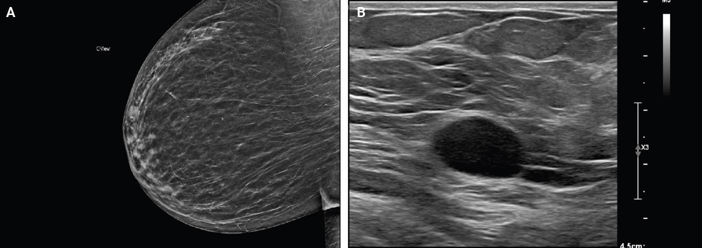
PDF - Schwannomas are benign peripheral nerve sheath tumors composed of Schwann cells, which uncommonly involve the breast. Most breast schwannomas are clinically present as a superficial palpable breast mass but may also be detected on screening mammography. Excision is the preferred treatment if symptomatic, and these are not known to recur. Histomorphology is similar to other anatomic sites: bland spindle cells with wavy nuclei, nuclear palisading (Verocay bodies), variably hypercellular (Antoni A) and hypocellular (Antoni B) areas, myxoid stroma, hyalinized vessels and variable cystic degeneration. Classic immunohistochemistry is diffuse and strong labeling for S100 and Sox10. Notable diagnostic pitfalls specific to the breast include myofibroblastoma, particularly the palisaded variant, and fascicular pseudoangiomatous stromal hyperplasia.
Case Report
- Soft Tissue Perineurioma.
- Yoon La Choi, Dae Soo Kim, Jai Hyang Go, Yeon Lim Suh
- Korean J Pathol. 1998;32(11):1028-1031.
- 2,240 View
- 10 Download
-
Abstract
- Perineurial cells, which normally surround the nerve fascicles within a nerve, can be distinguished from Schwann cells by their immunoreactivity for epithelial membrane antigen (EMA) and lack of reactivity for S-100 protein. Perineurioma is a form of benign peripheral nerve sheath tumor (PNST) almost exclusively composed of perineurial cells. It is often difficult to differentiate this tumor from the other benign PNSTs or ectopic meningioma by histology alone. Immunohistochemical and electron microscopic studies are helpful for differential diagnosis. We recently experienced a case of soft tissue perineurioma in a 14-year-old girl. This tumor was presented as a 5.6 cm sized subcutaneous movable mass in the elbow. The well encapsulated soft tissue tumor consisted of spindle cells which have whorling and storiform patterns within the collagenous stroma. The spindle cells were stained positive for EMA but negative for S-100 protein, chromogranin, neuron-specific enolase or Leu-7. Ultrastructurally, they possessed long cytoplasmic processes with incomplete basal lamina, primitive intercellular junction and occasional pinocytotic vesicles.
 First
First Prev
Prev


