 E-submission
Search
- Page Path
- HOME > Search
- Clinicopathological profile of high-grade differentiated thyroid carcinoma in an Indonesian tertiary hospital
- Novita , Agnes Stephanie Harahap, Maria Francisca Ham, Alfianto Widiono, Chan Kwon Jung
- J Pathol Transl Med. 2026;60(3):338-348. Published online April 23, 2026
- DOI: https://doi.org/10.4132/jptm.2026.01.15
- 2,139 View
- 72 Download
-
 Abstract
 PDF
PDF Supplementary Material
 - Background
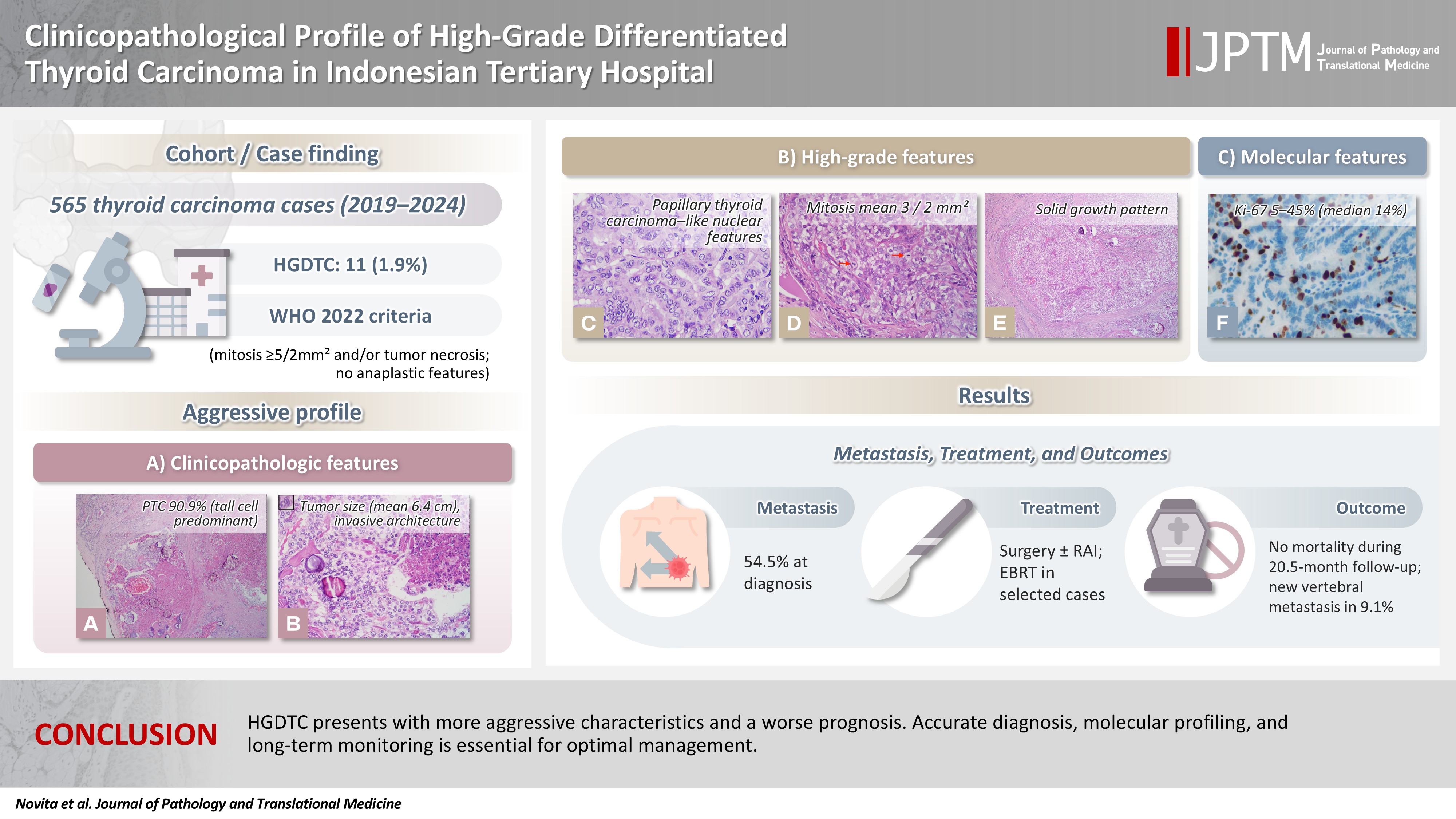
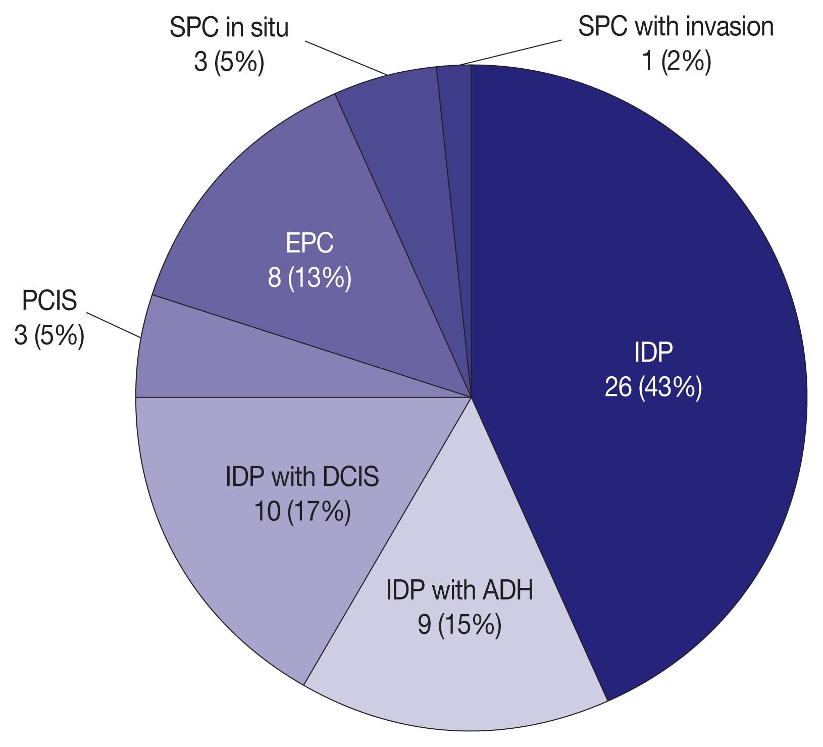
High-grade differentiated thyroid carcinoma (HGDTC) is a recently recognized entity in the 2022 World Health Organization classification, representing a more aggressive subtype of differentiated thyroid carcinoma. Previously, high-grade features such as increased mitotic activity and tumor necrosis were often overlooked, despite being important independent prognostic factors. Although rare, HGDTC carries significant diagnostic, prognostic, and therapeutic implications. Data remain limited in Indonesia. Methods: This retrospective descriptive study reviewed 565 thyroid carcinoma cases diagnosed at Cipto Mangunkusumo Hospital from 2019 to 2024. Eleven cases (1.9%) met HGDTC criteria. Clinicopathological characteristics, histologic subtypes, Ki-67 proliferation index, molecular alterations, treatment modalities, and clinical outcomes were analyzed. Results: Patients had a mean age of 54.6 years, with a female-to-male ratio of 2.7:1. Papillary thyroid carcinoma was the main type (90.9%), with the tall cell subtype predominating. Mean tumor size was 6.4 cm. Lymphatic invasion, vascular invasion, and extrathyroidal extension were present in 54.5%, 18.2%, and 45.5% of cases, respectively. All tumors showed necrosis. Mean mitotic count was 3 per 2 mm². The Ki-67 index ranged from 5% to 45% (median, 14%). BRAFV600E and TERT promoter mutations were detected in 18.2% and 36.4% of cases, respectively, with co-mutations in 18.2%. Six cases (54.5%) had metastases at time of diagnosis. During a mean follow-up of 20.5 months, one patient (9.1%) developed new vertebral metastases and all patients (100%) remained alive. Conclusions: HGDTC presents with more aggressive characteristics and a worse prognosis. Accurate diagnosis, molecular profiling, and long-term monitoring are essential for optimal management.
- The significance of papillary architecture in the follow-up biopsies of patients with progestin-treated atypical endometrial hyperplasia
- Wangpan J. Shi, Oluwole Fadare
- J Pathol Transl Med. 2026;60(1):58-68. Published online January 8, 2026
- DOI: https://doi.org/10.4132/jptm.2025.09.12
- 2,929 View
- 323 Download
-
Abstract
PDF
- Background
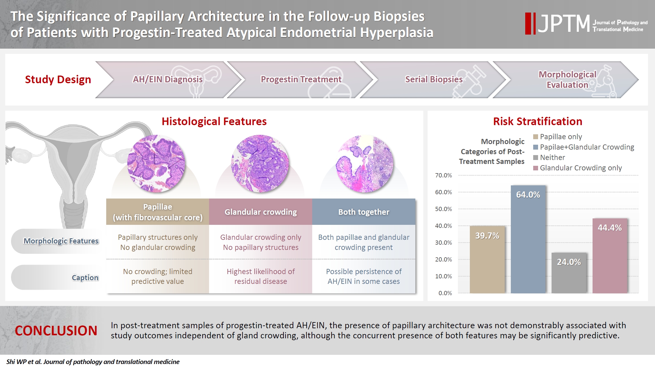
Follow-up biopsies in patients with progestin-treated atypical endometrial hyperplasia/endometrioid intraepithelial neoplasia (AH/EIN) may show papillary structures, the significance of which is unclear. Methods: The authors reviewed 253 serial specimens of 84 consecutive patients diagnosed with AH/EIN, inclusive of each patient's pre-progestin treatment sample and all post-treatment specimens. We assessed the predictive relationship between papillary architecture in a post-treatment biopsy and two study outcomes: AH/EIN or carcinoma in at least one sample subsequent to the one in which papillae were identified, and/or the last specimen received for that patient. Results: Papillae were identified in only 51.5% of pre-treatment samples but were present in at least one subsequent post-treatment sample for all patients. Post-treatment samples that exhibited papillae and no glandular crowding were associated with AH/EIN in at least one subsequent specimen in 39.7% (29/73) of cases, compared to 24.0% (6/25) in samples with neither papillae nor glandular crowding (p = .227) and 64.0% (16/25) in samples with concurrent gland crowding and papillae (p = .048). Univariate logistic regression analyses showed that the presence of papillae was not associated with study outcomes (odds ratio [OR], 0.99; 95% confidence interval [CI], 0.49 to 1.99; p = .985), as compared with gland crowding (OR, 1.54; 95% CI, 1.04 to 2.27; p = .031), or concurrent papillae and gland crowding (OR, 2.36; 95% CI, 1.01 to 5.52; p = .048). Conclusions: In post-treatment samples of progestin-treated AH/EIN, the presence of papillary architecture was not demonstrably associated with study outcomes independent of gland crowding, although the concurrent presence of both features may be significantly predictive.
- Low Ki-67 labeling index is a clinically useful predictive factor for recurrence-free survival in patients with papillary thyroid carcinoma
- Takashi Masui, Katsunari Yane, Ichiro Ota, Kennichi Kakudo, Tomoko Wakasa, Satoru Koike, Hirotaka Kinugawa, Ryuji Yasumatsu, Tadashi Kitahara
- J Pathol Transl Med. 2025;59(2):115-124. Published online February 18, 2025
- DOI: https://doi.org/10.4132/jptm.2024.11.08
- 7,966 View
- 278 Download
- 3 Web of Science
- 6 Crossref
-
Abstract
PDF
- Background
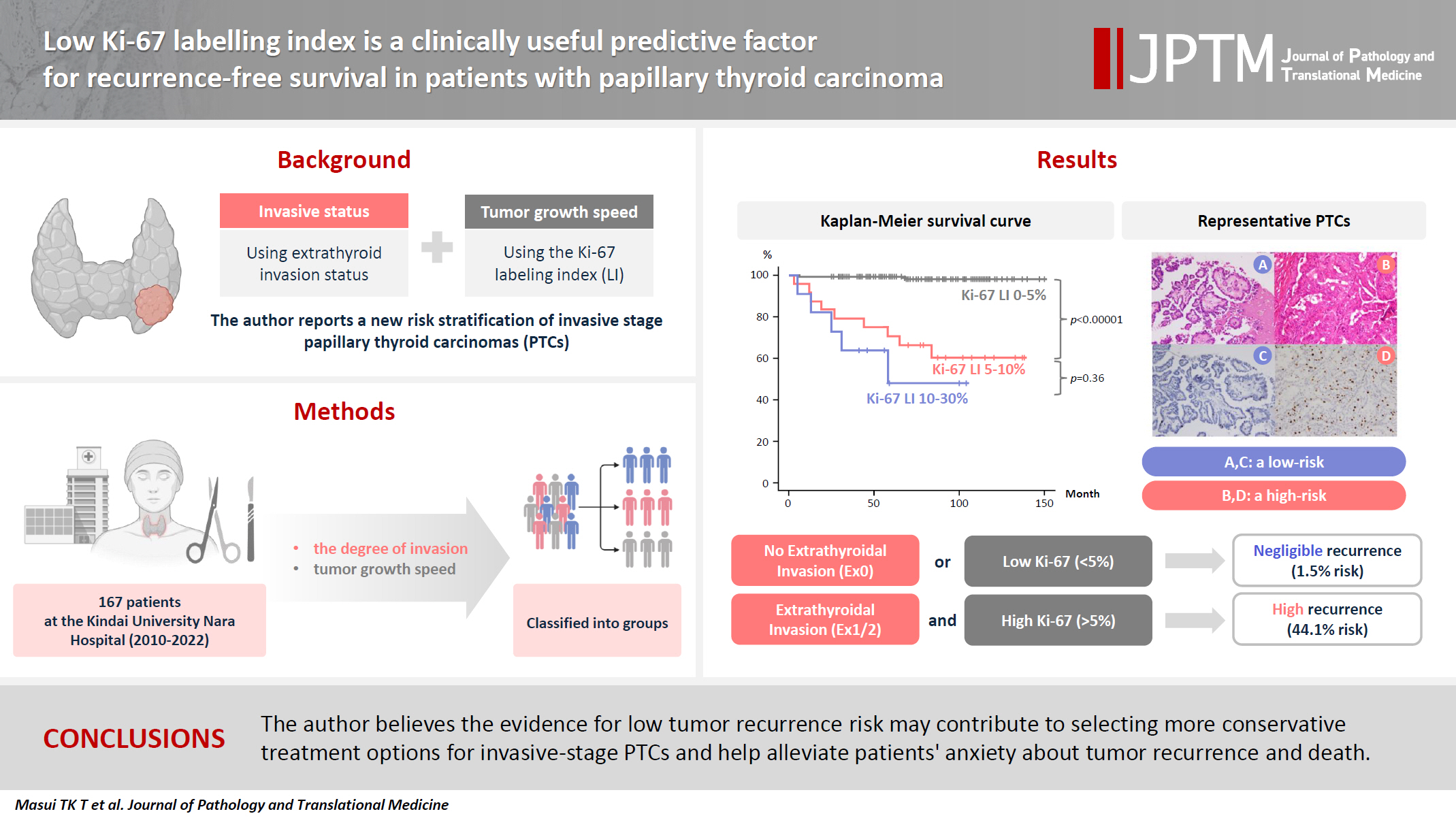
We report a new risk stratification of invasive stage papillary thyroid carcinomas (PTCs) by combining invasive status, using extrathyroid invasion (Ex) status, and tumor growth speed using the Ki-67 labeling index (LI). Methods: We examined tumor recurrence in 167 patients with PTC who were surgically treated at the Kindai University Nara Hospital between 2010 and 2022. The patients were classified according to the degree of invasion [negative (Ex0) or positive (Ex1, Ex2, and Ex3)] and tumor growth speed expressed with Ki-67 LI, as low (<5%) or high (>5%). This study confirmed previous findings that the disease-free survival (DFS) rate in PTCs significantly differed between patients with a high and low Ki-67 index. Results: When combining Ex status (negative or positive) and Ki-67 proliferation status (low or high), the DFS rate of invasion in the negative, low Ki-67 LI group was only 1.1%, while that of invasion in the positive, high Ki-67 LI was 44.1%. This study reports for the first time that recurrence risks can be stratified accurately when combining carcinoma’s essential two features of extrathyroid invasion status and tumor growth speed. Conclusions: We believe the evidence for low tumor recurrence risk may contribute to use of more conservative treatment options for invasive-stage PTCs and help alleviate patient anxiety about tumor recurrence and death. -
Citations
Citations to this article as recorded by
- Clinicopathological, proliferative, molecular, and prognostic characteristics of differentiated high-grade thyroid carcinoma: a multicenter retrospective study
Wenwen Cui, Lihang Xing, Zhenzhen Li, Xinjun Li, Junzhi Li
Frontiers in Oncology.2026;[Epub] CrossRef - Integrating Digital Ki-67 Labeling Index and K-TIRADS for Malignancy Risk Stratification in Thyroid Core Needle Biopsies
Yujin Cha, Sue Youn Kim, Chankyung Kim, Ja Seong Bae, Dong-Jun Lim, So-Lyung Jung, Chan Kwon Jung
Cancers.2026; 18(14): 2260. CrossRef - The Association of Interstitial Fibrosis and Ki67 Marker Expression in Papillary Thyroid Carcinoma with Histopathological Findings and Prognosis of Total Thyroidectomy
Osman Anıl Savaş, Selçuk Mercan, Amir Mahdi Akbari, Mehrdad Sheikhvatan
Journal of Clinical Medicine.2026; 15(14): 5703. CrossRef - Research Progress on the Correlation between Three Biomarkers, Ki-67, CAIX and VEGF and Clear Cell Renal Cell Carcinoma
锦容 马
Advances in Clinical Medicine.2025; 15(09): 326. CrossRef - Immunophenotypic Panel for Comprehensive Characterization of Aggressive Thyroid Carcinomas
Mihail Ceausu, Mihai Alin Publik, Dana Terzea, Carmen Adina Cristea, Dumitru Ioachim, Dana Manda, Sorina Schipor
Cells.2025; 14(19): 1554. CrossRef - High Ki-67 labeling index correlates with aggressive clinicopathological features in papillary thyroid carcinoma: a retrospective study
Defi Nurlia Erdian, Maria Francisca Ham, Dina Khoirunnisa, Agnes Stephanie Harahap
Thyroid Research.2025;[Epub] CrossRef
- Clinicopathological, proliferative, molecular, and prognostic characteristics of differentiated high-grade thyroid carcinoma: a multicenter retrospective study
- Cytologic hallmarks and differential diagnosis of papillary thyroid carcinoma subtypes
- Agnes Stephanie Harahap, Chan Kwon Jung
- J Pathol Transl Med. 2024;58(6):265-282. Published online November 7, 2024
- DOI: https://doi.org/10.4132/jptm.2024.10.11
- 23,607 View
- 829 Download
- 17 Web of Science
- 15 Crossref
-
Abstract
PDF
- Papillary thyroid carcinoma (PTC) is the most common thyroid malignancy, characterized by a range of subtypes that differ in their cytologic features, clinical behavior, and prognosis. Accurate cytologic evaluation of PTC using fine-needle aspiration is essential but can be challenging due to the morphologic diversity among subtypes. This review focuses on the distinct cytologic characteristics of various PTC subtypes, including the classic type, follicular variant, tall cell, columnar cell, hobnail, diffuse sclerosing, Warthin-like, solid/trabecular, and oncocytic PTCs. Each subtype demonstrates unique nuclear features, architectural patterns, and background elements essential for diagnosis and differentiation from other thyroid lesions. Recognizing these distinct cytologic patterns is essential for identifying aggressive subtypes like tall cell, hobnail, and columnar cell PTCs, which have a higher risk of recurrence, metastasis, and poorer clinical outcomes. Additionally, rare subtypes such as diffuse sclerosing and Warthin-like PTCs present unique cytologic profiles that must be carefully interpreted to avoid diagnostic errors. The review also highlights the cytologic indicators of lymph node metastasis and high-grade features, such as differentiated high-grade thyroid carcinoma. The integration of molecular testing can further refine subtype diagnosis by identifying specific genetic mutations. A thorough understanding of these subtype-specific cytologic features and molecular profiles is vital for accurate diagnosis, risk stratification, and personalized management of PTC patients. Future improvements in diagnostic techniques and standardization are needed to enhance cytologic evaluation and clinical decision-making in thyroid cancer.
-
Citations
Citations to this article as recorded by- Oncocytic Thyroid Tumours With Pathogenic FLCN Mutations Mimic Oncocytic Papillary Thyroid Carcinoma on Fine‐Needle Aspiration
Adeel M. Ashraf, Faisal Hassan, Adrian A. Dawkins, Julie C. Dueber, Derek B. Allison, Thèrése J. Bocklage
Cytopathology.2026; 37(1): 108. CrossRef - Using a new type of visible light-based emission fluorescence microscope to identify the benign and malignant nature of thyroid tissue during the surgical process: Analysis of diagnostic results
Yu Miao, Liu Xiaowei, Li Muyang, Gao Jian, Chen Lu
Photodiagnosis and Photodynamic Therapy.2026; 57: 105324. CrossRef - Clinical Behavior of Aggressive Variants of Papillary Thyroid Carcinoma: A Retrospective Case–Control Study
Jovan Ilic, Nikola Slijepcevic, Katarina Tausanovic, Bozidar Odalovic, Goran Zoric, Marija Milinkovic, Branislav Rovcanin, Milan Jovanovic, Matija Buzejic, Duska Vucen, Boban Stepanovic, Sara Ivanis, Milan Parezanovic, Milan Marinkovic, Vladan Zivaljevic
Cancers.2026; 18(2): 345. CrossRef - Advantages of thyroid core needle biopsy: an emerging selective first-line biopsy modality
Jae Ho Shin, Yeseul Kim, Min Kyoung Lee, Jung Hwan Baek, So Lyung Jung
Ultrasonography.2026; 45(3): 205. CrossRef - Clinicopathological profile of high-grade differentiated thyroid carcinoma in an Indonesian tertiary hospital
Novita, Agnes Stephanie Harahap, Maria Francisca Ham, Alfianto Widiono, Chan Kwon Jung
Journal of Pathology and Translational Medicine.2026; 60(3): 338. CrossRef - Interpretable SVM-Based Integrated Ultrasound Model for Preoperative Thyroid Nodule Subtype Classification: Improved Identification of Follicular Variant Papillary Thyroid Carcinoma
Ran Zheng, Zhen Wang, Yongxin Li, Yuanqing Zhang, Fang Nie
Diagnostics.2026; 16(13): 1950. CrossRef - Papillary thyroid carcinoma in thyroglossal duct cyst: a Peruvian case series
José Luis Paz-Ibarra, Marialejandra Delgado Rojas, Edward Paucar Holgado, Jenyfer María Fuentes-Mendoza, Luis Concepción-Urteaga, Juan Eduardo Quiroz-Aldave, Marcio José Concepción-Zavaleta, José Somocurcio Peralta
Endocrinology, Diabetes & Metabolism Case Reports.2026;[Epub] CrossRef - Single-cell reveals age-dependent epithelial reprogramming and EMT vulnerability in THCA
Qiankun Zhang, Wei Pan, Xiaohua Gong, Qi Zhou
Endocrine-Related Cancer.2026;[Epub] CrossRef - Nuclear pseudoinclusion is associated with BRAFV600E mutation: Analysis of nuclear features in papillary thyroid carcinoma
Agnes Stephanie Harahap, Dina Khoirunnisa, Salinah, Maria Francisca Ham
Annals of Diagnostic Pathology.2025; 75: 152434. CrossRef - 2025 Korean Thyroid Association Clinical Management Guideline on Active Surveillance for Low-Risk Papillary Thyroid Carcinoma
Eun Kyung Lee, Min Joo Kim, Seung Heon Kang, Bon Seok Koo, Kyungsik Kim, Mijin Kim, Bo Hyun Kim, Ji-hoon Kim, Shin Je Moon, Kyorim Back, Young Shin Song, Jong-hyuk Ahn, Hwa Young Ahn, Ho-Ryun Won, Won Sang Yoo, Min Kyoung Lee, Jeongmin Lee, Ji Ye Lee, Kyo
International Journal of Thyroidology.2025; 18(1): 30. CrossRef - Structure-based molecular screening and dynamic simulation of phytocompounds targeting VEGFR-2: a novel therapeutic approach for papillary thyroid carcinoma
Shuai Wang, Lingqian Zhang, Wenjun Zhang, Xiong Zeng, Jie Mei, Weidong Xiao, Lijie Yang
Frontiers in Pharmacology.2025;[Epub] CrossRef - 2025 Korean Thyroid Association Clinical Management Guideline on Active Surveillance for Low-Risk Papillary Thyroid Carcinoma
Eun Kyung Lee, Min Joo Kim, Seung Heon Kang, Bon Seok Koo, Kyungsik Kim, Mijin Kim, Bo Hyun Kim, Ji-hoon Kim, Shinje Moon, Kyorim Back, Young Shin Song, Jong-hyuk Ahn, Hwa Young Ahn, Ho-Ryun Won, Won Sang Yoo, Min Kyoung Lee, Jeongmin Lee, Ji Ye Lee, Kyon
Endocrinology and Metabolism.2025; 40(3): 307. CrossRef - A Case of Warthin-Like Variant of Papillary Thyroid Cancer
Amy Chow, Israa Laklouk
Cureus.2025;[Epub] CrossRef - Propensity score-matched analysis of the ‘2+2’ parathyroid strategy in total thyroidectomy with central neck dissection
Hao Gong, Simei Yao, Tianyuchen Jiang, Yi Yang, Yuhan Jiang, Zhujuan Wu, Anping Su
Frontiers in Endocrinology.2025;[Epub] CrossRef - Cytological Findings in Pediatric Thoracic Tumors: A Review of Diagnostic Insights and Pitfalls
Parikshaa Gupta, Pranab Dey
Acta Cytologica.2025; 70(3): 320. CrossRef
- Oncocytic Thyroid Tumours With Pathogenic FLCN Mutations Mimic Oncocytic Papillary Thyroid Carcinoma on Fine‐Needle Aspiration
- TERT mutations and aggressive histopathologic characteristics of radioiodine-refractory papillary thyroid cancer
- Ju Yeon Pyo, Yoon Jin Cha, SoonWon Hong
- J Pathol Transl Med. 2024;58(6):310-320. Published online September 12, 2024
- DOI: https://doi.org/10.4132/jptm.2024.07.29
- 7,287 View
- 362 Download
- 12 Web of Science
- 12 Crossref
-
Abstract
PDF
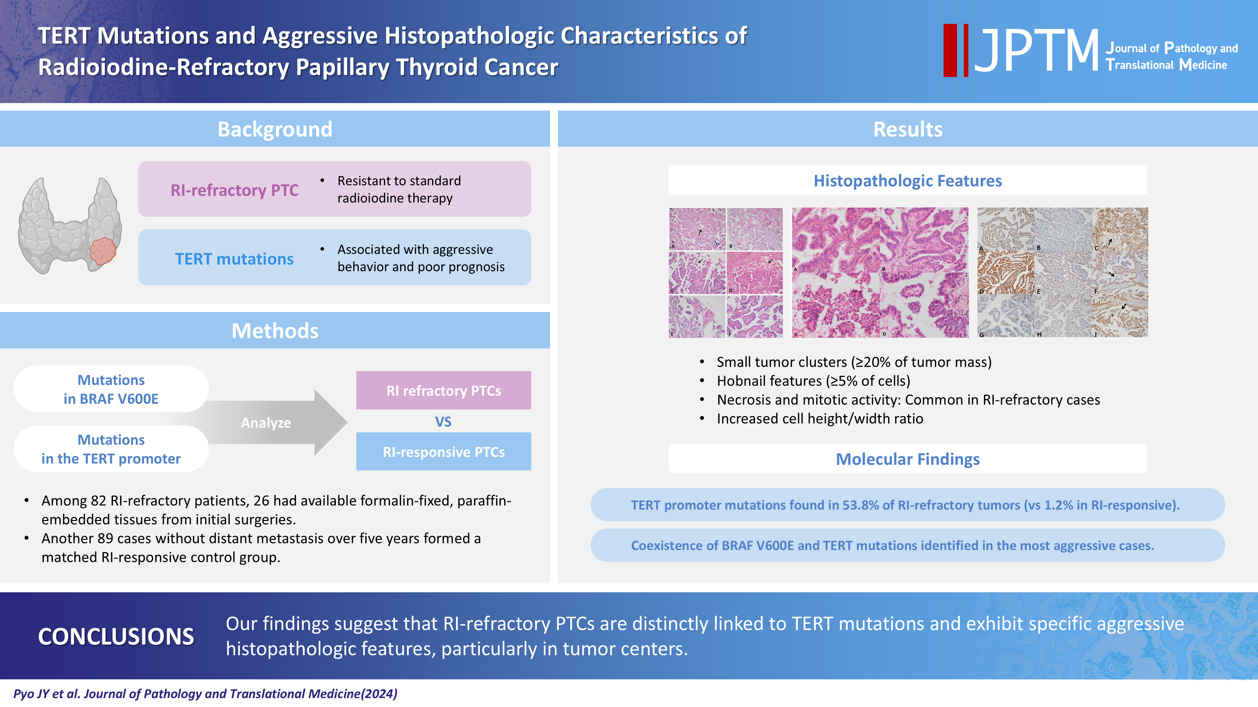
- Background
Radioiodine (RI) ablation following thyroid-stimulating hormone suppression is an effective treatment for papillary thyroid cancer (PTC), typically leading to favorable outcomes. However, RI-refractory tumors exhibit aggressive behavior and poor prognoses. Recent studies highlight the role of genetic abnormalities in PTC signaling pathways, including the activation of telomerase reverse transcriptase (TERT), and the correlation of mutations with adverse outcomes.
Methods
This study analyzed mutations in BRAF V600E and the TERT-promoter genes, comparing clinicopathological features between RI-refractory and RI-responsive PTCs. Among 82 RI-refractory patients, formalin-fixed, paraffin-embedded tissues from initial surgeries were available for 26. Another 89 without distant metastasis over 5 years formed a matched RI-responsive control group.
Results
Histopathologically, RI-refractory PTCs showed increased frequencies of small tumor clusters without fibrovascular cores, hobnail features, and a high height-to-width ratio of tumor cells. These tumors were more likely to exhibit necrosis, mitosis, lymph node metastasis, extrathyroidal extension, and involvement of resection margins. TERT-promoter mutations were statistically significantly associated with these aggressive clinicopathologic features. Immunohistochemically, decreased expression of sodium iodide symporter and thyroglobulin stimulating hormone receptor proteins was common in RI-refractory PTCs, along with lower levels of oncogenic proteins such as vascular endothelial cell growth factor, vascular endothelial cell growth factor receptor 2, and nuclear factor kappa-light-chain-enhancer of activated B cells. Total loss of PTEN expression was occasionally observed. In contrast, all cases tested positive for cytoplasmic β-catenin.
Conclusions
RI-refractory PTCs are linked to TERT mutations and exhibit specific aggressive histopathologic features, particularly in tumor centers. -
Citations
Citations to this article as recorded by- Calcifying nested stromal-epithelial tumor of the liver: Report of two cases revealing novel WT1 mutation and distinct epigenetic features
Andrea Strakova-Peterikova, Franco Fedeli, Boris Rychly, Jiri Soukup, Michael Michal, Petr Martinek, Marian Grendar, Elaheh Mosaieby, Nikola Ptakova, Maryna Slisarenko, Michal Michal, Kvetoslava Michalova
Virchows Archiv.2026; 488(4): 801. CrossRef - Characterizing thyroid carcinomas in the elderly: Histological subtypes and TERT promoter mutation analysis based on the latest WHO classification
Myoung Ju Koh, Songmi Noh, Jin Kyong Kim, Gi Jeong Kim
Annals of Diagnostic Pathology.2026; 80: 152578. CrossRef - Insulin resistance and metabolic dysfunction in thyroid nodules and differentiated thyroid cancer
Stefano Iuliano, Maria Mirabelli, Stefania Giuliano, Antonio Brunetti
Current Opinion in Oncology.2026; 38(1): 1. CrossRef - Differentiated high-grade thyroid carcinoma (DHGTC): clinicopathological analysis of a new entity in a chilean center
Marlín Solórzano, Ignacio Fuentes, José Miguel González, Nicole Lustig, Lorena Mosso, Joel Falcón, Catalina Ruiz, Joaquín Viñambres, Rodolfo Cabello, Hernán González, Pablo H Montero, Francisco Cruz, Rodrigo Jaimovich, Juan Carlos Quintana, Antonieta Sola
Endocrine.2026;[Epub] CrossRef - Characteristics and outcome of pediatric and adult differentiated thyroid cancer with distant metastases
Ali S. Alzahrani, Lulu Alobaid, Eman Albasri, Afnan Hadadi, Abdulrhman Hakami, Fayha Abothenain, Deema Alturki, Najla Ewain, Ali Howaidi, Hindi Alhindi, Ghada Alskait, Yasser Aljufan, Shatha Alghaihb, Azzam Alkhalifah, Leenah AlAyoubi, Amani Abualnaja
Frontiers in Endocrinology.2026;[Epub] CrossRef - TERT promoter–mutated thyroid carcinomas: prognostic and histologic insights according to the WHO 2022 classification
Seung Eun Lee, Bogyeong Han, Jung‐Sun Kim, Young Lyun Oh
Histopathology.2026; 89(2): 262. CrossRef - Clinicopathological profile of high-grade differentiated thyroid carcinoma in an Indonesian tertiary hospital
Novita, Agnes Stephanie Harahap, Maria Francisca Ham, Alfianto Widiono, Chan Kwon Jung
Journal of Pathology and Translational Medicine.2026; 60(3): 338. CrossRef - ARMS‐qPCR‐Based Detection of BRAF and TERT Promoter Mutations: A Cost‐Effective Strategy for Molecular Diagnosis of Papillary Thyroid Carcinoma
Yuanyuan Jia, Yajun Zhang, Dandan Sun, Jiabao Yang, Mengshi Zhou, Min Gao, Rui Li, Xiaoyu Song
International Journal of Endocrinology.2026;[Epub] CrossRef - Targeted therapy in thyroid cancer: molecular alterations and clinical management
YiHeng Yang, YeSheng Zhang, YongCan Xu, XiaoXin Gu, Neng Lou, GuoChao Ye
Frontiers in Endocrinology.2026;[Epub] CrossRef - How long should we follow up patients with papillary thyroid carcinoma?: A case report describing brain metastasis after 8 years
Hui Li, Yidong Zhu, Xiaozhen Zhao
Medicine.2026; 105(29): e49774. CrossRef - The ability of anexelekto (AXL) expression and TERT promoter mutation to predict radioiodine-refractory differentiated thyroid carcinoma
Hasrayati Agustina, Tutik Nur Ayni, Yohana Azhar, Erwin Affandi Soeriadi, Bethy Suryawathy Hernowo
Diagnostic Pathology.2025;[Epub] CrossRef - Clinicopathologic characteristics of papillary thyroid carcinoma, tall cell subtype and subtype with tall cell features, an institutional experience
Xueting Jin, Shunsuke Koga, Xiao Zhou, Niaz Z. Khan, Zubair W. Baloch
Human Pathology.2025; 161: 105867. CrossRef
- Calcifying nested stromal-epithelial tumor of the liver: Report of two cases revealing novel WT1 mutation and distinct epigenetic features
- Diagnostic challenges in the assessment of thyroid neoplasms using nuclear features and vascular and capsular invasion: a multi-center interobserver agreement study
- Agnes Stephanie Harahap, Mutiah Mutmainnah, Maria Francisca Ham, Dina Khoirunnisa, Abdillah Hasbi Assadyk, Husni Cangara, Aswiyanti Asri, Diah Prabawati Retnani, Fairuz Quzwain, Hasrayati Agustina, Hermawan Istiadi, Indri Windarti, Krisna Murti, Muhammad Takbir, Ni Made Mahastuti, Nila Kurniasari, Nungki Anggorowati, Pamela Abineno, Yulita Pundewi Setyorini, Kennichi Kakudo
- J Pathol Transl Med. 2024;58(6):299-309. Published online September 12, 2024
- DOI: https://doi.org/10.4132/jptm.2024.07.25
- Correction in: J Pathol Transl Med 2025;59(3):201
- 7,127 View
- 432 Download
- 2 Web of Science
- 1 Crossref
-
Abstract
PDFSupplementary Material
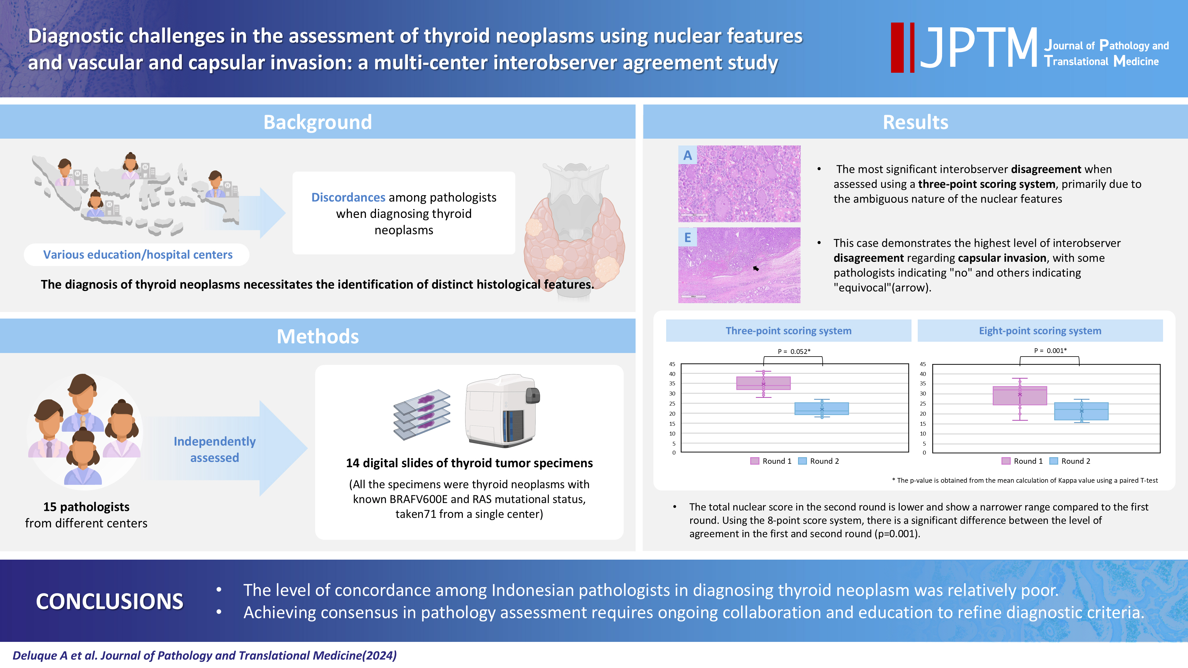
- Background
The diagnosis of thyroid neoplasms necessitates the identification of distinct histological features. Various education/hospital centers located in cities across Indonesia likely result in discordances among pathologists when diagnosing thyroid neoplasms.
Methods
This study examined the concordance among Indonesian pathologists in assessing nuclear features and capsular and vascular invasion of thyroid tumors. Fifteen pathologists from different centers independently assessed the same 14 digital slides of thyroid tumor specimens. All the specimens were thyroid neoplasms with known BRAFV600E and RAS mutational status, from a single center. We evaluated the pre- and post-training agreement using the Fleiss kappa. The significance of the training was evaluated using a paired T-test.
Results
Baseline agreement on nuclear features was slight to fair based on a 3-point scoring system (k = 0.14 to 0.28) and poor to fair based on an eight-point system (k = –0.02 to 0.24). Agreements on vascular (κ = 0.35) and capsular invasion (κ = 0.27) were fair, whereas the estimated molecular type showed substantial agreement (κ = 0.74). Following the training, agreement using the eight-point system significantly improved (p = 0.001).
Conclusions
The level of concordance among Indonesian pathologists in diagnosing thyroid neoplasm was relatively poor. Consensus in pathology assessment requires ongoing collaboration and education to refine diagnostic criteria. -
Citations
Citations to this article as recorded by- Nuclear pseudoinclusion is associated with BRAFV600E mutation: Analysis of nuclear features in papillary thyroid carcinoma
Agnes Stephanie Harahap, Dina Khoirunnisa, Salinah, Maria Francisca Ham
Annals of Diagnostic Pathology.2025; 75: 152434. CrossRef
- Nuclear pseudoinclusion is associated with BRAFV600E mutation: Analysis of nuclear features in papillary thyroid carcinoma
- Noninvasive follicular thyroid neoplasm with papillary-like nuclear features: its updated diagnostic criteria, preoperative cytologic diagnoses and impact on the risk of malignancy
- Hee Young Na, So Yeon Park
- J Pathol Transl Med. 2022;56(6):319-325. Published online November 9, 2022
- DOI: https://doi.org/10.4132/jptm.2022.09.29
- 14,795 View
- 333 Download
- 9 Web of Science
- 9 Crossref
-
Abstract
PDF
- Due to the extremely indolent behavior, a subset of noninvasive encapsulated follicular variant papillary thyroid carcinomas has been classified as “noninvasive follicular thyroid neoplasm with papillary-like nuclear features (NIFTP)” since 2016 and is no longer considered carcinoma. Since the introduction of this new terminology, changes and refinements have been made in diagnostic criteria. Initially, the incidence of NIFTP was estimated substantial. However, the reported incidence of NIFTP varies greatly among studies and regions, with higher incidence in North American and European countries than in Asian countries. Thus, the changes in the risk of malignancy (ROM) in the Bethesda System for Reporting Thyroid Cytopathology (TBSRTC) differ inevitably among regions. Because more conservative surgery is recommended for NIFTPs, distinguishing NIFTPs from papillary thyroid carcinomas in preoperative fine-needle aspiration cytology became one of the major concerns. This review will provide comprehensive overview of updates on diagnostic criteria, actual incidence and preoperative cytologic diagnoses of NIFTP, and its impact on the ROM in TBSRTC.
-
Citations
Citations to this article as recorded by- Diagnosis of invasive encapsulated follicular variant papillary thyroid carcinoma by protein-based machine learning
Truong Phan-Xuan Nguyen, Minh-Khang Le, Sittiruk Roytrakul, Shanop Shuangshoti, Nakarin Kitkumthorn, Somboon Keelawat
Journal of Pathology and Translational Medicine.2025; 59(1): 39. CrossRef - Papillae, psammoma bodies, and/or many nuclear pseudoinclusions are helpful criteria but should not be required for a definitive cytologic diagnosis of papillary thyroid carcinoma: An institutional experience of 207 cases with surgical follow up
Tarik M. Elsheikh, Matthew Thomas, Jennifer Brainard, Jessica Di Marco, Erica Manosky, Bridgette Springer, Dawn Underwood, Deborah J. Chute
Cancer Cytopathology.2024; 132(6): 348. CrossRef - ThyroSeq overview on indeterminate thyroid nodules: An institutional experience
Sam Sirotnikov, Christopher C. Griffith, Daniel Lubin, Chao Zhang, Nabil F. Saba, Dehong Li, Amanda Kornfield, Amy Chen, Qiuying Shi
Diagnostic Cytopathology.2024; 52(7): 353. CrossRef - Oncocytic Noninvasive Follicular Thyroid Neoplasm with Papillary-Like Nuclear Features: A Case Report
Kaveripakam Ajay Joseph, Sana Ahuja, Sufian Zaheer
Indian Journal of Surgical Oncology.2024; 15(S4): 606. CrossRef - Cytologic hallmarks and differential diagnosis of papillary thyroid carcinoma subtypes
Agnes Stephanie Harahap, Chan Kwon Jung
Journal of Pathology and Translational Medicine.2024; 58(6): 265. CrossRef - Preoperative evaluation of thyroid nodules – Diagnosis and management strategies
Tapoi Dana Antonia, Lambrescu Ioana Maria, Gheorghisan-Galateanu Ancuta-Augustina
Pathology - Research and Practice.2023; 246: 154516. CrossRef - Reevaluating diagnostic categories and associated malignancy risks in thyroid core needle biopsy
Chan Kwon Jung
Journal of Pathology and Translational Medicine.2023; 57(4): 208. CrossRef - Strategies for Treatment of Thyroid Cancer
Deepika Yadav, Pramod Kumar Sharma, Rishabha Malviya, Prem Shankar Mishra
Current Drug Targets.2023; 24(5): 406. CrossRef - Identification of NIFTP-Specific mRNA Markers for Reliable Molecular Diagnosis of Thyroid Tumors
So-Yeon Lee, Jong-Lyul Park, Kwangsoon Kim, Ja Seong Bae, Jae-Yoon Kim, Seon-Young Kim, Chan Kwon Jung
Endocrine Pathology.2023; 34(3): 311. CrossRef
- Diagnosis of invasive encapsulated follicular variant papillary thyroid carcinoma by protein-based machine learning
- Papillary and medullary thyroid carcinomas coexisting in the same lobe, first suspected based on fine-needle aspiration cytology: a case report
- Hyun Hee Koh, Young Lyun Oh
- J Pathol Transl Med. 2022;56(5):301-308. Published online September 13, 2022
- DOI: https://doi.org/10.4132/jptm.2022.08.03
- 7,708 View
- 120 Download
- 6 Crossref
-
Abstract
PDF
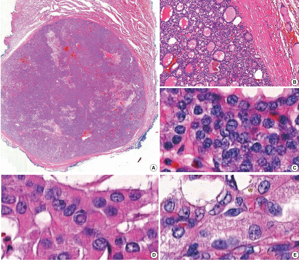
- Because different types of thyroid malignancies have distinct embryological origins, coexisting tumors are rarely observed. We describe a coexisting papillary thyroid carcinoma (PTC) and medullary thyroid carcinoma (MTC) first suspected by fine-needle aspiration cytology (FNAC). A 57-year-old female presented with an irregular mass in the right thyroid lobe. The cytopathologic findings of fine-needle aspiration showed two components: a papillary-like arrangement consisting of cells with pale enlarged nuclei indicative of PTC and loose clusters comprised of oval cells with granular chromatin indicative of MTC. The diagnosis of a coexisting PTC and MTC was initially confirmed by calcitonin immunocytochemistry and later after total thyroidectomy. Although some surgical case reports of PTC and MTC coexisting in either the same or different lobes have been documented, a case suspected by FNAC before the surgery has rarely been reported. Because appropriate treatment and prognosis of PTC and MTC are different, cytopathologists should be aware of this rare entity.
-
Citations
Citations to this article as recorded by- Evaluation of Diagnostic Accuracy of Medullary Thyroid Carcinoma Using Fine‐Needle Aspiration Cytology—Based on a Single Tertiary Centre Experience
Si‐Yi Chen, Dong‐Mei Gu
Cytopathology.2026; 37(3): 255. CrossRef - Synchronous Presence of Papillary, Medullary, and Anaplastic Thyroid Tumors in a Single Patient: A Rare Case Report
Mohammed Al Essa, Reema Aldawish, Abdullah Alkhaldi, Ghaidaa Aljbli, Thamer Althunayan, Abdullah Alkarni, Abdullah Alsalamah
American Journal of Case Reports.2026;[Epub] CrossRef - Synchronous papillary and medullary thyroid carcinoma with distinct genetic mutations: A case report
Huanyu Jiang, Lijuan Zhou, Gang Zou, Haidong Zhang, Zhenkun Yu
Oral Oncology.2025; 161: 107191. CrossRef - Coexisting papillary and medullary thyroid carcinomas in a 60 year old male: a case report
Allahdad Khan, Anam Malik, Abdul Ahad Riaz, Muhammad Hussnain Sadiq, Muhammad Shahzaib Arshad, Alka Rani, Ibrahim Nagmeldin Hassan
Annals of Medicine & Surgery.2025; 87(10): 6740. CrossRef - Dedifferentiated Leiomyosarcoma of the Uterine Corpus with Heterologous Component: Clinicopathological Analysis of Five Consecutive Cases from a Single Institution and Comprehensive Literature Review
Suyeon Kim, Hyunsik Bae, Hyun-Soo Kim
Diagnostics.2024; 14(2): 160. CrossRef - Coexisting Medullary and Papillary Thyroid Carcinomas: A Case of Dual Neoplasia With a High Risk of Misdiagnosis
Santiago Sierra Castillo, Maria A Henao Rincón, David Aristizabal Colorado, David Alexander Vernaza Trujillo, Alin Abreu Lomba
Cureus.2024;[Epub] CrossRef
- Evaluation of Diagnostic Accuracy of Medullary Thyroid Carcinoma Using Fine‐Needle Aspiration Cytology—Based on a Single Tertiary Centre Experience
- Correlation between myoferlin expression and lymph node metastasis in papillary thyroid carcinoma
- Ji Min Na, Dong Chul Kim, Dae Hyun Song, Hyo Jung An, Hyun Min Koh, Jeong-Hee Lee, Jong Sil Lee, Jung Wook Yang, Min Hye Kim
- J Pathol Transl Med. 2022;56(4):199-204. Published online May 11, 2022
- DOI: https://doi.org/10.4132/jptm.2022.03.19

- 5,906 View
- 180 Download
-
Abstract
PDF
- Background
Myoferlin is a multifunctional protein expressed in various normal and cancer cells, with novel oncogenic roles being newly discovered. Recently, correlations have been found between myoferlin expression and unfavorable prognosis in various carcinomas. This study investigated the prognostic role of myoferlin expression in papillary thyroid carcinoma (PTC), specifically that associated with nodal metastasis.
Methods
We collected clinicopathological data and PTC tissues from 116 patients who had been admitted to Gyeongsang National University Hospital in 2010. Immunohistochemical analysis was performed on surgical specimen-derived tissue microarray blocks. Myoferlin expression was graded, and the relationship between expression level and pathological features of tumors based on the American Joint Committee on Cancer staging system was evaluated.
Results
Of the 116 patient samples, 100 cases exhibited positive myoferlin expression. Higher grade of myoferlin expression was correlated with lower T category group (p = .010). Presence of lymph node metastasis was determined to be significantly correlated with low-grade myoferlin expression (p = .019), with no significant difference between pN1a and pN1b tumors.
Conclusions
Our study revealed an adverse correlation between myoferlin expression and pathological features of PTC, evidence of the potential prognostic role of myoferlin in PTC lymph node metastasis.
- A multicenter study of interobserver variability in pathologic diagnosis of papillary breast lesions on core needle biopsy with WHO classification
- Hye Ju Kang, Sun Young Kwon, Ahrong Kim, Woo Gyeong Kim, Eun Kyung Kim, Ae Ree Kim, Chungyeul Kim, Soo Kee Min, So Young Park, Sun Hee Sung, Hye Kyoung Yoon, Ahwon Lee, Ji Shin Lee, Hyang Im Lee, Ho Chang Lee, Sung Chul Lim, Sun Young Jun, Min Jung Jung, Chang Won Jung, Soo Youn Cho, Eun Yoon Cho, Hye Jeong Choi, So Yeon Park, Jee Yeon Kim, In Ae Park, Youngmee Kwon
- J Pathol Transl Med. 2021;55(6):380-387. Published online October 6, 2021
- DOI: https://doi.org/10.4132/jptm.2021.07.29
- 8,521 View
- 230 Download
- 4 Web of Science
- 5 Crossref
-
Abstract
PDFSupplementary Material
- Background
Papillary breast lesions (PBLs) comprise diverse entities from benign and atypical lesions to malignant tumors. Although PBLs are characterized by a papillary growth pattern, it is challenging to achieve high diagnostic accuracy and reproducibility. Thus, we investigated the diagnostic reproducibility of PBLs in core needle biopsy (CNB) specimens with World Health Organization (WHO) classification.
Methods
Diagnostic reproducibility was assessed using interobserver variability (kappa value, κ) and agreement rate in the pathologic diagnosis of 60 PBL cases on CNB among 20 breast pathologists affiliated with 20 medical institutions in Korea. This analysis was performed using hematoxylin and eosin (H&E) staining and immunohistochemical (IHC) staining for cytokeratin 5 (CK5) and p63. The pathologic diagnosis of PBLs was based on WHO classification, which was used to establish simple classifications (4-tier, 3-tier, and 2-tier).
Results
On WHO classification, H&E staining exhibited ‘fair agreement’ (κ = 0.21) with a 47.0% agreement rate. Simple classifications presented improvement in interobserver variability and agreement rate. IHC staining increased the kappa value and agreement rate in all the classifications. Despite IHC staining, the encapsulated/solid papillary carcinoma (EPC/SPC) subgroup (κ = 0.16) exhibited lower agreement compared to the non-EPC/SPC subgroup (κ = 0.35) with WHO classification, which was similar to the results of any other classification systems.
Conclusions
Although the use of IHC staining for CK5 and p63 increased the diagnostic agreement of PBLs in CNB specimens, WHO classification exhibited a higher discordance rate compared to any other classifications. Therefore, this result warrants further intensive consensus studies to improve the diagnostic reproducibility of PBLs with WHO classification. -
Citations
Citations to this article as recorded by- Beyond the benign: A rare case report of myxoid pleomorphic liposarcoma
Arslan Ahmad, Muhammad Ammar, Muhammad Hasnain Saleem Choudary, Muhammad Nouman Sadiq, Rana Uzair Ahmad, Nouman Aziz
Radiology Case Reports.2025; 20(5): 2500. CrossRef - Invasive papillary carcinoma of the breast
Shijing Wang, Qingfu Zhang, Xiaoyun Mao
Frontiers in Oncology.2024;[Epub] CrossRef - Recommendations for Performance Evaluation of Machine Learning in Pathology: A Concept Paper From the College of American Pathologists
Matthew G. Hanna, Niels H. Olson, Mark Zarella, Rajesh C. Dash, Markus D. Herrmann, Larissa V. Furtado, Michelle N. Stram, Patricia M. Raciti, Lewis Hassell, Alex Mays, Liron Pantanowitz, Joseph S. Sirintrapun, Savitri Krishnamurthy, Anil Parwani, Giovann
Archives of Pathology & Laboratory Medicine.2024; 148(10): e335. CrossRef - Encapsulated papillary carcinoma of the breast: A single institution experience
Liang Xu, Qixin Mao, Qiuming Liu, Yufeng Gao, Lihua Luo, Chungen Guo, Wei Qu, Ningning Yan, Yali Cao
Oncology Letters.2023;[Epub] CrossRef - High-risk and selected benign breast lesions diagnosed on core needle biopsy: Evidence for and against immediate surgical excision
Aparna Harbhajanka, Hannah L. Gilmore, Benjamin C. Calhoun
Modern Pathology.2022; 35(11): 1500. CrossRef
- Beyond the benign: A rare case report of myxoid pleomorphic liposarcoma
- Proto-oncogene Pokemon in thyroid cancer: a potential promoter of tumorigenesis in papillary thyroid carcinoma
- Kyungseek Chang, Sung-Im Do, Kyungeun Kim, Seoung Wan Chae, In-gu Do, Hyun Joo Lee, Dong Hoon Kim, Jin Hee Sohn
- J Pathol Transl Med. 2021;55(5):317-323. Published online August 9, 2021
- DOI: https://doi.org/10.4132/jptm.2021.06.28
- 7,083 View
- 132 Download
- 4 Web of Science
- 4 Crossref
-
Abstract
PDFSupplementary Material
- Background
Pokemon is an oncogenic transcription regulator that plays a critical role in cellular differentiation. Although it has been found to be overexpressed in several types of cancer involving different organs, its role in thyroid gland has yet to be reported. The objective of this study was to evaluate the expression of Pokemon in papillary thyroid carcinoma (PTC) based on clinicopathological parameters.
Methods
Tissue microarray samples derived from patients with PTC or benign thyroid disease were used to evaluate Pokemon expression based on immunohistochemical analysis. Correlations of its expression with various clinicopathological parameters were then analyzed.
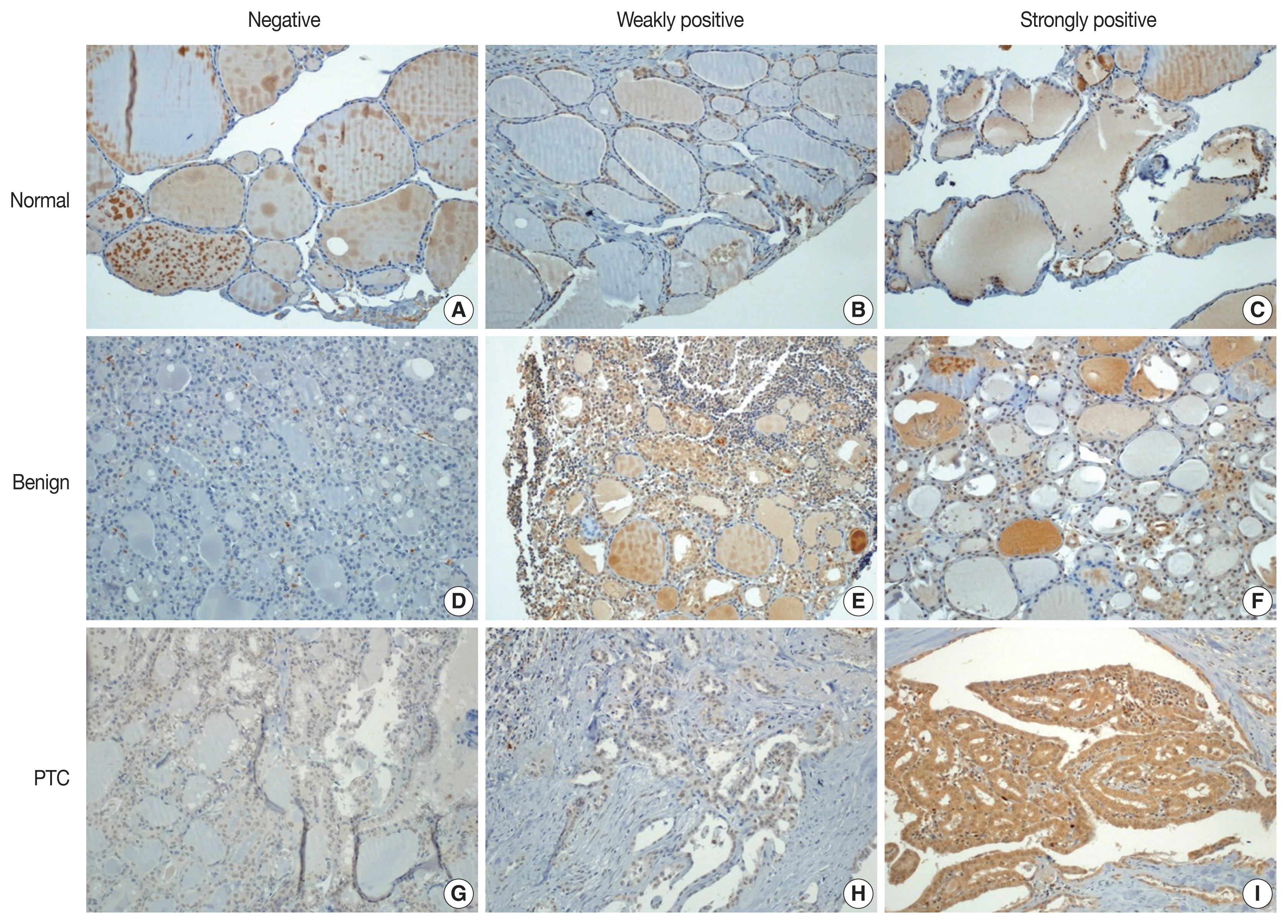
Results
Pokemon expression was observed in 22.0% of thyroid follicular cells from the normal group, 44.0% from the group with benign thyroid diseases, and 92.1% from the group with PTC (p < .001). The intensity of Pokemon expression was markedly higher in the PTC group. Pokemon expression level and PTC tumor size showed an inverse correlation. T1a tumors showed strong expression levels of Pokemon. However, larger tumors showed weak expression (p = .006).
Conclusions
Pokemon expression is associated with tumorigenesis of PTC, with expression showing an inverse correlation with PTC tumor size. This might be related to the negative regulation of aerobic glycolysis by Pokemon. -
Citations
Citations to this article as recorded by- Systems biology approach delineates critical pathways associated with papillary thyroid cancer: a multi-omics data analysis
Febby Payva, Santhy K. S., Remya James, Amrisa Pavithra E, Venketesh Sivaramakrishnan
Thyroid Research.2025;[Epub] CrossRef - Understanding the dysregulation of PURPL, a novel long intergenic noncoding RNA, in thyroid cancer progression
Mina Kazemzadeh, Reza Safaralizadeh, Amir Ali Mokhtarzadeh, Mohammad Ali Hosseinpour Feizi
Human Gene.2025; 46: 201499. CrossRef - ZBTB7A as a therapeutic target for cancer
Ying Zhou, Xisha Chen, Xuyu Zu
Biochemical and Biophysical Research Communications.2024; 736: 150888. CrossRef - Knockdown of FBI-1 Inhibits the Warburg Effect and Enhances the Sensitivity of Hepatocellular Carcinoma Cells to Molecular Targeted Agents via miR-3692/HIF-1α
Juan Liu, Chao Yang, Xiao-Mei Huang, Pan-Pan Lv, Ya-Kun Yang, Jin-Na Zhao, Si-Yuan Zhao, Wan-Jun Sun
Frontiers in Oncology.2021;[Epub] CrossRef
- Systems biology approach delineates critical pathways associated with papillary thyroid cancer: a multi-omics data analysis
- Highly prevalent BRAF V600E and low-frequency TERT promoter mutations underlie papillary thyroid carcinoma in Koreans
- Sue Youn Kim, Taeeun Kim, Kwangsoon Kim, Ja Seong Bae, Jeong Soo Kim, Chan Kwon Jung
- J Pathol Transl Med. 2020;54(4):310-317. Published online June 15, 2020
- DOI: https://doi.org/10.4132/jptm.2020.05.12
- 12,996 View
- 199 Download
- 30 Web of Science
- 33 Crossref
-
Abstract
PDF
- Background
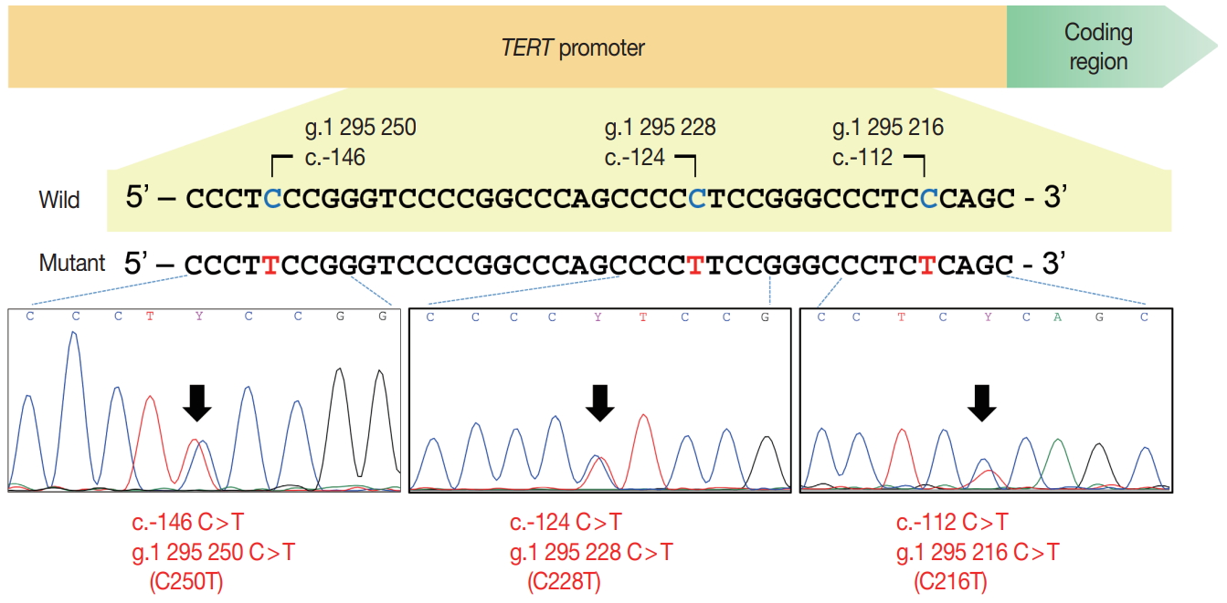
The presence of telomerase reverse transcriptase (TERT) promoter mutations have been associated with a poor prognosis in patients with papillary thyroid carcinomas (PTC). The frequency of TERT promoter mutations varies widely depending on the population and the nature of the study.
Methods
Data were prospectively collected in 724 consecutive patients who underwent thyroidectomy for PTC from 2018 to 2019. Molecular testing for BRAF V600E and TERT promoter mutations was performed in all cases.
Results
TERT promoter alterations in two hotspots (C228T and C250T) and C216T were found in 16 (2.2%) and 4 (0.6%) of all PTCs, respectively. The hotspot mutations were significantly associated with older age at diagnosis, larger tumor size, extrathyroidal extension, higher pathologic T category, lateral lymph node metastasis, and higher American Thyroid Association recurrence risk. The patients with C216T variant were younger and had a lower American Thyroid Association recurrence risk than those with hotspot mutations. Concurrent BRAF V600E was found in 19 of 20 cases with TERT promoter mutations. Of 518 microcarcinomas measuring ≤1.0 cm in size, hotspot mutations and C216T variants were detected in five (1.0%) and three (0.6%) cases, respectively.
Conclusions
Our study indicates a low frequency of TERT promoter mutations in Korean patients with PTC and supports previous findings that TERT promoter mutations are more common in older patients with unfavorable clinicopathologic features and BRAF V600E. TERT promoter mutations in patients with microcarcinoma are uncommon and may have a limited role in risk stratification. The C216T variant seems to have no clinicopathologic effect on PTC. -
Citations
Citations to this article as recorded by- Evaluation of BRAF V600E and TERT mutation analysis in differential thyroid cancers
Nigar Aktash, Ahmet Cem Dural, Husnu Aydin, Nuri Alper Sahbaz, Deniz Guzey, Serdar Altınay, Cevher Akarsu, Yasir Musa Kesgin, Sezer Bulut, Mehmet Karabulut
Updates in Surgery.2026; 78(2): 783. CrossRef - Summary and Analysis of Molecular Biological Changes, PD-L1 Immune Status and Clinicopathological Features of 78 Cases of Papillary Thyroid Carcinoma (<1 cm in Diameter) Combined With Lateral Cervical Lymph Node Metastasis
Xiaoteng Sun, Zhengyan He, Weijie Yu, Baoyuan Li, Xinmiao Xu, Xiaoqin Zhang, Minglong Yin
Applied Immunohistochemistry & Molecular Morphology.2026; 34(2): 95. CrossRef - Integrated clinicopathological and molecular profiling of papillary thyroid carcinoma identifies predominant BRAF and TERT promoter mutations: a cross-sectional study
Rawezh Qadir M. Salih, Dlnya A. Mohammed, Nizar M.T. Hamawandi
Journal of Biological Research - Bollettino della Società Italiana di Biologia Sperimentale.2026;[Epub] CrossRef - The impact of C216T and hot spot mutations of the TERT promoter on the clinicopathologic characteristics and S100A10 expression in papillary thyroid carcinoma: a comparative study
Ping Li, Chuqiang Huang, Xiaoling Liu, Huihui Gui, Jian Li
Diagnostic Pathology.2025;[Epub] CrossRef - Refining NTRK Fusion Detection in Papillary Thyroid Carcinoma Through Pan-TRK Immunohistochemistry and Histopathologic Features
Hyun Lee, Sue Youn Kim, Ji Min Park, Seung-Hyun Jung, Ozgur Mete, Chan Kwon Jung
Endocrine Pathology.2025;[Epub] CrossRef - Validation of Diagnostic Utility of Washout CYFRA 21-1 in Lymph Node Metastasis of Thyroid Cancer
Jeongmin Lee, Yuri Shin, Jeongun Kwak, Hye Lim Park, Sohee Lee, Mee Kyung Kim, Ja Seong Bae, Chan Kwon Jung, So Lyung Jung, Jung-Min Lee, Sang-Ah Chang, Dong-Jun Lim
Clinical Cancer Research.2025; 31(10): 1922. CrossRef - 2025 Korean Thyroid Association Clinical Management Guideline on Active Surveillance for Low-Risk Papillary Thyroid Carcinoma
Eun Kyung Lee, Min Joo Kim, Seung Heon Kang, Bon Seok Koo, Kyungsik Kim, Mijin Kim, Bo Hyun Kim, Ji-hoon Kim, Shin Je Moon, Kyorim Back, Young Shin Song, Jong-hyuk Ahn, Hwa Young Ahn, Ho-Ryun Won, Won Sang Yoo, Min Kyoung Lee, Jeongmin Lee, Ji Ye Lee, Kyo
International Journal of Thyroidology.2025; 18(1): 30. CrossRef - 2025 Korean Thyroid Association Clinical Management Guideline on Active Surveillance for Low-Risk Papillary Thyroid Carcinoma
Eun Kyung Lee, Min Joo Kim, Seung Heon Kang, Bon Seok Koo, Kyungsik Kim, Mijin Kim, Bo Hyun Kim, Ji-hoon Kim, Shinje Moon, Kyorim Back, Young Shin Song, Jong-hyuk Ahn, Hwa Young Ahn, Ho-Ryun Won, Won Sang Yoo, Min Kyoung Lee, Jeongmin Lee, Ji Ye Lee, Kyon
Endocrinology and Metabolism.2025; 40(3): 307. CrossRef - Active surveillance for adult low-risk papillary thyroid microcarcinoma—a review focused on the 30-year experience of Kuma Hospital—
Yasuhiro Ito, Akira Miyauchi, Makoto Fujishima, Masashi Yamamoto, Takahiro Sasaki
Endocrine Journal.2024; 71(1): 7. CrossRef - Diagnostic utilities of washout CYFRA 21-1 combined with washout thyroglobulin for metastatic lymph nodes in thyroid cancer: a prospective study
Joonseon Park, Solji An, Kwangsoon Kim, Jeong Soo Kim, Chan Kwon Jung, Ja Seong Bae
Scientific Reports.2024;[Epub] CrossRef - Korean Thyroid Association Guidelines on the Management of Differentiated Thyroid Cancers; Part I. Initial Management of Differentiated Thyroid Cancers - Chapter 5. Evaluation of Recurrence Risk Postoperatively and Initial Risk Stratification in Different
Eun Kyung Lee, Young Shin Song, Ho-Cheol Kang, Sun Wook Kim, Dong Gyu Na, Shin Je Moon, Dong-Jun Lim, Kyong Yeun Jung, Yun Jae Chung, Chan Kwon Jung, Young Joo Park
International Journal of Thyroidology.2024; 17(1): 68. CrossRef - Korean Thyroid Association Management Guidelines for Patients with Thyroid Nodules 2024
Young Joo Park, Eun Kyung Lee, Young Shin Song, Su Hwan Kang, Bon Seok Koo, Sun Wook Kim, Dong Gyu Na, Seung-Kuk Baek, So Won Oh, Min Kyoung Lee, Sang-Woo Lee, Young Ah Lee, Yong Sang Lee, Ji Ye Lee, Dong-Jun Lim, Leehi Joo, Yuh-Seog Jung, Chan Kwon Jung,
International Journal of Thyroidology.2024; 17(1): 208. CrossRef - PD-L1 Expression and Its Modulating Factors in Anaplastic Thyroid Carcinoma
Shipra Agarwal, Chan Kwon Jung, Pranitha Gaddam, Mitsuyoshi Hirokawa, Takuya Higashiyama, Jen-Fan Hang, Wei-An Lai, Somboon Keelawat, Zhiyan Liu, Hee Young Na, So Yeon Park, Junya Fukuoka, Shinya Satoh, Zhanna Mussazhanova, Masahiro Nakashima, Kennichi Ka
American Journal of Surgical Pathology.2024; 48(10): 1233. CrossRef - 2023 Korean Thyroid Association Management Guidelines for Patients with Thyroid Nodules
Young Joo Park, Eun Kyung Lee, Young Shin Song, Soo Hwan Kang, Bon Seok Koo, Sun Wook Kim, Dong Gyu Na, Seung-Kuk Baek, So Won Oh, Min Kyoung Lee, Sang-Woo Lee, Young Ah Lee, Yong Sang Lee, Ji Ye Lee, Dong-Jun Lim, Leehi Joo, Yuh-Seog Jung, Chan Kwon Jung
International Journal of Thyroidology.2023; 16(1): 1. CrossRef - Incidence and risk factors for occult lesions in low-risk papillary thyroid microcarcinoma patients with tumor characteristics appropriate for thermal ablation: A retrospective study
Langping Jin, Kaijun Zhu, Changliang Xu, Jiaying Lu, Liming Huang
Medicine.2023; 102(38): e34938. CrossRef - Identification of NIFTP-Specific mRNA Markers for Reliable Molecular Diagnosis of Thyroid Tumors
So-Yeon Lee, Jong-Lyul Park, Kwangsoon Kim, Ja Seong Bae, Jae-Yoon Kim, Seon-Young Kim, Chan Kwon Jung
Endocrine Pathology.2023; 34(3): 311. CrossRef - Risk factors and predictive model for recurrence in papillary thyroid carcinoma: a single-center retrospective cohort study based on 955 cases
Yin Li, Jiahe Tian, Ke Jiang, Zhongyu Wang, Songbo Gao, Keyang Wei, Ankui Yang, Qiuli Li
Frontiers in Endocrinology.2023;[Epub] CrossRef - BRAFV600E Positivity-Dependent Effect of Age on Papillary Thyroid Cancer Recurrence Risk
Joonseon Park, Solji An, Kwangsoon Kim, Ja Seong Bae, Jeong Soo Kim
Cancers.2023; 15(22): 5395. CrossRef - BRAFV600E mutation test on fine‐needle aspiration specimens of thyroid nodules: Clinical correlations for 4600 patients
Huang Chen, Aiping Song, Ye Wang, Yifan He, Jie Tong, Jinxi Di, Chun Li, Zhongren Zhou, Xiaopin Cai, Dingrong Zhong, Jiping Da
Cancer Medicine.2022; 11(1): 40. CrossRef - Clinicopathological indicators for TERT promoter mutation in papillary thyroid carcinoma
Hee Young Na, Hyeong Won Yu, Woochul Kim, Jae Hoon Moon, Chang Ho Ahn, Sang Il Choi, Yeo Koon Kim, June Young Choi, So Yeon Park
Clinical Endocrinology.2022; 97(1): 106. CrossRef - A Systematic Review and Meta‐analysis on the Occurrence of Biomarker Mutation in Colorectal Cancer among the Asian Population
Hafeez Afolabi, Salzihan Md Salleh, Zaidi Zakaria, Ch’ng Ewe Seng, Siti Norasikin Binti Mohd Nafil, Ahmad Aizat Bin Abdul Aziz, Yusuf Wada, Ahmad Irekeola, Syed Sameer Aga
BioMed Research International.2022;[Epub] CrossRef -
A Significance of Concomitant BRAF
V600E
and TERT Mutations in Polish Patients with Papillary Thyroid Microcarcinoma: A Retrospective Cohort Study Based on 430 Cases
Artur Kuchareczko, Janusz Kopczyński, Artur Kowalik, Kinga Hińcza-Nowak, Agnieszka Walczyk, Iwona Pałyga, Tomasz Trybek, Monika Szymonek, Danuta Gąsior-Perczak, Klaudia Gadawska-Juszczyk, Estera Mikina, Izabela Płachta, Agnieszka Suligowska, Agnieszka Płu
Thyroid®.2022; 32(11): 1372. CrossRef - Machine learning for identifying benign and malignant of thyroid tumors: A retrospective study of 2,423 patients
Yuan-yuan Guo, Zhi-jie Li, Chao Du, Jun Gong, Pu Liao, Jia-xing Zhang, Cong Shao
Frontiers in Public Health.2022;[Epub] CrossRef - TERT Promoter and BRAF V600E Mutations in Papillary Thyroid Cancer: A Single-Institution Experience in Korea
Min Jhi Kim, Jin Kyong Kim, Gi Jeong Kim, Sang-Wook Kang, Jandee Lee, Jong Ju Jeong, Woong Youn Chung, Daham Kim, Kee-Hyun Nam
Cancers.2022; 14(19): 4928. CrossRef - Frequency of TERT Promoter Mutations in Real-World Analysis of 2,092 Thyroid Carcinoma Patients (Endocrinol Metab 2022;37:652-63, Heera Yang et al.)
Sue Youn Kim, Chan Kwon Jung
Endocrinology and Metabolism.2022; 37(6): 947. CrossRef - Frequency of TERT Promoter Mutations in Real-World Analysis of 2,092 Thyroid Carcinoma Patients (Endocrinol Metab 2022;37:652-63, Heera Yang et al.)
Hyunju Park, Jae Hoon Chung
Endocrinology and Metabolism.2022; 37(6): 949. CrossRef - Molecular Pathology of Non-familial Follicular Epithelial–Derived Thyroid Cancer in Adults: From RAS/BRAF-like Tumor Designations to Molecular Risk Stratification
Paula Soares, Antónia Afonso Póvoa, Miguel Melo, João Vinagre, Valdemar Máximo, Catarina Eloy, José Manuel Cameselle-Teijeiro, Manuel Sobrinho-Simões
Endocrine Pathology.2021; 32(1): 44. CrossRef - Clinicopathological Characteristics and Recurrence-Free Survival of Rare Variants of Papillary Thyroid Carcinomas in Korea: A Retrospective Study
Mijin Kim, Sun Wook Cho, Young Joo Park, Hwa Young Ahn, Hee Sung Kim, Yong Joon Suh, Dughyun Choi, Bu Kyung Kim, Go Eun Yang, Il-Seok Park, Ka Hee Yi, Chan Kwon Jung, Bo Hyun Kim
Endocrinology and Metabolism.2021; 36(3): 619. CrossRef - Clinical Application of TERT Promoter Mutations in Urothelial Carcinoma
Yujiro Hayashi, Kazutoshi Fujita, George J. Netto, Norio Nonomura
Frontiers in Oncology.2021;[Epub] CrossRef - MicroRNA Profile for Diagnostic and Prognostic Biomarkers in Thyroid Cancer
Jong-Lyul Park, Seon-Kyu Kim, Sora Jeon, Chan-Kwon Jung, Yong-Sung Kim
Cancers.2021; 13(4): 632. CrossRef - Prospective Analysis of TERT Promoter Mutations in Papillary Thyroid Carcinoma at a Single Institution
Yun-Suk Choi, Seong-Woon Choi, Jin-Wook Yi
Journal of Clinical Medicine.2021; 10(10): 2179. CrossRef - Significance of telomerase reverse-transcriptase promoter mutations in differentiated thyroid cancer
Hung-Fei Lai, Chi-Yu Kuo, Shih-Ping Cheng
Formosan Journal of Surgery.2021; 54(5): 171. CrossRef - Early Diagnosis of Low-Risk Papillary Thyroid Cancer Results Rather in Overtreatment Than a Better Survival
Jolanta Krajewska, Aleksandra Kukulska, Malgorzata Oczko-Wojciechowska, Agnieszka Kotecka-Blicharz, Katarzyna Drosik-Rutowicz, Malgorzata Haras-Gil, Barbara Jarzab, Daria Handkiewicz-Junak
Frontiers in Endocrinology.2020;[Epub] CrossRef
- Evaluation of BRAF V600E and TERT mutation analysis in differential thyroid cancers
- A Multi-institutional Study of Prevalence and Clinicopathologic Features of Non-invasive Follicular Thyroid Neoplasm with Papillary-like Nuclear Features (NIFTP) in Korea
- Ja Yeong Seo, Ji Hyun Park, Ju Yeon Pyo, Yoon Jin Cha, Chan Kwon Jung, Dong Eun Song, Jeong Ja Kwak, So Yeon Park, Hee Young Na, Jang-Hee Kim, Jae Yeon Seok, Hee Sung Kim, Soon Won Hong
- J Pathol Transl Med. 2019;53(6):378-385. Published online October 21, 2019
- DOI: https://doi.org/10.4132/jptm.2019.09.18
- 10,133 View
- 346 Download
- 17 Web of Science
- 16 Crossref
-
Abstract
PDF
- Background
In the present multi-institutional study, the prevalence and clinicopathologic characteristics of non-invasive follicular thyroid neoplasm with papillary-like nuclear features (NIFTP) were evaluated among Korean patients who underwent thyroidectomy for papillary thyroid carcinoma (PTC).
Methods
Data from 18,819 patients with PTC from eight university hospitals between January 2012 and February 2018 were retrospectively evaluated. Pathology reports of all PTCs and slides of potential NIFTP cases were reviewed. The strict criterion of no papillae was applied for the diagnosis of NIFTP. Due to assumptions regarding misclassification of NIFTP as non-PTC tumors, the lower boundary of NIFTP prevalence among PTCs was estimated. Mutational analysis for BRAF and three RAS isoforms was performed in 27 randomly selected NIFTP cases.
Results
The prevalence of NIFTP was 1.3% (238/18,819) of all PTCs when the same histologic criteria were applied for NIFTP regardless of the tumor size but decreased to 0.8% (152/18,819) when tumors ≥1 cm in size were included. The mean follow-up was 37.7 months and no patient with NIFTP had evidence of lymph node metastasis, distant metastasis, or disease recurrence during the follow-up period. A difference in prevalence of NIFTP before and after NIFTP introduction was not observed. BRAFV600E mutation was not found in NIFTP. The mutation rate for the three RAS genes was 55.6% (15/27).
Conclusions
The low prevalence and indolent clinical outcome of NIFTP in Korea was confirmed using the largest number of cases to date. The introduction of NIFTP may have a small overall impact in Korean practice. -
Citations
Citations to this article as recorded by- Case report & review: Bilateral NIFTP harboring concomitant HRAS and KRAS mutation: Report of an unusual case and literature review
Marianna Rita Brogna, Francesca Collina, Maria Grazia Chiofalo, Debora De Bartolo, Angela Montone, Maria Rosaria Schiano, Michele Del Sesto, Nubia Pizza, Gerardo Ferrara
Molecular Carcinogenesis.2024; 63(12): 2273. CrossRef - Non-invasive follicular thyroid neoplasm with papillary-like nuclear features (NIFTP): what do we need to know?
Andrés Coca-Pelaz, Juan P. Rodrigo, Abbas Agaimy, Dana M. Hartl, Göran Stenman, Vincent Vander Poorten, Antti A. Mäkitie, Mark Zafereo, Karthik N. Rao, Gregory W. Randolph, Alessandra Rinaldo, Alfio Ferlito
Virchows Archiv.2024; 485(6): 977. CrossRef - Study of non-invasive follicular thyroid neoplasm: A borderline entity
Rupali Bavikar, Ruchi S. Randive, Anubhaw Verma, Madhuri Singh, Vidya Viswanathan, Arpana Dharwadkar
Journal of Cancer Research and Therapeutics.2024; 20(5): 1365. CrossRef - Analysis of a pre-2017 follicular variant papillary thyroid carcinoma cohort reclassified as noninvasive follicular thyroid neoplasm with papillary-like features (NIFTP): an 11-year retrospective single institution experience
Shaham Beg, Sana Irfan Khan, Isabella Cui, Theresa Scognamiglio, Rema Rao
Journal of the American Society of Cytopathology.2023; 12(2): 112. CrossRef - Noninvasive Follicular Thyroid Neoplasm With Papillary-Like Nuclear Features: What a Surgeon Should Know
Jabir Alharbi, Thamer Alraddadi, Haneen Sebeih, Mohammad A Alessa, Haddad H Alkaf, Ahmed Bahaj, Sherif K Abdelmonim
Cureus.2023;[Epub] CrossRef - NTRK Fusion in a Cohort of BRAF p. V600E Wild-Type Papillary Thyroid Carcinomas
Seung Eun Lee, Mi-Sook Lee, Heejin Bang, Mi Young Kim, Yoon-La Choi, Young Lyun Oh
Modern Pathology.2023; 36(8): 100180. CrossRef - A Comprehensive Study on the Diagnosis and Management of Noninvasive Follicular Thyroid Neoplasm with Papillary-Like Nuclear Features
Bayan A. Alzumaili, Lauren N. Krumeich, Reagan Collins, Timothy Kravchenko, Emad I. Ababneh, Adam S. Fisch, William C. Faquin, Vania Nosé, Maria Martinez-Lage, Gregory W. Randolph, Rajshri M. Gartland, Carrie C. Lubitz, Peter M. Sadow
Thyroid®.2023; 33(5): 566. CrossRef - Clinical-Pathological and Molecular Evaluation of 451 NIFTP Patients from a Single Referral Center
Paola Vignali, Agnese Proietti, Elisabetta Macerola, Anello Marcello Poma, Liborio Torregrossa, Clara Ugolini, Alessio Basolo, Antonio Matrone, Teresa Rago, Ferruccio Santini, Rossella Elisei, Gabriele Materazzi, Fulvio Basolo
Cancers.2022; 14(2): 420. CrossRef - Noninvasive follicular thyroid neoplasm with papillary-like nuclear features: its updated diagnostic criteria, preoperative cytologic diagnoses and impact on the risk of malignancy
Hee Young Na, So Yeon Park
Journal of Pathology and Translational Medicine.2022; 56(6): 319. CrossRef - SFE-AFCE-SFMN 2022 Consensus on the management of thyroid nodules : Follow-up: How and how long?
Sophie Leboulleux, Livia Lamartina, Emmanuelle Lecornet Sokol, Fabrice Menegaux, Laurence Leenhardt, Gilles Russ
Annales d'Endocrinologie.2022; 83(6): 407. CrossRef - Different Threshold of Malignancy for RAS-like Thyroid Tumors Causes Significant Differences in Thyroid Nodule Practice
Kennichi Kakudo
Cancers.2022; 14(3): 812. CrossRef - Clinicopathological parameters for predicting non-invasive follicular thyroid neoplasm with papillary features (NIFTP)
Eunju Jang, Kwangsoon Kim, Chan Kwon Jung, Ja Seong Bae, Jeong Soo Kim
Therapeutic Advances in Endocrinology and Metabolism.2021;[Epub] CrossRef - The Incidence of Noninvasive Follicular Thyroid Neoplasm with Papillary-Like Nuclear Features: A Meta-Analysis Assessing Worldwide Impact of the Reclassification
Chanchal Rana, Huy Gia Vuong, Thu Quynh Nguyen, Hoang Cong Nguyen, Chan Kwon Jung, Kennichi Kakudo, Andrey Bychkov
Thyroid.2021;[Epub] CrossRef - The Genomic Landscape of Thyroid Cancer Tumourigenesis and Implications for Immunotherapy
Amandeep Singh, Jeehoon Ham, Joseph William Po, Navin Niles, Tara Roberts, Cheok Soon Lee
Cells.2021; 10(5): 1082. CrossRef - Noninvasive follicular thyroid neoplasm with papillary-like nuclear features (NIFTP) is rare, benign lesion using modified stringent diagnostic criteria: Reclassification and outcome study
David Cubero Rego, Hwajeong Lee, Anne Boguniewicz, Timothy A. Jennings
Annals of Diagnostic Pathology.2020; 44: 151439. CrossRef - Noninvasive Follicular Thyroid Neoplasm with Papillary-Like Nuclear Features: From Echography to Genetic Profile
Francesca Maletta, Enrico Costantino Falco, Alessandro Gambella, Jasna Metovic, Mauro Papotti
The Tohoku Journal of Experimental Medicine.2020; 252(3): 209. CrossRef
- Case report & review: Bilateral NIFTP harboring concomitant HRAS and KRAS mutation: Report of an unusual case and literature review
- Prognostic Role of Metastatic Lymph Node Ratio in Papillary Thyroid Carcinoma
- Jung-Soo Pyo, Jin Hee Sohn, Kyungseek Chang
- J Pathol Transl Med. 2018;52(5):331-338. Published online August 30, 2018
- DOI: https://doi.org/10.4132/jptm.2018.08.07
- 10,703 View
- 132 Download
- 16 Web of Science
- 15 Crossref
-
Abstract
PDF
- Background
The aim of this study is to elucidate the clinicopathological significances, including the prognostic role, of metastatic lymph node ratio (mLNR) and tumor deposit diameter in papillary thyroid carcinoma (PTC) through a retrospective review and meta-analysis.
Methods
We categorized the cases into high (≥ 0.44) and low mLNR (< 0.44) and investigated the correlations with clinicopathological parameters in 64 PTCs with neck level VI lymph node (LN) metastasis. In addition, meta-analysis of seven eligible studies was used to investigate the correlation between mLNR and survival.
Results
Among 64 PTCs with neck level VI LN metastasis, high mLNR was found in 34 PTCs (53.1%). High mLNR was significantly correlated with macrometastasis (tumor deposit diameter ≥ 0.2 cm), extracapsular spread, and number of metastatic LNs. Based on linear regression test, mLNR was significantly increased by the largest LN size but not the largest metastatic LN (mLN) size. High mLNR was not correlated with nuclear factor κB or cyclin D1 immunohistochemical expression, Ki-67 labeling index, or other pathological parameters of primary tumor. Based on meta-analysis, high mLNR significantly correlated with worse disease-free survival at the 5-year and 10-year follow-up (hazard ratio [HR], 4.866; 95% confidence interval [CI], 3.527 to 6.714 and HR, 5.769; 95% CI, 2.951 to 11.275, respectively).
Conclusions
Our data showed that high mLNR significantly correlated with worse survival, macrometastasis, and extracapsular spread of mLNs. Further cumulative studies for more detailed criteria of mLNR are needed before application in daily practice. -
Citations
Citations to this article as recorded by- The application of a clinical-multimodal ultrasound radiomics model for predicting cervical lymph node metastasis of thyroid papillary carcinoma
Chang Liu, Shangjie Yang, Tian Xue, Qian Zhang, Yanjing Zhang, Yufang Zhao, Guolin Yin, Xiaohui Yan, Ping Liang, Liping Liu
Frontiers in Oncology.2025;[Epub] CrossRef - The Predictive Value of a Nomogram Based on Ultrasound Radiomics, Clinical Factors, and Enhanced Ultrasound Features for Central Lymph Node Metastasis in Papillary Thyroid Microcarcinoma
Lei Gao, Xiuli Wen, Guanghui Yue, Hui Wang, Ziqing Lu, Beibei Wu, Zhihong Liu, Yuming Wu, Dongmei Lin, Shijian Yi, Wei Jiang, Yi Hao
Ultrasonic Imaging.2025; 47(2): 93. CrossRef - Lymph Node Metastasis Ratio: Prognostic Significance in Papillary Thyroid Cancer
Ana Rita Ferreira, Diogo Ramalho, Daniela Martins, Andreia Amado, Susana Graça, Carlos Soares, Bela Pereira, Maria João Oliveira, Manuel Oliveira, Antónia Póvoa
Indian Journal of Surgery.2025; 87(6): 1047. CrossRef - CD105 (Endoglin) Expression as a Prognostic Marker in Aggressive Papillary Thyroid Carcinoma
İlker Çordan, Tuğba Günler
Clinical Endocrinology.2025; 103(4): 596. CrossRef - Application and subgroup analysis of competing risks model based on different lymph node staging systems in differentiated thyroid cancer
Zhe Xu Cao, Jiang Sheng Huang, Ming Ming Wang
Updates in Surgery.2024; 76(5): 1927. CrossRef - Цитологічне прогнозування агресії раку щитоподібної залози як новий перспективний напрямок у клінічній тиреоїдології
H.V. Zelinska
Endokrynologia.2024; 29(4): 363. CrossRef - Thyroglobulin expression, Ki-67 index, and lymph node ratio in the prognostic assessment of papillary thyroid cancer
Helene Lindfors, Marie Karlsen, Ellinor Karlton, Jan Zedenius, Catharina Larsson, Catharina Ihre Lundgren, C. Christofer Juhlin, Ivan Shabo
Scientific Reports.2023;[Epub] CrossRef - Incidental Node Metastasis as an Independent Factor of Worse Disease-Free Survival in Patients with Papillary Thyroid Carcinoma
Renan Aguera Pinheiro, Ana Kober Leite, Beatriz Godoi Cavalheiro, Evandro Sobroza de Mello, Luiz Paulo Kowalski, Leandro Luongo Matos
Cancers.2023; 15(3): 943. CrossRef - A High-Quality Nomogram for Predicting Lung Metastasis in Newly Diagnosed Stage IV Thyroid Cancer: A Population-Based Study
WenYi Wang, JiaJing Liu, XiaoFan Xu, LiQun Huo, XuLin Wang, Jun Gu
Technology in Cancer Research & Treatment.2023;[Epub] CrossRef - Lymph Node Ratio Predicts Recurrence in Patients with Papillary Thyroid Carcinoma with Low Lymph Node Yield
Il Ku Kang, Joonseon Park, Ja Seong Bae, Jeong Soo Kim, Kwangsoon Kim
Cancers.2023; 15(11): 2947. CrossRef - Superiority of metastatic lymph node ratio over number of node metastases and TNM/AJCC N classification in predicting cancer‐specific survival in medullary thyroid cancer
Andreas Machens, Kerstin Lorenz, Frank Weber, Henning Dralle
Head & Neck.2022; 44(12): 2717. CrossRef - Value of Combining Clinical Factors, Conventional Ultrasound, and Contrast-Enhanced Ultrasound Features in Preoperative Prediction of Central Lymph Node Metastases of Different Sized Papillary Thyroid Carcinomas
Yanfang Wang, Fang Nie, Guojuan Wang, Ting Liu, Tiantian Dong, Yamin Sun
Cancer Management and Research.2021; Volume 13: 3403. CrossRef - Atypical Histiocytoid Cells and Multinucleated Giant Cells in Fine-Needle Aspiration Cytology of the Thyroid Predict Lymph Node Metastasis of Papillary Thyroid Carcinoma
Ji Eun Choi, Ja Seong Bae, Dong-Jun Lim, So Lyung Jung, Chan Kwon Jung
Cancers.2019; 11(6): 816. CrossRef - Patients Aged ≥55 Years With Stage T1-2N1M1 Differentiated Thyroid Cancer Should Be Downstaged in the Eighth Edition AJCC/TNM Cancer Staging System
Zeming Liu, Sichao Chen, Yihui Huang, Di Hu, Min Wang, Wei Wei, Chao Zhang, Wen Zeng, Liang Guo
Frontiers in Oncology.2019;[Epub] CrossRef - Prognostic Implication of Metastatic Lymph Node Ratio in Colorectal Cancers: Comparison Depending on Tumor Location
Jung-Soo Pyo, Young-Min Shin, Dong-Wook Kang
Journal of Clinical Medicine.2019; 8(11): 1812. CrossRef
- The application of a clinical-multimodal ultrasound radiomics model for predicting cervical lymph node metastasis of thyroid papillary carcinoma
- Cytologic Diagnosis of Noninvasive Follicular Thyroid Neoplasm with Papillary-like Nuclear Features and Its Impact on the Risk of Malignancy in the Bethesda System for Reporting Thyroid Cytopathology: An Institutional Experience
- Milim Kim, Joung Eun Kim, Hyun Jeong Kim, Yul Ri Chung, Yoonjin Kwak, So Yeon Park
- J Pathol Transl Med. 2018;52(3):171-178. Published online April 3, 2018
- DOI: https://doi.org/10.4132/jptm.2018.04.03
- 12,903 View
- 210 Download
- 24 Web of Science
- 19 Crossref
-
Abstract
PDF
- Background
This study was performed to analyze cytologic diagnosis of noninvasive follicular thyroid neoplasm with papillary-like nuclear features (NIFTP) and its impact on the risk of malignancy (ROM) in the Bethesda System for Reporting Thyroid Cytopathology (TBSRTC).
Methods
Five thousand five hundred and forty-nine cases of thyroid fine-needle aspiration cytology (FNAC) diagnosed between 2012 and 2014 were included in this study. Diagnostic categories based on TBSRTC were compared with final surgical diagnoses, and the ROM in each category was calculated both when NIFTP was included in malignant lesions and when excluded from malignant lesions.
Results
Of the 5,549 thyroid FNAC cases, 1,891 cases underwent surgical resection. In final diagnosis, 1,700 cases were revealed as papillary thyroid carcinoma (PTC), and 25 cases were reclassified as NIFTP. The cytologic diagnoses of NIFTP were non-diagnostic in one, benign in five, atypia of undetermined significance (AUS) in 14, follicular neoplasm in two, and suspicious for malignancy in three cases. Collectively, NIFTP/encapsulated follicular variant of PTC (EFVPTC) were more frequently classified as benign, AUS, or follicular neoplasm and less frequently categorized as malignant compared to conventional PTCs. Exclusion of NIFTP from malignant diagnoses resulted in a slight decrease in malignancy rates in non-diagnostic, benign, AUS, follicular neoplasm, and suspicious for malignancy categories without any statistical significance.
Conclusions
The decrease in the ROM was not significant when NIFTP was excluded from malignant lesions. In thyroid FNACs, NIFTP/EFVPTCs were mostly classified into indeterminate categories. Therefore, it might be feasible to separate NIFTP/EFVPTC from conventional PTC on FNAC to guide clinicians to conservative management for patients with NIFTP/EFVPTC. -
Citations
Citations to this article as recorded by- High Rates of Unnecessary Surgery for Indeterminate Thyroid Nodules in the Absence of Molecular Test and the Cost-Effectiveness of Utilizing Molecular Test in an Asian Population: A Decision Analysis
Man Him Matrix Fung, Ching Tang, Gin Wai Kwok, Tin Ho Chan, Yan Luk, David Tak Wai Lui, Carlos King Ho Wong, Brian Hung Hin Lang
Thyroid®.2025; 35(2): 166. CrossRef - Spatial transcriptomics reveals prognosis‐associated cellular heterogeneity in the papillary thyroid carcinoma microenvironment
Kai Yan, Qing‐Zhi Liu, Rong‐Rong Huang, Yi‐Hua Jiang, Zhen‐Hua Bian, Si‐Jin Li, Liang Li, Fei Shen, Koichi Tsuneyama, Qing‐Ling Zhang, Zhe‐Xiong Lian, Haixia Guan, Bo Xu
Clinical and Translational Medicine.2024;[Epub] CrossRef - Cytological Features of “Non-invasive Follicular Tumour with Papillary Like Nuclear Features” – A Single Institutional Experience in India
K Amita, HB Rakshitha, M Sanjay, Prashantha Kalappa
Journal of Cytology.2023; 40(1): 28. CrossRef - Detailed fine needle aspiration cytopathology findings of noninvasive follicular thyroid neoplasm with papillary‐like nuclear features with nuclear grading correlated to that of biopsy and Bethesda category and systematic review
Sevgiye Kaçar Özkara, Gupse Turan
Diagnostic Cytopathology.2023; 51(12): 758. CrossRef - Non-Invasive Follicular Thyroid Neoplasm with Papillary-Like Nuclear Features Is Not a Cytological Diagnosis, but It Influences Cytological Diagnosis Outcomes: A Systematic Review and Meta-Analysis
Elina Haaga, David Kalfert, Marie Ludvíková, Ivana Kholová
Acta Cytologica.2022; 66(2): 85. CrossRef - Noninvasive follicular thyroid neoplasm with papillary-like nuclear features: its updated diagnostic criteria, preoperative cytologic diagnoses and impact on the risk of malignancy
Hee Young Na, So Yeon Park
Journal of Pathology and Translational Medicine.2022; 56(6): 319. CrossRef - Usage and Diagnostic Yield of Fine-Needle Aspiration Cytology and Core Needle Biopsy in Thyroid Nodules: A Systematic Review and Meta-Analysis of Literature Published by Korean Authors
Soon-Hyun Ahn
Clinical and Experimental Otorhinolaryngology.2021; 14(1): 116. CrossRef - Comprehensive DNA Methylation Profiling Identifies Novel Diagnostic Biomarkers for Thyroid Cancer
Jong-Lyul Park, Sora Jeon, Eun-Hye Seo, Dong Hyuck Bae, Young Mun Jeong, Yourha Kim, Ja Seong Bae, Seon-Kyu Kim, Chan Kwon Jung, Yong Sung Kim
Thyroid®.2020; 30(2): 192. CrossRef - Differences in surgical resection rate and risk of malignancy in thyroid cytopathology practice between Western and Asian countries: A systematic review and meta‐analysis
Huy Gia Vuong, Hanh Thi Tuyet Ngo, Andrey Bychkov, Chan Kwon Jung, Trang Huyen Vu, Kim Bach Lu, Kennichi Kakudo, Tetsuo Kondo
Cancer Cytopathology.2020; 128(4): 238. CrossRef - Noninvasive follicular neoplasm with papillary like nuclear features: A comprehensive analysis with a diagnostic algorithm
Chanchal Rana, Shreyamsa Manjunath, Pooja Ramakant, Kulranjan Singh, Suresh Babu, Anand Mishra
Diagnostic Cytopathology.2020; 48(4): 330. CrossRef - Noninvasive follicular thyroid neoplasm with papillary‐like nuclear features and the risk of malignancy in The Bethesda System for the Reporting of Thyroid Cytopathology
Danielle Elliott Range, Xiaoyin “Sara” Jiang
Diagnostic Cytopathology.2020; 48(6): 531. CrossRef - Did Introducing a New Category of Thyroid Tumors (Non-invasive Follicular Thyroid Neoplasm with Papillary-like Nuclear Features) Decrease the Risk of Malignancy for the Diagnostic Categories in the Bethesda System for Reporting Thyroid Cytopathology?
Janusz Kopczyński, Agnieszka Suligowska, Kornelia Niemyska, Iwona Pałyga, Agnieszka Walczyk, Danuta Gąsior-Perczak, Artur Kowalik, Kinga Hińcza, Ryszard Mężyk, Stanisław Góźdź, Aldona Kowalska
Endocrine Pathology.2020; 31(2): 143. CrossRef - High risk of malignancy in cases with atypia of undetermined significance on fine needle aspiration of thyroid nodules even after exclusion of NIFTP
Sevgiye Kaçar Özkara, Büşra Yaprak Bayrak, Gupse Turan
Diagnostic Cytopathology.2020; 48(11): 986. CrossRef - The importance of risk of neoplasm as an outcome in cytologic‐histologic correlation studies on thyroid fine needle aspiration
Yu‐Hsin Chen, Kristen L. Partyka, Rae Dougherty, Harvey M. Cramer, Howard H. Wu
Diagnostic Cytopathology.2020; 48(12): 1237. CrossRef - Preoperative diagnostic categories of fine needle aspiration cytology for histologically proven thyroid follicular adenoma and carcinoma, and Hurthle cell adenoma and carcinoma: Analysis of cause of under- or misdiagnoses
Hee Young Na, Jae Hoon Moon, June Young Choi, Hyeong Won Yu, Woo-Jin Jeong, Yeo Koon Kim, Ji-Young Choe, So Yeon Park, Paula Soares
PLOS ONE.2020; 15(11): e0241597. CrossRef - How is noninvasive follicular thyroid neoplasm with papillary-like nuclear features (NIFTP) shaping the way we interpret thyroid cytology?
Michiya Nishino
Journal of the American Society of Cytopathology.2019; 8(1): 1. CrossRef - Cytological Diagnoses Associated with Noninvasive Follicular Thyroid Neoplasms with Papillary-Like Nuclear Features According to the Bethesda System for Reporting Thyroid Cytopathology: A Systematic Review and Meta-Analysis
Massimo Bongiovanni, Luca Giovanella, Francesco Romanelli, Pierpaolo Trimboli
Thyroid.2019; 29(2): 222. CrossRef - Preoperative Diagnostic Categories of Noninvasive Follicular Thyroid Neoplasm with Papillary-Like Nuclear Features in Thyroid Core Needle Biopsy and Its Impact on Risk of Malignancy
Hee Young Na, Ji Won Woo, Jae Hoon Moon, June Young Choi, Woo-Jin Jeong, Yeo Koon Kim, Ji-Young Choe, So Yeon Park
Endocrine Pathology.2019; 30(4): 329. CrossRef - Papillary Thyroid Microcarcinoma: Reclassification to Non-Invasive Follicular Thyroid Neoplasm with Papillary-Like Nuclear Features (NIFTP): a Retrospective Clinicopathologic Study
Khurram Shafique, Virginia A. LiVolsi, Kathleen Montone, Zubair W. Baloch
Endocrine Pathology.2018; 29(4): 339. CrossRef
- High Rates of Unnecessary Surgery for Indeterminate Thyroid Nodules in the Absence of Molecular Test and the Cost-Effectiveness of Utilizing Molecular Test in an Asian Population: A Decision Analysis
- Preoperative Cytologic Diagnosis of Warthin-like Variant of Papillary Thyroid Carcinoma
- Jisup Kim, Beom Jin Lim, Soon Won Hong, Ju Yeon Pyo
- J Pathol Transl Med. 2018;52(2):105-109. Published online February 12, 2018
- DOI: https://doi.org/10.4132/jptm.2017.12.26
- 10,326 View
- 156 Download
- 10 Web of Science
- 10 Crossref
-
Abstract
PDF
- Background
Warthin-like variant of papillary thyroid carcinoma (WLV-PTC) is a relatively rare variant of papillary thyroid carcinoma with favorable prognosis. However, preoperative diagnosis using fine-needle aspiration (FNA) specimens is challenging especially with lymphocytic thyroiditis characterized by Hürthle cells and lymphocytic background. To determine a helpful cytological differential point, we compared WLV-PTC FNA findings with conventional papillary thyroid carcinoma with lymphocytic thyroiditis (PTC-LT) and conventional papillary thyroid carcinoma without lymphocytic thyroiditis (PTC) regarding infiltrating inflammatory cells and their distribution. Preoperative diagnosis or potential for WLV-PTC will be helpful for surgeons to decide the scope of operation.
Methods
Of the 8,179 patients treated for papillary thyroid carcinoma between January 2007 and December 2012, 16 patients (0.2%) were pathologically confirmed as WLV-PTC and four cases were available for cytologic review. For comparison, we randomly selected six PTC-LT cases and five PTC cases during the same period. The number of intratumoral and background lymphocytes, histiocytes, neutrophils, and the presence of giant cells were evaluated and compared using conventional smear and ThinPrep preparations.
Results
WLV-PTC showed extensive lymphocytic smear with incorporation of thyroid follicular tumor cell clusters and frequent histiocytes. WLV-PTC was associated with higher intratumoral and background lymphocytes and histiocytes compared with PTC-LT or PTC. The difference was more distinct in liquid-based cytology.
Conclusions
The lymphocytic smear pattern and the number of inflammatory cells of WLV-PTC are different from those of PTC-LT or PTC and will be helpful for the differential diagnosis of WLV-PTC in preoperative FNA. -
Citations
Citations to this article as recorded by- Clinicopathologic Features of Warthin-Like Subtype of Papillary Thyroid Carcinoma
Lixin Wang, Depeng Hu, Bugao Guan, Rongrong Sun, Xiaohong Li
Applied Immunohistochemistry & Molecular Morphology.2026;[Epub] CrossRef - An Algorithmic Approach to Defining Variants of Papillary Thyroid Carcinoma: Accuracy of Fine Needle Aspiration Cytology
Neha Nigam, Neha Kumari, Rishabh Sahai, Nandita Chaudhary, Sabaretnam Mayilvaganan, Pallavi Prasad, Prabhakar Mishra
Journal of Cytology.2025; 42(1): 27. CrossRef - A Case of Warthin-Like Variant of Papillary Thyroid Cancer
Amy Chow, Israa Laklouk
Cureus.2025;[Epub] CrossRef - Warthin-like variant of papillary thyroid carcinoma: a rare case report
Rafif E Mattar, Osama Almubadel, Areej Bokhari
Journal of Surgical Case Reports.2025;[Epub] CrossRef - Warthin-like variant of papillary thyroid carcinoma with lymph node metastases: a case report and review of the literature
Andrii Hryshchyshyn, Andrii Bahrii, Pavlina Botsun, Volodymyr Chuba
Journal of Medical Case Reports.2024;[Epub] CrossRef - Cytologic hallmarks and differential diagnosis of papillary thyroid carcinoma subtypes
Agnes Stephanie Harahap, Chan Kwon Jung
Journal of Pathology and Translational Medicine.2024; 58(6): 265. CrossRef - Warthin-like papillary thyroid carcinoma: a case report and comprehensive review of the literature
Abdel Mouhaymen Missaoui, Fatma Hamza, Wafa Belabed, Manel Mellouli, Mohamed Maaloul, Slim Charfi, Issam Jardak, Tahya Sellami-Boudawara, Nabila Rekik, Mohamed Abid
Frontiers in Endocrinology.2023;[Epub] CrossRef - The Warthin-like variant of papillary thyroid carcinomas: a clinicopathologic analysis report of two cases
Xing Zhao, Yijia Zhang, Pengyu Hao, Mingzhen Zhao, Xingbin Shen
Oncologie.2023; 25(5): 581. CrossRef - Challenges in Cytology Specimens With Hürthle Cells
Eleni Thodou, Sule Canberk, Fernando Schmitt
Frontiers in Endocrinology.2021;[Epub] CrossRef - Warthin-like variant of Papillary thyroid carcinoma—Case report of an uncommon tumour with review of literature
Pradyumna Kumar Sahoo, Rashmi Patnayak, Perwez Alam Khan, Amitabh Jena
International Journal of Surgery Case Reports.2020; 77(C): 9. CrossRef
- Clinicopathologic Features of Warthin-Like Subtype of Papillary Thyroid Carcinoma
- The Significance of TROP2 Expression in Predicting BRAF Mutations in Papillary Thyroid Carcinoma
- Joon Seog Kong, Hyeon Jin Kim, Min-Jung Kim, Areumnuri Kim, Dalnim Lee, Kanghee Han, Sunhoo Park, Jae Soo Koh, Jae Kyung Myung
- J Pathol Transl Med. 2018;52(1):14-20. Published online December 11, 2017
- DOI: https://doi.org/10.4132/jptm.2017.10.17
- 11,873 View
- 251 Download
- 17 Web of Science
- 17 Crossref
-
Abstract
PDF
- Background
Trophoblast antigen 2 (TROP2) is a human trophoblast cell-surface glycoprotein that is overexpressed in several types of epithelial cancers, and is suggested to be associated with an unfavorable prognosis. BRAF mutations are the most common genetic alteration in papillary thyroid carcinoma (PTC). We evaluated the correlation between TROP2 expression and BRAF mutation in PTC.
Methods
First, we carried out pyrosequencing for BRAF mutations and immunohistochemistry for TROP2 expression with a tissue microarray consisting of 52 PTC cases. Membranous staining in at least 5% of tumor cells was designated as positive staining and we analyzed the relationship between TROP2 expression and diverse clinicopathological factors, including BRAF mutation. Second, we tested TROP2 mRNA expression in three thyroid cancer cell lines with BRAF mutations (BCPAP, SNU790, and 8505C) and a normal thyroid cell line. Additionally, we checked TROP2 protein levels in a normal thyroid cell line after introduction of the BRAF V600E mutation.
Results
In this study, 21 of 26 cases with BRAF mutation showed TROP2 immunoreactivity, whereas all 26 cases without BRAF mutation showed no immunoreactivity for TROP2 with a statistically significant difference (p<.001). Upregulation of TROP2 mRNA was observed in all three thyroid cancer cell lines, but not in the normal thyroid cell line. Interestingly, however, the TROP2 expression was increased in the normal thyroid cell line after introduction of the BRAF V600E mutation.
Conclusions
Based on these results, we concluded that TROP2 expression is significantly associated with BRAF mutation and that TROP2 immunohistochemistry could be used for predicting BRAF mutations or diagnosing papillary thyroid carcinoma. -
Citations
Citations to this article as recorded by- Trop2-Targeted [18F]AlF-RESCA-RT4 ImmunoPET/CT in Guiding Clinical Surgeries of Thyroid Cancers: A Proof-of-Concept Study
Xinlu Yin, Wenzhi Jia, Le Xu, Wenjie Zheng, Yiqing Gao, Linglin Tang, You Zhang, Qianyun Wu, Dongsheng Xu, Shuxian An, Weijun Wei, Jianjun Liu, Qinyi Zhou, Jialin Feng, Jun Chen
Molecular Pharmaceutics.2026; 23(3): 1696. CrossRef - TROP-2 Immunohistochemistry: The Evaluation of TROP-2 in the Diagnosis of Thyroid Neoplasms
Apeksha Sharma, Sushama Gurwale, Charusheela Gore, Bhaskar Bhardwaj, Archana Buch
Journal of the Indian Medical Association.2026; 124(6): 22. CrossRef - Pan-cancer multi-omic integration of Trop2 reveals biological determinants and translational implications for ADC therapy
Giulia Notini, Barbara Galbardi, Giulia Viale, Matteo M. Naldini, Carlo Bosi, Giacomo De Micheli, Luca Licata, Marco Mariani, Marta Piras, Carmen Criscitiello, Marco Barreca, Maurizio Callari, Caterina Zanibelli, Francesca Patanè, Antonella Chiavassa, Jos
npj Precision Oncology.2026;[Epub] CrossRef - Association between Clinicopathological Features and BRAF V600E Mutation in Papillary Thyroid Carcinoma: A Meta-Analysis and Systematic Review
Tao Fu, Jiahao Chen, Xiaxia Jin, Yingzheng Gao, Weidong Du
Oncology.2026; : 1. CrossRef - Evolution of TROP2: Biological insights and clinical applications
Xinyue Zhang, Hong Xiao, Fangjian Na, Jingqi Sun, Qi Guan, Ruotong Liu, Lutong Cai, Heming Li, Mingfang Zhao
European Journal of Medicinal Chemistry.2025; 296: 117863. CrossRef - Diagnostic and prognostic utility of TROP-2, SLP-2, and CXCL12 expression in papillary thyroid carcinoma
Amany Selim Attia, Samia Hussein, Hend Sameh, Amr Khalil, Ahmad Barakat Waley, Ihab Matar, Reham Sameh
Cancer Biomarkers.2024; 39(3): 211. CrossRef - BRAF mutation, selected miRNAs and genes expression in primary papillary thyroid carcinomas and local lymph node metastases
David Kalfert, Marie Ludvikova, Martin Pesta, Tommi Hakala, Lucie Dostalova, Hana Grundmannova, Jindra Windrichova, Katerina Houfkova, Tereza Knizkova, Jaroslav Ludvik, Jiri Polivka, Ivana Kholova
Pathology - Research and Practice.2024; 258: 155319. CrossRef - Unveiling the placental secrets: Exploring histopathological changes and TROP2 expression in intrahepatic cholestasis of pregnancy
Cezmi Baran Ozalp, Sozdar Akdogan, Dilan Cetinavci, Melike Nur Akin, Hulya Elbe, Burcu Kasap
Placenta.2024; 154: 201. CrossRef - Trop2-Targeted Molecular Imaging in Solid Tumors: Current Advances and Future Outlook
Yongshun Liu, Wenpeng Huang, Rachel J. Saladin, Jessica C. Hsu, Weibo Cai, Lei Kang
Molecular Pharmaceutics.2024; 21(12): 5909. CrossRef - TROP2 is a Good Indicator for Infiltrative Nature of Carcinoma Rather than Diagnosing Malignancy in Thyroid
E. Kılınc, P. Gunes, A. Doganer
Indian Journal of Otolaryngology and Head & Neck Surgery.2022; 74(S2): 2560. CrossRef - Advances in Trop2-targeted therapy: Novel agents and opportunities beyond breast cancer
Xinlin Liu, Junwen Deng, Yang Yuan, Wujun Chen, Wenshe Sun, Yanhong Wang, Haiming Huang, Bing Liang, Tao Ming, Jialian Wen, Binghuan Huang, Dongming Xing
Pharmacology & Therapeutics.2022; 239: 108296. CrossRef - Knockdown of Trop2 inhibits proliferation and migration and induces apoptosis of endometrial cancer cells via AKT/β‐catenin pathway
Xiaotong Sun, Guangyang Xing, Cui Zhang, Kun Lu, Yuqiong Wang, Xiyan He
Cell Biochemistry and Function.2020; 38(2): 141. CrossRef - Can TROP2 be used as a prognostic marker in endometrioid endometrial carcinoma?
SerkanY Celik, Özgürİlhan Çelik
Indian Journal of Pathology and Microbiology.2020; 63(3): 418. CrossRef - Diagnostic Value of TROP-2 and CK19 Expression in Papillary Thyroid Carcinoma in Both Surgical and Cytological Specimens
Asmaa Gaber Abdou, Mohammed Shabaan, Rania Abdallha, Nehal Nabil
Clinical Pathology.2019;[Epub] CrossRef - Evaluation of Diagnostic Utility of Immunohistochemistry Markers of TROP-2 and HBME-1 in the Diagnosis of Thyroid Carcinoma
Nooshin Zargari, Maral Mokhtari
European Thyroid Journal.2019; 8(1): 1. CrossRef - The diagnostic value of TROP-2, SLP-2 and CD56 expression in papillary thyroid carcinoma
Xueyang Yang, Yifang Hu, He Shi, Chengzhou Zhang, Zhixiao Wang, Xiaoyun Liu, Huanhuan Chen, Lijuan Zhang, Dai Cui
European Archives of Oto-Rhino-Laryngology.2018; 275(8): 2127. CrossRef - TROP2 promotes cell proliferation and migration in osteosarcoma through PI3K/AKT signaling
Qing‑Zhi Gu, Abulimiti Nijiati, Xing Gao, Kai‑Liang Tao, Cheng‑Duo Li, Xue‑Peng Fan, Zheng Tian
Molecular Medicine Reports.2018;[Epub] CrossRef
- Trop2-Targeted [18F]AlF-RESCA-RT4 ImmunoPET/CT in Guiding Clinical Surgeries of Thyroid Cancers: A Proof-of-Concept Study
- Programmed Death-Ligand 1 Expression and Its Correlation with Lymph Node Metastasis in Papillary Thyroid Carcinoma
- Hyo Jung An, Gyung Hyuck Ko, Jeong-Hee Lee, Jong Sil Lee, Dong Chul Kim, Jung Wook Yang, Min Hye Kim, Jin Pyeong Kim, Eun Jung Jung, Dae Hyun Song
- J Pathol Transl Med. 2018;52(1):9-13. Published online October 3, 2017
- DOI: https://doi.org/10.4132/jptm.2017.07.26
- 11,974 View
- 284 Download
- 22 Web of Science
- 19 Crossref
-
Abstract
PDF
- Background
The immunotherapeutic role of programmed death-ligand 1 (PD-L1) in life expectancy in many cancers has been highlighted. However, data regarding PD-L1 expression in papillary thyroid carcinoma (PTC) are limited. In this study, we describe the PD-L1 and programmed cell death protein 1 (PD-1) expressions in PTC and analyze their correlation with lymph node (LN) metastasis.
Methods
Clinicopathological data were obtained from 116 patients with PTC who were treated in Gyeongsang National University Hospital, Jinju, Korea in 2009. Tissue microarray blocks were made using representative paraffin blocks of classical PTCs excluding follicular variants. Two pathologists graded the proportion and intensity of PD-L1 and PD-1 expression in both tumor and inflammatory cells. According to their proportions, positive PTC cells were scored as negative (0%), grade 1 (1%–50%), and grade 2 (51%–100%). Similarly, positive inflammatory cells were graded as negative (0%), grade 1 (1%–10%), and grade 2 (11%–20%). The intensity of each protein expression was simplified as positive or negative.
Results
A statistically significant correlation exists between the proportions of PD-1 and PD-L1 expression both in papillary carcinoma (p=.001) and peritumoral lymphoid cells in the thyroid (p<.001). In addition, the proportion of PD-L1 expression in PTC cells was closely related to metastatic LNs (p=.036).
Conclusions
PD-L1 is a valuable predictive marker for LN metastasis in PTC. Immunomodulating therapies that inhibit PD-L1 might be an option for patients with LN metastasis. -
Citations
Citations to this article as recorded by- Study of PD-L1 Expression in Papillary Thyroid Carcinoma and Its Correlation to the Clinicopathologic Characteristics
Asmaa Gamal Mohamed El Sayed, Dina Ragab Diab Ibrahim, Mahmoud Mahmoud El-Leithy, Mai Mohamed Ali Ezz El Din, Hoda Hassan Abou Gabal, Reham Mohamed Faheim
Indian Journal of Medical and Paediatric Oncology.2026;[Epub] CrossRef - Summary and Analysis of Molecular Biological Changes, PD-L1 Immune Status and Clinicopathological Features of 78 Cases of Papillary Thyroid Carcinoma (<1 cm in Diameter) Combined With Lateral Cervical Lymph Node Metastasis
Xiaoteng Sun, Zhengyan He, Weijie Yu, Baoyuan Li, Xinmiao Xu, Xiaoqin Zhang, Minglong Yin
Applied Immunohistochemistry & Molecular Morphology.2026; 34(2): 95. CrossRef - Prognostic characterization of papillary thyroid carcinoma: Insights into the role of PD-L1 and mutational landscape in disease aggressiveness and outcome
Chanchal Rana, Prabhakar Mishra, Isha Makkar, Kulranjan Singh, Pooja Ramakant, Anand Mishra, Gagan Chhabra
American Journal of Clinical Pathology.2026;[Epub] CrossRef - Immunotyping of thyroid cancer for clinical outcomes and implications
Jin Xu, Zhen Luo, Dayong Xu, Mujing Ke, Cheng Tan
Cancer Immunology, Immunotherapy.2025;[Epub] CrossRef - Evaluation of PD-L1, TERT promoter mutations, and BRAFV600E mutation in poorly differentiated, differentiated high grade thyroid carcinoma and anaplastic carcinoma of the thyroid: our institutional experience
Alessia Piermattei, Giuseppe Migliara, Angela Feraco, Carmine Bruno, Luisa Cioni, Qianqian Zhang, Belen Padial-Urtueta, Elisabetta Merenda, Guido Fadda, Marco Raffaelli, Luigi Maria Larocca, Antonino Mule, Alfredo Pontecorvi, Esther Diana Rossi
Virchows Archiv.2025; 487(3): 605. CrossRef - Chronic Lymphocytic Thyroiditis with Oncocytic Metaplasia Influences PD-L1 Expression in Papillary Thyroid Carcinoma
Vitor Barreto Santana, Vitória Machado Krüger, Maria Cristina Yunes Abrahão, Pietru Lentz Martins Cantú, Rosicler Luzia Brackmann, Gisele Moroni Pandolfi, Liane Scheffler Marisco, Gabriela Remonatto, Luciana Adolfo Ferreira, Marcia Silveira Graudenz
Head and Neck Pathology.2024;[Epub] CrossRef - Exploring markers of immunoresponsiveness in papillary thyroid carcinoma and future treatment strategies
Atish Mohanty, Michelle Afkhami, Amanda Reyes, Rebecca Pharaon, Holly Yin, Haiqing Li, Dana Do, Diana Bell, Arin Nam, Sue Chang, Thomas Gernon, Robert Kang, Arya Amini, Sagus Sampath, Prakash Kulkarni, Raju Pillai, Vicky Villaflor, Ravi Salgia, Ellie Magh
Journal for ImmunoTherapy of Cancer.2024; 12(7): e008505. CrossRef - Update regarding the role of PD-L1 in oncocytic thyroid lesions on cytological samples
Marco Dell'Aquila, Pietro Tralongo, Alessia Granitto, Maurizio Martini, Sara Capodimonti, Mariangela Curatolo, Vincenzo Fiorentino, Alfredo Pontecorvi, Guido Fadda, Celestino Pio Lombardi, Maco Raffaelli, Liron Pantanowitz, Luigi Maria Larocca, Esther Dia
Journal of Clinical Pathology.2023; 76(10): 671. CrossRef - Analysis of anti‐apoptotic PVT1 oncogene and apoptosis‐related proteins (p53, Bcl2, PD‐1, and PD‐L1) expression in thyroid carcinoma
Afaf T. Ibrahiem, Amin K. Makhdoom, Khalid S. Alanazi, Abdulaziz M. Alanazi, Abdulaziz M. Mukhlef, Saad H. Elshafey, Eman A. Toraih, Manal S. Fawzy
Journal of Clinical Laboratory Analysis.2022;[Epub] CrossRef - EphA10 drives tumor progression and immune evasion by regulating the MAPK/ERK cascade in lung adenocarcinoma
Wenyue Zhao, Lu Liu, Xuehao Li, Shun Xu
International Immunopharmacology.2022; 110: 109031. CrossRef - Hashimoto’s Thyroiditis Minimizes Lymph Node Metastasis in BRAF Mutant Papillary Thyroid Carcinomas
Peter P. Issa, Mahmoud Omar, Yusef Buti, Chad P. Issa, Bert Chabot, Christopher J. Carnabatu, Ruhul Munshi, Mohammad Hussein, Mohamed Aboueisha, Mohamed Shama, Ralph L. Corsetti, Eman Toraih, Emad Kandil
Biomedicines.2022; 10(8): 2051. CrossRef - Expression of β-Catenin in Thyroid Neoplasms (Histopathological and Immunohistochemical Study)
Mohamed Sherif Ismail, Amr Mousa Abdel Gawad Mousa, Mohammed Faisal Darwish, M. Mostafa Salem, Randa Said
Open Access Macedonian Journal of Medical Sciences.2022; 10(A): 1565. CrossRef - Identification and validation of an immune-related prognostic signature and key gene in papillary thyroid carcinoma
Rujia Qin, Chunyan Li, Xuemin Wang, Zhaoming Zhong, Chuanzheng Sun
Cancer Cell International.2021;[Epub] CrossRef - PD‐L1 and thyroid cytology: A possible diagnostic and prognostic marker
Marco Dell’Aquila, Alessia Granitto, Maurizio Martini, Sara Capodimonti, Alessandra Cocomazzi, Teresa Musarra, Vincenzo Fiorentino, Alfredo Pontecorvi, Celestino Pio Lombardi, Guido Fadda, Liron Pantanowitz, Luigi Maria Larocca, Esther Diana Rossi
Cancer Cytopathology.2020; 128(3): 177. CrossRef - Programmed Death-Ligand 1 (PD-L1) Is a Potential Biomarker of Disease-Free Survival in Papillary Thyroid Carcinoma: a Systematic Review and Meta-Analysis of PD-L1 Immunoexpression in Follicular Epithelial Derived Thyroid Carcinoma
Ilaria Girolami, Liron Pantanowitz, Ozgur Mete, Matteo Brunelli, Stefano Marletta, Chiara Colato, Pierpaolo Trimboli, Anna Crescenzi, Massimo Bongiovanni, Mattia Barbareschi, Albino Eccher
Endocrine Pathology.2020; 31(3): 291. CrossRef - Regression of Papillary Thyroid Cancer during Nivolumab for Renal Cell Cancer
Andrea Palermo, Andrea Napolitano, Daria Maggi, Anda Mihaela Naciu, Gaia Tabacco, Silvia Manfrini, Anna Crescenzi, Chiara Taffon, Francesco Pantano, Bruno Vincenzi, Guiseppe Tonini, Daniele Santini
European Thyroid Journal.2020; 9(3): 157. CrossRef - A potential biomarker hsa-miR-200a-5p distinguishing between benign thyroid tumors with papillary hyperplasia and papillary thyroid carcinoma
Xian Wang, Shan Huang, Xiaocan Li, Dongrui Jiang, Hongzhen Yu, Qiang Wu, Chaobing Gao, Zhengsheng Wu, Yi-Hsien Hsieh
PLOS ONE.2018; 13(7): e0200290. CrossRef - Papillary Thyroid Carcinoma Emerging from Hashimoto Thyroiditis Demonstrates Increased PD-L1 Expression, Which Persists with Metastasis
Daniel Lubin, Ezra Baraban, Amanda Lisby, Sahar Jalali-Farahani, Paul Zhang, Virginia Livolsi
Endocrine Pathology.2018; 29(4): 317. CrossRef - Chemotherapeutic Treatments Increase PD-L1 Expression in Esophageal Squamous Cell Carcinoma through EGFR/ERK Activation
Hoi Yan Ng, Jian Li, Lihua Tao, Alfred King-Yin Lam, Kwok Wah Chan, Josephine Mun Yee Ko, Valen Zhuoyou Yu, Michael Wong, Benjamin Li, Maria Li Lung
Translational Oncology.2018; 11(6): 1323. CrossRef
- Study of PD-L1 Expression in Papillary Thyroid Carcinoma and Its Correlation to the Clinicopathologic Characteristics
- Prognostic Significance of a Micropapillary Pattern in Pure Mucinous Carcinoma of the Breast: Comparative Analysis with Micropapillary Carcinoma
- Hyun-Jung Kim, Kyeongmee Park, Jung Yeon Kim, Guhyun Kang, Geumhee Gwak, Inseok Park
- J Pathol Transl Med. 2017;51(4):403-409. Published online June 9, 2017
- DOI: https://doi.org/10.4132/jptm.2017.03.18
- 10,622 View
- 205 Download
- 20 Web of Science
- 21 Crossref
-
Abstract
PDF
- Background
Mucinous carcinoma of the breast is an indolent tumors with a favorable prognosis; however, micropapillary features tend to lead to aggressive behavior. Thus, mucinous carcinoma and micropapillary carcinoma exhibit contrasting biologic behaviors. Here, we review invasive mucinous carcinoma with a focus on micropapillary features and correlations with clinicopathological factors.
Methods
A total of 64 patients with invasive breast cancer with mucinous or micropapillary features were enrolled in the study. Of 36 pure mucinous carcinomas, 17 (47.2%) had micropapillary features and were termed mucinous carcinoma with micropapillary features (MUMPC), and 19 (52.8%) had no micropapillary features and were termed mucinous carcinoma without micropapillary features. MUMPC were compared with 15 invasive micropapillary carcinomas (IMPC) and 13 invasive ductal and micropapillary carcinomas (IDMPC).
Results
The clinicopathological factors of pure mucinous carcinoma and MUMPC were not significantly different. In contrast to IMPC and IDMPC, MUMPC had a low nuclear grade, lower mitotic rate, higher expression of hormone receptors, negative human epidermal growth factor receptor 2 (HER2) status, lower Ki-67 proliferating index, and less frequent lymph node metastasis (p < .05). According to univariate analyses, progesterone receptor, HER2, T-stage, and lymph node metastasis were significant risk factors for overall survival; however, only T-stage remained significant in a multivariate analysis (p < .05).
Conclusions
In contrast to IMPC and IDMPC, the micropapillary pattern in mucinous carcinoma does not contribute to aggressive behavior. However, further analysis of a larger series of patients is required to clarify the prognostic significance of micropapillary patterns in mucinous carcinoma of the breast. -
Citations
Citations to this article as recorded by- Mucinous Micropapillary Carcinoma of Breast in a Developing World Setting: Case Series With Clinicopathological Insights and Survival Analysis From a Tertiary Care Hospital
Saba Anjum, Mehwish Mooghal, Naila Kayani, Abida Sattar, Lubna Vohra, Sana Zeeshan, Romana Idress, Suraiya Saleem
Case Reports in Pathology.2026;[Epub] CrossRef - Mucinous carcinoma of the breast: morphological spectrum, diagnostic pitfalls and classification challenges
Emad A Rakha, Bara Wazwaz, Stephen B Fox
Journal of Clinical Pathology.2026; 79(7): 433. CrossRef - Establishment and Molecular Characterization of a Short‐Term Primary Culture Derived From Invasive Micropapillary Carcinoma of the Breast
Mamta Gurav, Omshree Shetty, Shalaka Joshi, Seema Gulia, Rohan Chaubal, Sudeep Gupta, Tanuja Shet
Cell Biology International.2026;[Epub] CrossRef - Pure mucinous adenocarcinoma of the breast with the rare lymphoplasmacytic infiltration: A case report with review of literature
Yash Hasmukhbhai Prajapati, Vishal Bhabhor, Kahan Samirkumar Mehta, Mithoon Barot, Husen Boriwala, Mohamed Omar
Clinical Case Reports.2024;[Epub] CrossRef - Clinicopathological features and prognosis of mucinous breast carcinoma with a micropapillary structure
Beibei Yang, Menglu Shen, Bo Sun, Jing Zhao, Meng Wang
Thoracic Cancer.2024; 15(36): 2530. CrossRef - Expression of autocrine motility factor receptor (AMFR) in human breast and lung invasive micropapillary carcinomas
Jing Xu, Hongfei Ma, Qi Wang, Hui Zhang
International Journal of Experimental Pathology.2023; 104(1): 43. CrossRef - The Spectrum of Mucinous Lesions of the Breast
Upasana Joneja, Juan Palazzo
Archives of Pathology & Laboratory Medicine.2023; 147(1): 19. CrossRef - Pure Mucinous Carcinoma of the Breast: Radiologic-Pathologic Correlation
Cherie M Kuzmiak, Benjamin C Calhoun
Journal of Breast Imaging.2023; 5(2): 180. CrossRef - On Ultrasonographic Features of Mucinous Carcinoma with Micropapillary Pattern
Wei-Sen Yang, Yang Li, Ya Gao
Breast Cancer: Targets and Therapy.2023; Volume 15: 473. CrossRef - Micropapillary Breast Carcinoma: From Molecular Pathogenesis to Prognosis
Georgios-Ioannis Verras, Levan Tchabashvili, Francesk Mulita, Ioanna Maria Grypari, Sofia Sourouni, Evangelia Panagodimou, Maria-Ioanna Argentou
Breast Cancer: Targets and Therapy.2022; Volume 14: 41. CrossRef - Mucinous carcinoma of the breast: distinctive histopathologic and genetic characteristics
Minjung Jung
Kosin Medical Journal.2022; 37(3): 176. CrossRef - Triple-Positive Breast Carcinoma: Histopathologic Features and Response to Neoadjuvant Chemotherapy
Jennifer Zeng, Marcia Edelweiss, Dara S. Ross, Bin Xu, Tracy-Ann Moo, Edi Brogi, Timothy M. D'Alfonso
Archives of Pathology & Laboratory Medicine.2021; 145(6): 728. CrossRef - HER2 positive mucinous carcinoma of breast with micropapillary features: Report of a case and review of literature
Dinesh Chandra Doval, Rupal Tripathi, Sunil Pasricha, Pankaj Goyal, Chaturbhuj Agrawal, Anurag Mehta
Human Pathology: Case Reports.2021; 25: 200531. CrossRef - Sonographic Features of Pure Mucinous Breast Carcinoma With Micropapillary Pattern
Wu Zhou, Yong-Zhong Li, Li-Min Gao, Di-Ming Cai
Frontiers in Oncology.2021;[Epub] CrossRef - Clinicopathologic characteristics of HER2-positive pure mucinous carcinoma of the breast
Yunjeong Jang, Hera Jung, Han-Na Kim, Youjeong Seo, Emad Alsharif, Seok Jin Nam, Seok Won Kim, Jeong Eon Lee, Yeon Hee Park, Eun Yoon Cho, Soo Youn Cho
Journal of Pathology and Translational Medicine.2020; 54(1): 95. CrossRef - Mucinous carcinoma with micropapillary features is morphologically, clinically and genetically distinct from pure mucinous carcinoma of breast
Peng Sun, Zaixuan Zhong, Qianyi Lu, Mei Li, Xue Chao, Dan Chen, Wenyan Hu, Rongzhen Luo, Jiehua He
Modern Pathology.2020; 33(10): 1945. CrossRef - Micropapillary pattern in pure mucinous carcinoma of the breast – does it matter or not?
Xiaoli Xu, Rui Bi, Ruohong Shui, Baohua Yu, Yufan Cheng, Xiaoyu Tu, Wentao Yang
Histopathology.2019; 74(2): 248. CrossRef - An Update of Mucinous Lesions of the Breast
Beth T. Harrison, Deborah A. Dillon
Surgical Pathology Clinics.2018; 11(1): 61. CrossRef - The clinicopathological significance of micropapillary pattern in colorectal cancers
Jung-Soo Pyo, Mee Ja Park, Dong-Wook Kang
Human Pathology.2018; 77: 159. CrossRef - The sonographic findings of micropapillary pattern in pure mucinous carcinoma of the breast
Heqing Zhang, Li Qiu, Yulan Peng
World Journal of Surgical Oncology.2018;[Epub] CrossRef - Diagnostic dilemma of micropapillary variant of mucinous breast cancer
Geok Hoon Lim, Zhiyan Yan, Mihir Gudi
BMJ Case Reports.2018; 2018: bcr-2018-225775. CrossRef
- Mucinous Micropapillary Carcinoma of Breast in a Developing World Setting: Case Series With Clinicopathological Insights and Survival Analysis From a Tertiary Care Hospital
- Do Helper T Cell Subtypes in Lymphocytic Thyroiditis Play a Role in the Antitumor Effect?
- Seok Woo Yang, Seong-Ho Kang, Kyung Rae Kim, In Hong Choi, Hang Seok Chang, Young Lyun Oh, Soon Won Hong
- J Pathol Transl Med. 2016;50(5):377-384. Published online September 15, 2016
- DOI: https://doi.org/10.4132/jptm.2016.07.25
- 11,064 View
- 108 Download
- 3 Web of Science
- 4 Crossref
-
Abstract
PDF
- Background
Papillary thyroid carcinoma (PTC) is frequently accompanied by lymphocytic thyroiditis (LT). Some reports claim that Hashimoto’s thyroiditis (the clinical form of LT) enhances the likelihood of PTC; however, others suggest that LT has antitumor activity. This study was aimed to find out the relationship between the patterns of helper T cell (Th) cytokines in thyroid tissue of PTC with or without LT and the clinicopathological manifestation of PTC.
Methods
Fresh surgical samples of PTC with (13 cases) or without (10 cases) LT were used. The prognostic parameters (tumor size, extra-thyroidal extension of PTC, and lymph node metastasis) were analyzed. The mRNA levels of two subtypes of Th cytokines, Th1 (tumor necrosis factor α [TNF-α], interferon γ [IFN-γ ], and interleukin [IL] 2) and Th2 (IL-4 and IL-10), were analyzed. Because most PTC cases were microcarcinomas and recent cases without clinical follow-up, negative or faint p27 immunoreactivity was used as a surrogate marker for lymph node metastasis.
Results
PTC with LT cases showed significantly higher expression of TNF-α (p = .043), IFN-γ (p < .010), IL-4 (p = .015) than those without LT cases. Although the data were not statistically significant, all analyzed cytokines (except for IL-4) were highly expressed in the cases with higher expression of p27 surrogate marker.
Conclusions
These results indicate that mixed Th1 (TNF-α, IFN-γ , and IL-2) and Th2 (IL-10) immunity might play a role in the antitumor effect in terms of lymph node metastasis. -
Citations
Citations to this article as recorded by- Papillary thyroid carcinoma with Hashimoto’s thyroiditis: impact and correlation
Shengpeng Yao, Hong Zhang
Frontiers in Endocrinology.2025;[Epub] CrossRef - Obesity and Thyroid Cancer Risk: An Update
Fabiana Franchini, Giuseppe Palatucci, Annamaria Colao, Paola Ungaro, Paolo Emidio Macchia, Immacolata Cristina Nettore
International Journal of Environmental Research and Public Health.2022; 19(3): 1116. CrossRef - Association between Hashimoto thyroiditis and clinical outcomes of papillary thyroid carcinoma: A meta-analysis
Qizhi Tang, Weiyu Pan, Liangyue Peng, Francis Moore
PLOS ONE.2022; 17(6): e0269995. CrossRef - The Heat Shock Protein Story—From Taking mTORC1,2 and Heat Shock Protein Inhibitors as Therapeutic Measures for Treating Cancers to Development of Cancer Vaccines
Peter Chin Wan Fung, Regina Kit Chee Kong
Journal of Cancer Therapy.2017; 08(11): 962. CrossRef
- Papillary thyroid carcinoma with Hashimoto’s thyroiditis: impact and correlation
- Difference of the Nuclear Green Light Intensity between Papillary Carcinoma Cells Showing Clear Nuclei and Non-neoplastic Follicular Epithelia in Papillary Thyroid Carcinoma
- Hyekyung Lee, Tae Hwa Baek, Meeja Park, Seung Yun Lee, Hyun Jin Son, Dong Wook Kang, Joo Heon Kim, Soo Young Kim
- J Pathol Transl Med. 2016;50(5):355-360. Published online August 22, 2016
- DOI: https://doi.org/10.4132/jptm.2016.05.19
- 9,199 View
- 90 Download
-
Abstract
PDF
- Background
There is subjective disagreement regarding nuclear clearing in papillary thyroid carcinoma. In this study, using digital instruments, we were able to quantify many ambiguous pathologic features and use numeric data to express our findings.
Methods
We examined 30 papillary thyroid carcinomas. For each case, we selected representative cancer cells showing clear nuclei and surrounding non-neoplastic follicular epithelial cells and evaluated objective values of green light intensity (GLI) for quantitative analysis of nuclear clearing in papillary thyroid carcinoma.
Results
From 16,274 GLI values from 600 cancer cell nuclei and 13,752 GLI values from 596 non-neoplastic follicular epithelial nuclei, we found a high correlation of 94.9% between GLI and clear nuclei. GLI between the cancer group showing clear nuclei and non-neoplastic follicular epithelia was statistically significant. The overall average level of GLI in the cancer group was over two times higher than the non-neoplastic group despite a wide range of GLI. On a polygonal line graph, there was a fluctuating unique difference between both the cancer and non-neoplastic groups in each patient, which was comparable to the microscopic findings.
Conclusions
Nuclear GLI could be a useful factor for discriminating between carcinoma cells showing clear nuclei and non-neoplastic follicular epithelia in papillary thyroid carcinoma.
- A Rare Case of Recurrent Metastatic Solid Pseudopapillary Neoplasm of the Pancreas
- Hye Seung Lee, Han Kyeom Kim, Bong Kyung Shin, Jin Hyuk Choi, Yoo Jin Choi, Ha Yeon Kim
- J Pathol Transl Med. 2017;51(1):87-91. Published online August 6, 2016
- DOI: https://doi.org/10.4132/jptm.2016.06.16
- 13,726 View
- 234 Download
- 19 Web of Science
- 17 Crossref
-
Abstract
PDF
- A 61-year-old woman visited our hospital for bilateral multiple lung nodules and a mass in her thorax. She had a long history of multiple metastatic recurrences of solid pseudopapillary neoplasm (SPN); 24 years previously, the patient had undergone pylorus-preserving pancreaticoduodenectomy for a 9.9 × 8.6 cm mass in the pancreatic head. The tumor was diagnosed as an SPN. Nine years later, metastatic nodules were found on computed tomography in the patient’s liver and peritoneum and were excised. She subsequently underwent an additional eight metastatectomy procedures in diverse organs. For the presented event, the lung nodules were removed. The prevalence of malignant SPN in the general population is 5%–15%. However, multiple metastatic recurrence of malignant SPN is rare; the lung is a particularly rare site of metastasis, found in only three cases in the literature. Here, we describe this exceptional case and provide a literature review.
-
Citations
Citations to this article as recorded by- Solid Pseudopapillary Neoplasms (SPNs) of the Pancreas: Appraisal of Contemporary Clinicopathologic Features ‐ Clinical Implications and Surgical Outcomes of Patients Treated and Followed at a Single Institution
Subhadra Acharya, Luke T. Meredith, Zachary Kaplan, Rehana Persaud, David Moskal, Brandon Wummer, Wei Jiang, Richard Zheng, Haresh Naringrekar, Harish Lavu, Charles J. Yeo, Avinoam Nevler, Wilbur B. Bowne
Journal of Surgical Oncology.2026; 133(8): 1039. CrossRef - A rare case of a large solid pseudopapillary neoplasm with extensive liver metastasis
Jun Hyung Kim, Hyung Sun Kim, Jung Min Lee, Ji Hae Nahm, Joon Seong Park
Annals of Hepato-Biliary-Pancreatic Surgery.2025; 29(1): 83. CrossRef - Curative Resection for Metastatic Solid Pseudopapillary Neoplasm of Pancreas—a Case Series
Aparna M. Jagannathan, Manbha L. Rymbai, Abhilasha Anand, Anoop Paul, Borna Das, Thomas Alex Kodiatte, Frederick L. Vyas, Ravish Sanghi Raju, Philip Joseph
Indian Journal of Surgical Oncology.2024; 15(S2): 232. CrossRef - Malignant Solid Pseudopapillary Neoplasm of the Pancreas: An Orthogonal Analysis
Andrew M. Fleming, Leah E. Hendrick, Danny Yakoub, Hafeez Abdelhafeez, Jeremiah L. Deneve, Max R. Langham, Evan S. Glazer, Andrew M. Davidoff, Nipun B. Merchant, Paxton V. Dickson, Andrew J. Murphy
Annals of Surgical Oncology.2024; 31(1): 475. CrossRef - A Unique Presentation of Solid Pseudopapillary Neoplasm of the Pancreas Requiring Pancreaticoduodenectomy Without Pancreatojejunostomy: A Case Report and Literature Review
Alexis L Carmona, Sameh A Fayek
Cureus.2024;[Epub] CrossRef - Case report: Clinical analysis and literature review of five cases of metastatic solid pseudopapillary tumor of the pancreas
Run Hu, Renjie Gui, Xi Nie, Huaxin Duan
Frontiers in Oncology.2024;[Epub] CrossRef - Case of Treatment of Solid Pseudopapillary Pancreatic Tumor
F. S. Rakhimova, N. D. Mamashev, O. A. Shimkina, B. Kh. Bebezov
Creative surgery and oncology.2023; 13(2): 178. CrossRef - Diagnosis and treatment of solid pseudopapillary neoplasm of the pancreas in children: A report of 18 cases
Ayiguzaili Maimaijiang, Haiyun Wang, Wanfu Li, Yaqi Wang
Frontiers in Pediatrics.2022;[Epub] CrossRef - Large tumor size, lymphovascular invasion, and synchronous metastasis are associated with the recurrence of solid pseudopapillary neoplasms of the pancreas
Goeun Lee, You-Na Sung, Sung Joo Kim, Jae Hoon Lee, Ki-Byung Song, Dae Wook Hwang, Jihun Kim, Sang Soo Lee, Song Cheol Kim, Seung-Mo Hong
HPB.2021; 23(2): 220. CrossRef - Solid-Pseudopapillary Neoplasm of the Pancreas: A 63-Case Analysis of Clinicopathologic and Immunohistochemical Features and Risk Factors of Malignancy
Hongchun Chen, Yuchen Huang, Ningning Yang, Wentian Yan, Ruxue Yang, Shan Zhang, Panpan Yang, Nan Li, Zhenzhong Feng
Cancer Management and Research.2021; Volume 13: 3335. CrossRef - Repeated Resection for Recurrent Metastatic Solid Pseudopapillary Neoplasm of the Pancreas
Kentaro Ichimura, Takahiro Uesaka, Hironobu Kikuchi, Koji Okuda, Masao Sunahara, Takahiro Oshima, Kazuhito Misawa
American Journal of Case Reports.2021;[Epub] CrossRef - Minimally Invasive Approach for Pancreatic Solid Pseudopapillary Neoplasm and Initially Undiagnosed Slowly Progressing Liver Tumor
Shohei Takaichi, Yoshifumi Iwagami, Shogo Kobayashi, Yoshito Tomimaru, Hirofumi Akita, Tadafumi Asaoka, Takehiro Noda, Kunihito Gotoh, Masaki Mori, Yuichiro Doki, Hidetoshi Eguchi
Pancreas.2020; 49(8): e70. CrossRef - Rare Solid Tumor of the Exocrine Pancreas: A Pictorial Review
Marco Dioguardi Burgio, Maxime Ronot, Valérie Vilgrain
Seminars in Ultrasound, CT and MRI.2019; 40(6): 483. CrossRef - An updated review of pancreatic cystic lesions
Pradhyumna Muralidharan, Jeffrey Pradeep Raj, Pavani Nathala
Medical Studies.2019; 35(2): 153. CrossRef - The Stomach: a Rare Site for Metastatic Solid Pseudopapillary Neoplasm of the Pancreas
Prajwala S. Prakash, Dexter Yak Seng Chan, Krishnakumar Madhavan
Journal of Gastrointestinal Surgery.2018; 22(4): 759. CrossRef - European evidence-based guidelines on pancreatic cystic neoplasms
Gut.2018; 67(5): 789. CrossRef - Liver Transplantation for a Metastatic Pancreatic Solid-Pseudopapillary Tumor (Frantz Tumor): A Case Report
Michał Wójciak, Jolanta Gozdowska, Marek Pacholczyk, Wojciech Lisik, Maciej Kosieradzki, Andrzej Cichocki, Olga Tronina, Magdalena Durlik
Annals of Transplantation.2018; 23: 520. CrossRef
- Solid Pseudopapillary Neoplasms (SPNs) of the Pancreas: Appraisal of Contemporary Clinicopathologic Features ‐ Clinical Implications and Surgical Outcomes of Patients Treated and Followed at a Single Institution
- Detection of Tumor Multifocality Is Important for Prediction of Tumor Recurrence in Papillary Thyroid Microcarcinoma: A Retrospective Study and Meta-Analysis
- Jung-Soo Pyo, Jin Hee Sohn, Guhyun Kang
- J Pathol Transl Med. 2016;50(4):278-286. Published online June 6, 2016
- DOI: https://doi.org/10.4132/jptm.2016.03.29
- 13,113 View
- 112 Download
- 23 Web of Science
- 26 Crossref
-
Abstract
PDF
- Background
The clinicopathological characteristics and conclusive treatment modality for multifocal papillary thyroid microcarcinoma (mPTMC) have not been fully established.
Methods
A retrospective study, systematic review, and meta-analysis were conducted to elucidate the clinicopathological significance of mPTMC. We investigated the multiplicity of 383 classical papillary thyroid microcarcinomas (PTMCs) and the clinicopathological significance of incidental mPTMCs. Correlation between tumor recurrence and multifocality in PTMCs was evaluated through a systematic review and meta-analysis.
Results
Tumor multifocality was identified in 103 of 383 PTMCs (26.9%). On linear regression analysis, primary tumor diameter was significantly correlated with tumor number (R2=0.014, p=.021) and supplemental tumor diameter (R2=0.117, p=.023). Of 103 mPTMCs, 61 (59.2%) were non-incidental, with tumor detected on preoperative ultrasonography, and 42 (40.8%) were diagnosed (incidental mPTMCs) on pathological examination. Lymph node metastasis and higher tumor stage were significantly correlated with tumor multifocality. However, there was no difference in nodal metastasis or tumor stage between incidental and non-incidental mPTMCs. On meta-analysis, tumor multifocality was significantly correlated with tumor recurrence in PTMCs (odds ratio, 2.002; 95% confidence interval, 1.475 to 2.719, p<.001).
Conclusions
Our results show that tumor multifocality in PTMC, regardless of manner of detection, is significantly correlated with aggressive tumor behavior. -
Citations
Citations to this article as recorded by- Risk Stratification in Papillary Thyroid Microcarcinoma: Clinical Features Predicting Multifocality, Lymph Node Metastasis, and Recurrence – A Retrospective Cohort Study
Chih-Chieh Hsu, Chun-Yi Tsai, Li-Ching Lin, Shang-Yu Wang, Chun-Nan Yeh, Miaw-Jene Liou, Szu-Tah Chen
Cancer Management and Research.2026; Volume 18: 1. CrossRef - Clinical Impact of Multifocality and Bilaterality on Lymph Node Metastasis in Papillary Thyroid Microcarcinoma
Merima Goran, Marko Buta, Srdjan Nikolic, Nada Santrac, Nikola Jeftic, Nevena Savkovic, Jovan Raketic, Zoran Kozomara, Natasa Medic-Milijic, Ana Cvetkovic, Saska Pavlovic, Ivan Markovic
Diagnostics.2026; 16(2): 208. CrossRef - A machine learning model utilizing Delphian lymph node characteristics to predict contralateral central lymph node metastasis in papillary thyroid carcinoma: a prospective multicenter study
Jia-ling He, Yu-zhao Yan, Yan Zhang, Jin-sui Li, Fei Wang, Yi You, Wei Liu, Ying Hu, Ming-Hao Wang, Qing-wen Pan, Yan Liang, Ming-shijing Ren, Zi-wei Wu, Kai You, Yi Zhang, Jun Jiang, Peng Tang
International Journal of Surgery.2025; 111(1): 360. CrossRef - Individual risk factors for recurrence after hemithyroidectomy and thyroidectomy in patients with papillary thyroid microcarcinoma in the presence of autoimmune thyroiditis
E.V. Ryabchenko
P.A. Herzen Journal of Oncology.2023; 12(3): 20. CrossRef - The value of total tumor diameter in unilateral multifocal papillary thyroid carcinoma: a propensity score matching analysis
Zhu-juan Wu, Bao-ying Xia, Zi-wei Chen, Hao Gong, Munire Abuduwaili, Zhi-chao Xing, An-ping Su
Frontiers in Endocrinology.2023;[Epub] CrossRef - Negative Histopathological Prognostic Factors Affecting Morbidity in T1 Differentiated Thyroid Carcinoma
Mine Araz, Elgin Özkan, Pınar Gunduz, Cigdem Soydal, N. Özlem Küçük, K. Metin Kır
Cancer Biotherapy and Radiopharmaceuticals.2022; 37(1): 56. CrossRef - Total tumor diameter is a better indicator of multifocal papillary thyroid microcarcinoma: A propensity score matching analysis
Ke-cheng Jiang, Bei Lin, Yu Zhang, Ling-qian Zhao, Ding-cun Luo
Frontiers in Endocrinology.2022;[Epub] CrossRef - Retrospective comparison of individual risk factors hemithyroidectomy and thyroidectomy in patients with papillary carcinoma of the thyroid gland in combination with autoimmune thyroiditis
E. V. Ryabchenko
Head and neck tumors (HNT).2022; 12(4): 71. CrossRef - The relationship between lipotoxicity and risk of extrathyroidal extension in papillary thyroid microcarcinoma
Changlin Li, Haixia Guan, Qiao He, Yishen Zhao, Nan liang, Jiao Zhang, Gianlorenzo Dionigi, Hui Sun
Endocrine.2021; 74(3): 646. CrossRef - Multifocality and Progression of Papillary Thyroid Microcarcinoma During Active Surveillance
Ryuta Nagaoka, Aya Ebina, Kazuhisa Toda, Tomoo Jikuzono, Marie Saitou, Masaomi Sen, Hiroko Kazusaka, Mami Matsui, Keiko Yamada, Hiroki Mitani, Iwao Sugitani
World Journal of Surgery.2021; 45(9): 2769. CrossRef - Correlation of ThyroSeq Results with Surgical Histopathology in Cytologically Indeterminate Thyroid Nodules
Patrick D. Chin, Catherine Y. Zhu, Dipti P. Sajed, Gregory A. Fishbein, Michael W. Yeh, Angela M. Leung, Masha J. Livhits
Endocrine Pathology.2020; 31(4): 377. CrossRef - Application of Ultrasonography in the Diagnosis and Management of Papillary Thyroid Microcarcinoma
Huang, MD Kun, Liu, MD Ji-Bin
ADVANCED ULTRASOUND IN DIAGNOSIS AND THERAPY.2020; 4(4): 284. CrossRef - Analysis of Malignant Thyroid Neoplasms with a Striking Rise of Papillary Microcarcinoma in an Endemic Goiter Region
Alka Mary Mathai, K. Preetha, S. Valsala Devi, Sam Vicliph, Raja Pradeep, Aqib Shaick
Indian Journal of Otolaryngology and Head & Neck Surgery.2019; 71(S1): 121. CrossRef - The first postoperative-stimulated serum thyroglobulin is a prognostic factor for thyroid microcarcinomas
Isabela de Oliveira Amui, José Vicente Tagliarini, Emanuel C. Castilho, Mariângela de Alencar Marques, Yoshio Kiy, José Eduardo Corrente, Gláucia M.F.S. Mazeto
Brazilian Journal of Otorhinolaryngology.2019; 85(1): 37. CrossRef - Surgical management of follicular thyroid carcinoma in children and adolescents: A study of 30 cases
Claudio Spinelli, Leonardo Rallo, Riccardo Morganti, Valentina Mazzotti, Alessandro Inserra, Giovanni Cecchetto, Maura Massimino, Paola Collini, Silvia Strambi
Journal of Pediatric Surgery.2019; 54(3): 521. CrossRef - The role of prophylactic central compartment lymph node dissection in elderly patients with differentiated thyroid cancer: a multicentric study
Claudio Gambardella, Renato Patrone, Francesco Di Capua, Chiara Offi, Claudio Mauriello, Guglielmo Clarizia, Claudia Andretta, Andrea Polistena, Alessandro Sanguinetti, Pietrogiorgio Calò, Giovanni Docimo, Nicola Avenia, Giovanni Conzo
BMC Surgery.2019;[Epub] CrossRef - Cervical Lymph Node Metastases of Papillary Thyroid Carcinoma, in the Central and Lateral Compartments, in Children and Adolescents: Predictive Factors
C. Spinelli, F. Tognetti, S. Strambi, R. Morganti, M. Massimino, P. Collini
World Journal of Surgery.2018; 42(8): 2444. CrossRef - Active Surveillance for Papillary Thyroid Microcarcinoma: Challenges and Prospects
Shuai Xue, Peisong Wang, Zachary A. Hurst, Yi Seok Chang, Guang Chen
Frontiers in Endocrinology.2018;[Epub] CrossRef - A comparison of lobectomy and total thyroidectomy in patients with papillary thyroid microcarcinoma: a retrospective individual risk factor-matched cohort study
Hyemi Kwon, Min Ji Jeon, Won Gu Kim, Suyeon Park, Mijin Kim, Dong Eun Song, Tae-Yon Sung, Jong Ho Yoon, Suck Joon Hong, Tae Yong Kim, Young Kee Shong, Won Bae Kim
European Journal of Endocrinology.2017; 176(4): 371. CrossRef - Total thyroidectomy may be more reasonable as initial surgery in unilateral multifocal papillary thyroid microcarcinoma: a single-center experience
Shuai Xue, Peisong Wang, Jia Liu, Guang Chen
World Journal of Surgical Oncology.2017;[Epub] CrossRef - Lack of Efficacy of Radioiodine Remnant Ablation for Papillary Thyroid Microcarcinoma: Verification Using Inverse Probability of Treatment Weighting
Hyemi Kwon, Min Ji Jeon, Won Gu Kim, Suyeon Park, Mijin Kim, Tae Yong Kim, Minkyu Han, Dong Eun Song, Tae-Yon Sung, Jong Ho Yoon, Suck Joon Hong, Jin-Sook Ryu, Young Kee Shong, Won Bae Kim
Annals of Surgical Oncology.2017; 24(9): 2596. CrossRef - Radioactive Iodine Ablation Decrease Recurrences in Papillary Thyroid Microcarcinoma with Lateral Lymph Node Metastasis in Chinese Patients
Shuai Xue, Peisong Wang, Jia Liu, Guang Chen
World Journal of Surgery.2017; 41(12): 3139. CrossRef - The Prognostic Value of Tumor Multifocality in Clinical Outcomes of Papillary Thyroid Cancer
Fei Wang, Xiaolong Yu, Xiaopei Shen, Guangwu Zhu, Yueye Huang, Rengyun Liu, David Viola, Rossella Elisei, Efisio Puxeddu, Laura Fugazzola, Carla Colombo, Barbara Jarzab, Agnieszka Czarniecka, Alfred K Lam, Caterina Mian, Federica Vianello, Linwah Yip, Gar
The Journal of Clinical Endocrinology & Metabolism.2017; 102(9): 3241. CrossRef - Evaluation of surgical results of micropapillary thyroid cancers according to tumor size and focality
Bekir Uçan, Muhammed Erkam Sencar, Muhammed Kızılgül, Mustafa Özbek, İlknur Öztürk Ünsal, Erman Çakal
Ortadoğu Tıp Dergisi.2017; 9(3): 123. CrossRef - Contrastive study of two screening criteria for active surveillance in patients with low-risk papillary thyroid microcarcinoma: a retrospective analysis of 1001 patients
Kai Qian, Kai Guo, Xiaoke Zheng, Tuanqi Sun, Duanshu Li, Yi Wu, Qinghai Ji, Zhuoying Wang
Oncotarget.2017; 8(39): 65836. CrossRef - 10.1016/j.bjorlp.2018.01.005
CrossRef Listing of Deleted DOIs.2000;[Epub] CrossRef
- Risk Stratification in Papillary Thyroid Microcarcinoma: Clinical Features Predicting Multifocality, Lymph Node Metastasis, and Recurrence – A Retrospective Cohort Study
- Parafibromin Staining Characteristics in Urothelial Carcinomas and Relationship with Prognostic Parameters
- Serap Karaarslan, Banu Yaman, Hakan Ozturk, Banu Sarsik Kumbaraci
- J Pathol Transl Med. 2015;49(5):389-395. Published online September 15, 2015
- DOI: https://doi.org/10.4132/jptm.2015.08.10
- 10,058 View
- 47 Download
- 7 Web of Science
- 5 Crossref
-
Abstract
PDF
- Background
Parafibromin is a recently defined tumor suppressor gene. The aim of our study was to determine the relationships of parafibromin expression in urothelial carcinomas (UCs) with prognostic parameters and to evaluate the use of parafibromin as a potential marker of UC. Methods: Parafibromin expression was assessed in 49 UC specimens using immunohistochemistry. The correlations between parafibromin expression and clinical and pathologic parameters were investigated. Results: Of the patients, 42 (85.7%) were male, and the mean age was 69.6 ± 8.2 years (range, 54 to 88 years). Morphologically, the UCs were divided into two groups: papillary (n = 27) and non-papillary (n = 22). There were seven low-grade (14.3%) and 42 high-grade (85.7%) tumors. Parafibromin was negative in 13 tumors (26.5%), partially positive in 19 tumors (38.8%), and positive in 17 tumors (34.7%). Parafibromin expression was more negative in UCs from upper urinary locations (n=17) and with muscularis propria invasion (n=28), which was statistically significant (p = .009 and p = .007, respectively). There was no statistically significant relationship between parafibromin expression and gender, age, tumor grade, survival, or disease-free survival. Conclusions: We found that UC cases with parafibromin positivity had less of a tendency to show muscularis propria invasion and were more commonly located in the lower urinary system. These results need to be confirmed with studies based on larger case series. -
Citations
Citations to this article as recorded by- The roles of the tumor suppressor parafibromin in cancer
Hua-chuan Zheng, Hang Xue, Cong-yu Zhang
Frontiers in Cell and Developmental Biology.2022;[Epub] CrossRef - Prognostic role of parafibromin staining and CDC73 mutation in patients with parathyroid carcinoma: A systematic review and meta‐analysis based on individual patient data
Ruizhe Zhu, Zixing Wang, Ya Hu
Clinical Endocrinology.2020; 92(4): 295. CrossRef - Thein vitroandin vivoeffects of nuclear and cytosolic parafibromin expression on the aggressive phenotypes of colorectal cancer cells: a search of potential gene therapy target
Hua-chuan Zheng, Jia-jie Liu, Jing Li, Ji-cheng Wu, Lei Yang, Gui-feng Zhao, Xin Zhao, Hua-mao Jiang, Ke-qiang Huang, Zhi-jie Li
Oncotarget.2017; 8(14): 23603. CrossRef - The clinicopathological and prognostic significances ofCDC73expression in cancers: a bioinformatics analysis
Hua-Chuan Zheng, Bao-Cheng Gong, Shuang Zhao
Oncotarget.2017; 8(56): 95270. CrossRef - Significance of Parafibromin Expression in Laryngeal Squamous Cell Carcinomas
Inju Cho, Mija Lee, Sharon Lim, Ran Hong
Journal of Pathology and Translational Medicine.2016; 50(4): 264. CrossRef
- The roles of the tumor suppressor parafibromin in cancer
- Fallopian Metaplastic Papillary Tumour: An Atypical Transdifferentiation of the Tubal Epithelium?
- Miguel Fdo. Salazar, Isaías Estrada Moscoso, Lorena Troncoso Vázquez, Nubia Leticia López García, Paola Andrea Escalante Abril
- J Pathol Transl Med. 2015;49(2):148-155. Published online March 12, 2015
- DOI: https://doi.org/10.4132/jptm.2014.10.15
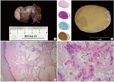
- 11,367 View
- 60 Download
- 3 Web of Science
- 3 Crossref
-
Abstract
PDF
- A metaplastic papillary tumor of the Fallopian tube is an extremely uncommon condition, with odd and confusing features that make it difficult to categorize as benign or borderline. Here, we summarize all the published cases to date and document the case of a 41-year-old woman diagnosed with this alteration after her last childbirth and ensuing tubal ligation. One of the tubes was bulky and filled with a caramel-like substance encircling a blurry spot. Light microscopy detailed a slender stalk covered by eosinophilic, columnar plump cells, showing atypical nuclei and focal budding. Mitotic figures were absent. The immunohistochemistry panel was positive for pan-cytokeratin, epithelial membrane antigen, cyclin D1, and hormone receptors. Additionally, a proliferation index of less than 5% was rated using Ki-67. The true nature of this tumor (reactive vs neoplastic) is uncertain. Nonetheless, its association with pregnancy suggests an adaptive change, likely similar to the atypical transdifferentiation proposed for Arias-Stella reaction.
-
Citations
Citations to this article as recorded by- Ungewöhnliche Proliferation des Eileiters
Angelina Vlaški, Vanessa Neukunft, Andrea Maria Gassel, Frederick Klauschen, Doris Mayr
Die Pathologie.2025; 46(1): 56. CrossRef - Fallopian tube papilloma
Shashank Mishra, Prerna Guleria
Indian Journal of Pathology and Microbiology.2021; 64(3): 608. CrossRef - Metaplastic papillary tumour of the fallopian tube, a rare entity, analysed by next‐generation sequencing
Sandra Sunitsch, Julia Reisinger, Luca Abete, Karl Kashofer, Peter Regitnig
Histopathology.2020; 76(6): 923. CrossRef
- Ungewöhnliche Proliferation des Eileiters
- The Limitations of Endoscopic Ultrasound-Guided Fine Needle Aspiration Cytology in the Diagnosis of Pancreatic Serous Cystadenoma: A Brief Case Report
- Heae Surng Park, Sun Och Yoon, Beom Jin Lim, Joo Hee Kim, Soon Won Hong
- Korean J Pathol. 2014;48(5):405-408. Published online October 27, 2014
- DOI: https://doi.org/10.4132/KoreanJPathol.2014.48.5.405
- 9,170 View
- 63 Download
- Pulmonary Hodgkin Lymphoma in a Patient with Crohn’s Disease
- Jae-Young Park, Juhie Lee
- Korean J Pathol. 2014;48(5):387-389. Published online October 27, 2014
- DOI: https://doi.org/10.4132/KoreanJPathol.2014.48.5.387
- 8,683 View
- 35 Download
- 2 Crossref
-
PDF
-
Citations
Citations to this article as recorded by- The Complex Relationship between Mechanisms Underlying Inflammatory Bowel Disease, Its Treatment, and the Risk of Lymphomas: A Comprehensive Review
Katarzyna Stasik, Rafał Filip
International Journal of Molecular Sciences.2024; 25(8): 4241. CrossRef - Extra-intestinal malignancies in inflammatory bowel diseases: An update with emphasis on MDCT and MR imaging features
A. Dohan, S.A. Faraoun, M. Barral, Y. Guerrache, M. Boudiaf, X. Dray, C. Hoeffel, M. Allez, O. Farges, L. Beaugerie, T. Aparicio, P. Marteau, E.K. Fishman, O. Lucidarme, C. Eveno, M. Pocard, R. Dautry, P. Soyer
Diagnostic and Interventional Imaging.2015; 96(9): 871. CrossRef
- The Complex Relationship between Mechanisms Underlying Inflammatory Bowel Disease, Its Treatment, and the Risk of Lymphomas: A Comprehensive Review
- Recurrent Thyroid Papillary Carcinoma in Children Under Ten Years Old: Report of Two Cases and Literature Review
- Byeong-Joo Noh, Ji-Youn Sung, Youn-Wha Kim, Yong-Koo Park
- Korean J Pathol. 2014;48(4):297-301. Published online August 26, 2014
- DOI: https://doi.org/10.4132/KoreanJPathol.2014.48.4.297
- 9,944 View
- 50 Download
- 2 Crossref
-
Abstract
PDF
Papillary thyroid carcinoma (PTC) in children under ten years old is very rare. To date, 18 cases of PTC in children under ten years old (including our two cases) have been reported in Korea. Here, we describe two cases of recurrent PTC with follicular variant and conventional type in an 8-year-old boy and a 7-year-old boy, respectively, and discuss clinicopathologic and molecular characteristics that differ in pediatric patients from adults.
-
Citations
Citations to this article as recorded by- Intentional Recurrent Laryngeal Nerve Sacrifice with Remarkable Vocal Compensation in Pediatric Papillary Thyroid Carcinoma: A Case Report
Shaheen Ayesha Nasirudeen, Balaji Ranganathan, Sriprakash Duraisamy, Jeenat Malawat
Indian Journal of Surgery.2026;[Epub] CrossRef - Extremely Well-Differentiated Papillary Thyroid Carcinoma Resembling Adenomatous Hyperplasia Can Metastasize to the Skull: A Case Report
Ju Yeon Pyo, Jisup Kim, Sung-eun Choi, Eunah Shin, Seok-Woo Yang, Cheong Soo Park, Seok-Mo Kim, SoonWon Hong
Yonsei Medical Journal.2017; 58(1): 255. CrossRef
- Intentional Recurrent Laryngeal Nerve Sacrifice with Remarkable Vocal Compensation in Pediatric Papillary Thyroid Carcinoma: A Case Report
- Well-Differentiated Papillary Mesothelioma of the Tunica Vaginalis: A Case Study and Review of the Literature
- Seyda Erdogan, Arbil Acikalin, Handan Zeren, Gulfılız Gonlusen, Suzan Zorludemir, Volkan Izol
- Korean J Pathol. 2014;48(3):225-228. Published online June 26, 2014
- DOI: https://doi.org/10.4132/KoreanJPathol.2014.48.3.225
- 11,393 View
- 73 Download
- 14 Crossref
-
Abstract
PDF
Well-differentiated papillary mesothelioma is an uncommon tumor of the testes that usually presents as a hydrocele. Here, we present the case of one patient who did not have a history of asbestos exposure. The tumor was localized in the tunica vaginalis and was composed of three pedunculated masses macroscopically. Microscopically, branching papillary structures with focal coagulative necrosis were present. In addition to immunohistochemistry, simian virus 40 DNA was also tested by polymerase chain reaction. This report presents one case of this rare entity, its clinical and macroscopic features, and follow-up results.
-
Citations
Citations to this article as recorded by- Factors predictive of progression in lesions categorised as well-differentiated papillary mesothelial tumour of the pleura, tunica vaginalis and peritoneum: a scoping review
Sarita Prabhakaran, Harry James Gaffney, Yazad Irani, Ashleigh J. Hocking, David Roder, Sonja Klebe
Cancer Treatment and Research Communications.2026; 47: 101137. CrossRef - Testicular/paratesticular mesothelial tumours: Uncommon histopathologic entities in a very complex anatomical site
Francesca Pagliuca, Stefano Lucà, Marco De Sio, Davide Arcaniolo, Gaetano Facchini, Marco De Martino, Francesco Esposito, Ferdinando DE Vita, Paolo Chieffi, Renato Franco
Pathology - Research and Practice.2024; 253: 155069. CrossRef - Well-differentiated Papillary Mesothelial Tumour of the Tunica Vaginalis Testis – A Rare Lesion, but one Pathologists Should Know About Two Patient Reports and a Review of the Literature
Johannes Kläger, Felicitas Oberndorfer, Cristophe Brunel, Julian Veser, Eva Compérat
International Journal of Surgical Pathology.2023; 31(6): 1126. CrossRef - A diagnostic approach to paratesticular lesions with tubulopapillary architecture: a series of 16 serous borderline tumors/low-grade serous carcinoma and 14 well-differentiated papillary mesothelial tumors and mesothelioma
Rabia Zafar, Lacey J. Schrader, John C. Cheville, J. Kenneth Schoolmeester, Anja C. Roden, Marie-Christine Aubry, Eunhee S. Yi, Aditya Raghunathan, Loren Herrera-Hernandez, R. Houston Thompson, Stephen A. Boorjian, Bradley C. Leibovich, Gary L. Keeney, Ra
Human Pathology.2022; 128: 31. CrossRef - Mesothelioma subtypes of the tunica vaginalis: a rare case report and review of histological criteria
Cutts Rebecca, Martin J Connor, Luxi Sun, Thomas Johnston, Rachel Gooch, John McLoughlin
Journal of Surgical Case Reports.2019;[Epub] CrossRef - Well‐differentiated papillary mesothelioma of tunica vaginalis testis of unknown malignant potential: Sonographic appearance
K.W.S. Ko, K.S. Tse, K.W. Shek, M.N. Hau, S.H. Ting
Journal of Clinical Ultrasound.2018; 46(5): 364. CrossRef - Tunica Vaginalis Thickening, Hemorrhagic Infiltration and Inflammatory Changes in 8 Children with Primary Hydrocele; Reactive Mesothelial Hyperplasia? A Prospective Clinical Study
Ioannis Patoulias, Evangelia Rachmani, Maria Kalogirou, Kyriakos Chatzopoulos, Dimitrios Patoulias
Acta Medica (Hradec Kralove, Czech Republic).2018; 61(2): 41. CrossRef - A 45-year-old man presenting with chest pain
Sheetu Singh, Arpita Jindal
Lung India.2018; 35(2): 171. CrossRef - Two Case Reports of Benign Testicular Mesothelioma and Review of the Literature
Cristobal Ramirez Sevilla, Carme Admella Salvador, Josep Feliu Canaleta, Juan Llopis Manzanera, Miguel Angel Barranco Sanz, Juan Antoni Romero Martin, Sergi Bernal Salguero, Francesco A. Mauri
Case Reports in Oncological Medicine.2017;[Epub] CrossRef - Well-differentiated Papillary Mesothelioma of the Tunica Vaginalis
Wei Keith Tan, Mae-Yen Tan, Hui Meng Tan, Rajadurai Pathmanathan, Wei Phin Tan
Urology.2016; 90: e7. CrossRef - Well-Differentiated Papillary Mesothelioma of the Tunica Vaginalis: Case Report and Systematic Review of Literature
Wei Keith Tan, Mae-Yen Tan, Wei Shen Tan, Soon Ching Gan, Rajadurai Pathmanathan, Hui Meng Tan, Wei Phin Tan
Clinical Genitourinary Cancer.2016; 14(4): e435. CrossRef - The legacy of the F344 rat as a cancer bioassay model (a retrospective summary of three common F344 rat neoplasms)
Robert R. Maronpot, Abraham Nyska, Jennifer E. Foreman, Yuval Ramot
Critical Reviews in Toxicology.2016; 46(8): 641. CrossRef - Malignant Mesothelioma of the Tunica Vaginalis: A Rare Neoplasm—Case Report and Literature Review
Manuel Segura-González, Jorge Urias-Rocha, Jorge Castelán-Pedraza
Clinical Genitourinary Cancer.2015; 13(6): e401. CrossRef - In vivo Optical Coherence Tomography Imaging of the Mesothelium Using Developed Window Models
Yeh-Chan Ahn, Yu-Gyeong Chae, Sang Seok Hwang, Bong-Kwon Chun, Maan Hong Jung, Sung Jin Nam, Hae-Young Lee, Jae Min Chung, Chulho Oak, Eun-Kee Park
Journal of the Optical Society of Korea.2015; 19(1): 69. CrossRef
- Factors predictive of progression in lesions categorised as well-differentiated papillary mesothelial tumour of the pleura, tunica vaginalis and peritoneum: a scoping review
- Tumor Sprouting in Papillary Thyroid Carcinoma Is Correlated with Lymph Node Metastasis and Recurrence
- Eunjung Lee, Wonkyung Jung, Jeong-Soo Woo, Jae Bok Lee, Bong Kyung Shin, Han Kyeom Kim, Aeree Kim, Baek-hui Kim
- Korean J Pathol. 2014;48(2):117-125. Published online April 28, 2014
- DOI: https://doi.org/10.4132/KoreanJPathol.2014.48.2.117
- 14,389 View
- 68 Download
- 9 Crossref
-
Abstract
PDF
Background Identification of poor prognostic factors in papillary thyroid carcinoma (PTC) patients is important for the patients' care and follow-up. We can sometimes see small tumor clusters without desmoplasia and no evidence of lymphatic emboli around the main tumor mass of PTC. We termed this form of tumor clustering, 'tumor sprouting,' and determined whether these tumors correlate with lymphovascular invasion, lymph node metastasis, and recurrence.
Methods We analyzed a total of 204 cases of papillary thyroid macrocarcinoma. Number, size and distance from the main tumor of the tumor sprouting were observed and analyzed with clinicopathologic characteristics.
Results Tumor sprouting was observed in 101 patients. Presence of tumor sprouting was significantly associated with positive resection margin (p=.002), lymphovascular invasion (p=.001), lymph node metastasis (p<.001), and recurrence (p=.004). Univariate analysis of recurrence-free survival revealed that tumor multiplicity (p=.037), positive resection margin (p=.007), lymphovascular invasion (p=.004), lymph node metastasis (p<.001), and tumor sprouting (p=.004) were poor prognostic factors. In multivariate analysis, positive resection margin was an independent poor prognostic factor of recurrence.
Conclusions In conclusion, tumor sprouting is significantly correlated with lymph node metastasis and recurrence. Evaluation of tumor sprouting in PTC patients could be helpful in predicting tumor recurrence or lymph node metastasis.
-
Citations
Citations to this article as recorded by- Deep learning habitat radiomics based on ultrasound for predicting preoperative locally progression and postoperative recurrence risk of thyroid cancer: a multicenter study
Lu Wenwu, Zhang Di, Wei Wei, Ding Wenbo, Wu Xin, Xie Xiang, Cheng Weibo, Wang Tao, Hu Song, Liu Boyuan, Zhou Wang, Zhang Chaoxue
International Journal of Surgery.2026; 112(3): 6814. CrossRef - The Initial Risk Stratification System for Differentiated Thyroid Cancer: Key Updates in the 2024 Korean Thyroid Association Guideline
Shinje Moon, Young Shin Song, Kyong Yeun Jung, Eun Kyung Lee, Jeongmin Lee, Dong-Jun Lim, Chan Kwon Jung, Young Joo Park
Endocrinology and Metabolism.2025; 40(3): 357. CrossRef - Korean Thyroid Association Guidelines on the Management of Differentiated Thyroid Cancers; Part I. Initial Management of Differentiated Thyroid Cancers - Chapter 5. Evaluation of Recurrence Risk Postoperatively and Initial Risk Stratification in Different
Eun Kyung Lee, Young Shin Song, Ho-Cheol Kang, Sun Wook Kim, Dong Gyu Na, Shin Je Moon, Dong-Jun Lim, Kyong Yeun Jung, Yun Jae Chung, Chan Kwon Jung, Young Joo Park
International Journal of Thyroidology.2024; 17(1): 68. CrossRef - Significance of Lymphovascular Invasion as a Prognostic Factor in Patients with Papillary Thyroid Cancer: a Systematic Review and Meta-Analysis
Ho-Ryun Won, Bon Seok Koo
International Journal of Thyroidology.2023; 16(2): 157. CrossRef - Peripheral Versus Intraparenchymal Papillary Thyroid Microcarcinoma: Different Morphologies and PD-L1 Expression
Bozidar Kovacevic, Dragana Vucevic, Snezana Cerovic, Catarina Eloy
Head and Neck Pathology.2022; 16(1): 200. CrossRef - Lymphovascular invasion and risk of recurrence in papillary thyroid carcinoma
Katy Wagner, Earl Abraham, Bryan Tran, David Roshan, James Wykes, Peter Campbell, Ardalan Ebrahimi
ANZ Journal of Surgery.2020; 90(9): 1727. CrossRef - The Predictors of Multicentricity in Well-Differentiated Thyroid Cancer
Mohamed Hegazi, Waleed El Nahas, Mohamed Elmetwally, Amr Hassan, Waleed Gado , Islam Abdou, Ahmed Senbel, Mohamed Samir Abou-Sheishaa
Journal of Analytical Oncology.2018; 7(4): 65. CrossRef - Prognostic impact of vascular invasion in differentiated thyroid carcinoma: a systematic review and meta-analysis
Huy Gia Vuong, Tetsuo Kondo, Uyen N P Duong, Thong Quang Pham, Naoki Oishi, Kunio Mochizuki, Tadao Nakazawa, Lewis Hassell, Ryohei Katoh
European Journal of Endocrinology.2017; 177(2): 207. CrossRef - Detection of Tumor Multifocality Is Important for Prediction of Tumor Recurrence in Papillary Thyroid Microcarcinoma: A Retrospective Study and Meta-Analysis
Jung-Soo Pyo, Jin Hee Sohn, Guhyun Kang
Journal of Pathology and Translational Medicine.2016; 50(4): 278. CrossRef
- Deep learning habitat radiomics based on ultrasound for predicting preoperative locally progression and postoperative recurrence risk of thyroid cancer: a multicenter study
- P2X7 Receptor Expression in Coexistence of Papillary Thyroid Carcinoma with Hashimoto's Thyroiditis
- Ji Hyun Kwon, Eun Sook Nam, Hyung Sik Shin, Seong Jin Cho, Hye Rim Park, Mi Jung Kwon
- Korean J Pathol. 2014;48(1):30-35. Published online February 25, 2014
- DOI: https://doi.org/10.4132/KoreanJPathol.2014.48.1.30
- 12,124 View
- 56 Download
- 21 Crossref
-
Abstract
PDF
Background This study was aimed at investigating the relation of P2X7 receptor (P2X7R) expression with the clinicopathological features of papillary thyroid carcinoma (PTC) coexisting with Hashimoto's thyroiditis (HT).
Methods We examined 170 patients (84, PTC with HT; 86, PTC without HT). P2X7R expression was examined by immunohistochemical methods. The staining intensity and patterns were evaluated and scored using a semi-quantitative method.
Results The PTC with HT group was more likely to contain women and had less extrathyroid extension, lymph node (LN) metastasis, lymphovascular invasion, and recurrence than the PTC without HT group. Patients positive for P2X7R had significantly higher frequencies of lymphovascular invasion, extrathyroid extension, LN metastasis, and absence of HT. As shown by multivariate analysis, the expression of P2X7R was significantly higher if HT was absent and extrathyroid extension was present. In the PTC with HT group, the expression of P2X7R was significantly higher in patients with tumor multifocality, lymphovascular invasion, and extrathyroid extension. In the PTC without HT group, the expression of P2X7R was significantly higher in women and those having tumor multifocality.
Conclusions Coexistence of PTC with HT is associated with good prognostic factors, and P2X7R expression in PTC was correlated with poor prognostic factors and the absence of HT.
-
Citations
Citations to this article as recorded by- Research Progress in the Relationship Between P2X7R and Cervical Cancer
Yiqing Tang, Cuicui Qiao, Qianqian Li, Xiaodi Zhu, Ronglan Zhao, Xiaoxiang Peng
Reproductive Sciences.2023; 30(3): 823. CrossRef - Warthin-like papillary thyroid carcinoma: a case report and comprehensive review of the literature
Abdel Mouhaymen Missaoui, Fatma Hamza, Wafa Belabed, Manel Mellouli, Mohamed Maaloul, Slim Charfi, Issam Jardak, Tahya Sellami-Boudawara, Nabila Rekik, Mohamed Abid
Frontiers in Endocrinology.2023;[Epub] CrossRef - Mapping the path towards novel treatment strategies: a bibliometric analysis of Hashimoto’s thyroiditis research from 1990 to 2023
Manping Guo, Qingna Li, Xingfang Liu, Yiming Wang, Qiaoning Yang, Rui Li, Yang Zhao, Chenfei Li, Song Sheng, Hangkun Ma, Zhenghong Li, Rui Gao
Frontiers in Endocrinology.2023;[Epub] CrossRef - Association between Hashimoto thyroiditis and clinical outcomes of papillary thyroid carcinoma: A meta-analysis
Qizhi Tang, Weiyu Pan, Liangyue Peng, Francis Moore
PLOS ONE.2022; 17(6): e0269995. CrossRef - Hashimoto’s Thyroiditis Minimizes Lymph Node Metastasis in BRAF Mutant Papillary Thyroid Carcinomas
Peter P. Issa, Mahmoud Omar, Yusef Buti, Chad P. Issa, Bert Chabot, Christopher J. Carnabatu, Ruhul Munshi, Mohammad Hussein, Mohamed Aboueisha, Mohamed Shama, Ralph L. Corsetti, Eman Toraih, Emad Kandil
Biomedicines.2022; 10(8): 2051. CrossRef - Overexpression of PD-L1 in Papillary Carcinoma and Its Association with Clinicopathological Variables
Servet KOCAÖZ, Gülay TURAN
Düzce Tıp Fakültesi Dergisi.2021; 23(3): 252. CrossRef - Effect of P2X7 receptor on tumorigenesis and its pharmacological properties
Wen-jun Zhang, Ce-gui Hu, Zheng-ming Zhu, Hong-liang Luo
Biomedicine & Pharmacotherapy.2020; 125: 109844. CrossRef - P2X7 in Cancer: From Molecular Mechanisms to Therapeutics
Romain Lara, Elena Adinolfi, Catherine A. Harwood, Mike Philpott, Julian A. Barden, Francesco Di Virgilio, Shaun McNulty
Frontiers in Pharmacology.2020;[Epub] CrossRef - The P2X7 Receptor in Inflammatory Diseases: Angel or Demon?
Luiz E. B. Savio, Paola de Andrade Mello, Cleide Gonçalves da Silva, Robson Coutinho-Silva
Frontiers in Pharmacology.2018;[Epub] CrossRef - Patients with Oncocytic Variant Papillary Thyroid Carcinoma Have a Similar Prognosis to Matched Classical Papillary Thyroid Carcinoma Controls
Azadeh A. Carr, Tina W.F. Yen, Diana I. Ortiz, Bryan C. Hunt, Gilbert Fareau, Becky L. Massey, Bruce H. Campbell, Kara L. Doffek, Douglas B. Evans, Tracy S. Wang
Thyroid.2018; 28(11): 1462. CrossRef - Extracellular purines, purinergic receptors and tumor growth
F Di Virgilio, E Adinolfi
Oncogene.2017; 36(3): 293. CrossRef - Multifaceted Effects of Extracellular Adenosine Triphosphate and Adenosine in the Tumor–Host Interaction and Therapeutic Perspectives
Paola de Andrade Mello, Robson Coutinho-Silva, Luiz Eduardo Baggio Savio
Frontiers in Immunology.2017;[Epub] CrossRef - Potential role of P2X7R in esophageal squamous cell carcinoma proliferation
André A Santos, Angélica R Cappellari, Fernanda O de Marchi, Marina P Gehring, Aline Zaparte, Caroline A Brandão, Tiago Giuliani Lopes, Vinicius D da Silva, Luis Felipe Ribeiro Pinto, Luiz Eduardo Baggio Savio, Aline Cristina Abreu Moreira-Souza, Robson C
Purinergic Signalling.2017; 13(3): 279. CrossRef - Potential relationship between Hashimoto's thyroiditis and BRAFV600E mutation status in papillary thyroid cancer
Rui‐chao Zeng, Lang‐ping Jin, En‐dong Chen, Si‐yang Dong, Ye‐feng Cai, Guan‐li Huang, Quan Li, Chun Jin, Xiao‐hua Zhang, Ou‐chen Wang
Head & Neck.2016;[Epub] CrossRef - Papillary thyroid carcinoma in Hashimoto’s thyroiditis as an incidental finding
Al Mogrampi Saad, A. Krexi, N. Papoulidis, M. Verroiotou, D. Michalakis, I. Fardellas
Hellenic Journal of Surgery.2016; 88(1): 47. CrossRef - Association of Hashimoto's thyroiditis and thyroid cancer
Salem I. Noureldine, Ralph P. Tufano
Current Opinion in Oncology.2015; 27(1): 21. CrossRef - Cribriform-morular variant of papillary thyroid carcinoma: a study of 3 cases featuring the PIK3CA mutation
Mi Jung Kwon, Young-Soo Rho, Jin Cheol Jeong, Hyung Sik Shin, Jong Seok Lee, Seong Jin Cho, Eun Sook Nam
Human Pathology.2015; 46(8): 1180. CrossRef - Understanding the roles of the P2X7 receptor in solid tumour progression and therapeutic perspectives
Sébastien Roger, Bilel Jelassi, Isabelle Couillin, Pablo Pelegrin, Pierre Besson, Lin-Hua Jiang
Biochimica et Biophysica Acta (BBA) - Biomembranes.2015; 1848(10): 2584. CrossRef - Purinergic signaling pathways in endocrine system
Ivana Bjelobaba, Marija M. Janjic, Stanko S. Stojilkovic
Autonomic Neuroscience.2015; 191: 102. CrossRef - P2X7 receptor stimulates breast cancer cell invasion and migration via the AKT pathway
JIYI XIA, XIAOLAN YU, LI TANG, GANG LI, TAO HE
Oncology Reports.2015; 34(1): 103. CrossRef - Regulation of the P2X7R by microRNA-216b in human breast cancer
Luming Zheng, Xukui Zhang, Feng Yang, Jian Zhu, Peng Zhou, Fang Yu, Lei Hou, Lei Xiao, Qingqing He, Baocheng Wang
Biochemical and Biophysical Research Communications.2014; 452(1): 197. CrossRef
- Research Progress in the Relationship Between P2X7R and Cervical Cancer
- Cytological Findings of the Micropapillary Variant of Urothelial Carcinoma: A Comparison with Typical High-Grade Urothelial Carcinoma
- Kyu-Ho Kim, Chang-Hwan Choi, Jee-Young Han, Lucia Kim, Suk-Jin Choi, In-Suh Park, Joon-Mee Kim, Young-Chae Chu
- Korean J Pathol. 2013;47(4):365-371. Published online August 26, 2013
- DOI: https://doi.org/10.4132/KoreanJPathol.2013.47.4.365
- 9,320 View
- 53 Download
- 4 Crossref
-
Abstract
PDF
Background Micropapillary variant of urothelial carcinoma (MPUC) showed distinct pathologic features and aggressive behavior. The cytologic findings of MPUC are still indistinct. In this study, we evaluated the cytological findings of MPUC compared with those of high-grade urothelial carcinoma (HGUC).
Methods The voided urine cytology of 8 cases of MPUC and 8 cases of HGUC was reviewed. Following cytological parameters were evaluated: cellularity, background, number of small, tight papillary clusters, small acinar structure, scattered single cells, cytoplasmic features, nuclear-to-cytoplasmic ratio, nuclear pleomorphism, nuclear membrane irregularity, hyperchromasia, chromatin pattern and nucleoli.
Results Compared to that of HGUC, cytology of MPUC showed large numbers of small, tight papillary clusters, small acinar structure, few numbers of single cells, and hyperchromatic nuclei. Other parameters were similar between the two groups; both groups showed similar cellularity, dense or vacuolated cytoplasm, moderate to severe nuclear pleomorphism, irregular nuclear membrane, coarse granular chromatin, and small and prominent nucleoli.
Conclusions The urine cytology of MPUCs showed smaller and tighter papillary cell clusters, more small acinar structures, fewer numbers of scattered single cells, and more hyperchromatic nuclei than that of HGUC. These features can help to distinguish MPUC and HGUC and offer an early cytological diagnosis of MPUC.
-
Citations
Citations to this article as recorded by- Cytologic features of micropapillary variant urothelial carcinoma in urinary tract cytology: Case series and review of literature
Nancy Y. Greenland, Yue Peng, Poonam Vohra, Z. Laura Tabatabai
Diagnostic Cytopathology.2022;[Epub] CrossRef - Cyto-histo correlations of plasmacytoid and micropapillary variants of high-grade urothelial carcinoma: do they fit well in The Paris System for reporting urinary cytology?
Liye Suo, Ivonne Vega, Michael Thrall
Journal of the American Society of Cytopathology.2021; 10(1): 20. CrossRef - A case report of urothelial carcinoma with combined micropapillary and plasmacytoid morphology in the urinary bladder
Sanghui Park, Min‐Sun Cho, Kwang Hyun Kim
Diagnostic Cytopathology.2016; 44(2): 124. CrossRef - Two cases with the micropapillary variant of urothelial carcinoma of the urinary bladder
Akiko KAGOTANI, Mitsuaki ISHIDA, Muneo IWAI, Nozomi IWAMOTO, Nozomi KASUGA, Yuji HAYASHI, Yoshimitsu MIYAHIRA, Ryoji KUSHIMA
The Journal of the Japanese Society of Clinical Cytology.2016; 55(3): 165. CrossRef
- Cytologic features of micropapillary variant urothelial carcinoma in urinary tract cytology: Case series and review of literature
- A Case of Multifocal Papillary Thyroid Carcinoma Consisting of One Encapsulated Follicular Variant with
BRAF K601E Mutation and Three Conventional Types withBRAF V600E Mutation - Wook Youn Kim, Young Sin Ko, Tae Sook Hwang, Hye Seung Han, So Dug Lim, Wan Seop Kim, Seo Young Oh
- Korean J Pathol. 2013;47(3):293-298. Published online June 25, 2013
- DOI: https://doi.org/10.4132/KoreanJPathol.2013.47.3.293
- 11,470 View
- 53 Download
- 7 Crossref
-
Abstract
PDF
Multifocal papillary thyroid carcinoma (mPTC) comprises about 20-30% of PTC. In mPTC, individual tumor foci can be identical or frequently composed of different histological types including follicular, solid, tall-cell or conventional patterns. We report a case of mPTC consisting of one encapsulated follicular variant of papillary thyroid carcinoma (FVPTC) and three conventional PTCs in a 44-year-old woman. This case genetically demonstrates unique features including the simultaneous presence of the
BRAF V600E (T1799A) mutation and theBRAF K601E (A1801G) mutation in conventional PTC and FVPTC, respectively.-
Citations
Citations to this article as recorded by- BRAF K601E Mutation in Oncocytic Carcinoma of the Thyroid: A Case Report and Literature Review
Antonio Matrone, Fabrizia Citro, Carla Gambale, Alessandro Prete, Elisa Minaldi, Raffaele Ciampi, Teresa Ramone, Gabriele Materazzi, Liborio Torregrossa, Rossella Elisei
Journal of Clinical Medicine.2023; 12(22): 6970. CrossRef - Case of aggressive metastatic follicular variant papillary thyroid carcinoma with BRAF K601E and BCORL1 mutations
Doaa Attia, Alexander Lurie, Qihui Zhai, Thomas Mesko, Robert Smallridge
BMJ Case Reports.2020; 13(6): e234208. CrossRef - BRAF gene: From human cancers to developmental syndromes
Muhammad Ramzan Manwar Hussain, Mukhtiar Baig, Hussein Sheik Ali Mohamoud, Zaheer Ulhaq, Daniel C. Hoessli, Ghaidaa Siraj Khogeer, Ranem Radwan Al-Sayed, Jumana Yousuf Al-Aama
Saudi Journal of Biological Sciences.2015; 22(4): 359. CrossRef - Clinical significance of BRAF V600E mutation in 154 patients with thyroid nodules
LINGYING YU, LIZHEN MA, QIAOFENG TU, YI ZHANG, YUEMING CHEN, DAOJUN YU, SHAOYU YANG
Oncology Letters.2015; 9(6): 2633. CrossRef - Clinicopathological Features of Rare BRAF Mutations in Korean Thyroid Cancer Patients
Uiju Cho, Woo Jin Oh, Ja Seong Bae, Sohee Lee, Young Sub Lee, Gyeong Sin Park, Youn Soo Lee, Chan Kwon Jung
Journal of Korean Medical Science.2014; 29(8): 1054. CrossRef - Recurrent Thyroid Papillary Carcinoma in Children Under Ten Years Old: Report of Two Cases and Literature Review
Byeong-Joo Noh, Ji-Youn Sung, Youn-Wha Kim, Yong-Koo Park
Korean Journal of Pathology.2014; 48(4): 297. CrossRef - Anaplastic Transformation of Papillary Thyroid Carcinoma in a Young Man: A Case Study with Immunohistochemical andBRAFAnalysis
Ji Hye Park, Hyeong Ju Kwon, Cheong Soo Park, SoonWon Hong
Korean Journal of Pathology.2014; 48(3): 234. CrossRef
- BRAF K601E Mutation in Oncocytic Carcinoma of the Thyroid: A Case Report and Literature Review
- Papillary Carcinoma of the Thyroid Gland with Nodular Fasciitis-like Stroma
- Ki Yong Na, Hyun-Soo Kim, Ji-Youn Sung, Won Seo Park, Youn Wha Kim
- Korean J Pathol. 2013;47(2):167-171. Published online April 24, 2013
- DOI: https://doi.org/10.4132/KoreanJPathol.2013.47.2.167
- 11,365 View
- 54 Download
- 14 Crossref
-
Abstract
PDF
Papillary thyroid carcinoma with nodular fasciitis-like stroma (PTC-NFS) is a rare variant of PTC. The term 'PTC with fibromatosis-like stroma' has been used as a synonym to describe this variant. It is characterized by extensive proliferation of fibroblasts and myofibroblasts in the tumor stroma, which occurs in up to 80% of the tumors. We herein describe a case of PTC-NFS which developed in a 49-year-old woman with the demonstration of findings of ultrasonography, fine needle aspiration cytology and histological examination of the lesion. To characterize the stromal components, we investigated the expression of several immunohistochemical markers which have been shown to be expressed differently in nodular fasciitis (NF) and fibromatosis (FM). The immunostaining results demonstrated nuclear and cytoplasmic accumulation of β-catenin, cytoplasmic transforming growth factor-β expression and nuclear Smad expression in the stromal cells, suggesting that the stromal cells in this case have similar molecular profiles to those of FM rather than NF.
-
Citations
Citations to this article as recorded by- The Association of Interstitial Fibrosis and Ki67 Marker Expression in Papillary Thyroid Carcinoma with Histopathological Findings and Prognosis of Total Thyroidectomy
Osman Anıl Savaş, Selçuk Mercan, Amir Mahdi Akbari, Mehrdad Sheikhvatan
Journal of Clinical Medicine.2026; 15(14): 5703. CrossRef - Papillary thyroid carcinoma with nodular fasciitis-like stroma: An intriguing encounter
Pranjal Kalita, Biswajit Dey, Vandana Raphael, Gauranga Handique, Sungjemla Longkumer, Nikhil Choudhary
Thyroid Research and Practice.2025; 21(1): 41. CrossRef - Comparative Evaluation of Machine Learning Algorithms with Data Balancing Approach and Hyperparameter Tuning in Predicting Thyroid Disorder Recurrence
Darnell Ignasius, Rhyan David Levandra, Ramadhan Rakhmat Sani, Ika Novita Dewi
Jurnal Masyarakat Informatika.2025; 16(2): 284. CrossRef - Papillary thyroid carcinoma with desmoid-type fibromatosis: the clinicopathological features with characteristic imaging and molecular correlation requiring comprehensive treatment
Haining Huang, Lei Li, Xiaolong Liu, Lihua Zhao, Zhihong Cui, Renya Zhang, Shuai Chen
Human Pathology.2023; 136: 84. CrossRef - Papillary Thyroid Carcinoma with Desmoid-Type Fibromatosis: Review of Published Cases
Abdallah Roukain, Stefano La Rosa, Massimo Bongiovanni, Marie Nicod Lalonde, Valérie Cristina, Michael Montemurro, Stephane Cochet, Alexandra Luquain, Peter A. Kopp, Gerasimos P. Sykiotis
Cancers.2021; 13(17): 4482. CrossRef - Case of medullary thyroid carcinoma with desmoid‐type fibromatosis
Yoon Ah Cho, Young Lyun Oh
Pathology International.2020; 70(6): 364. CrossRef - SOX11 expression in a case of papillary thyroid carcinoma with fibromatosis/fasciitis-like stroma containing BRAF c.1799_1801delTGA and CTNNB1 c.133T>C mutations
Soon Boon Justin Wong, Min En Nga, Michal Michal, Tomas Vanecek, Ju Ee Seet, Fredrik Petersson
Virchows Archiv.2019; 475(4): 519. CrossRef - Papillary thyroid cancer with extrathyroidal extension of desmoid-type fibromatosis. A case report of an aggressive presentation of an uncommon pathologic entity
Eve M. Roth, Courtney E. Barrows, Michiya Nishino, Barry Sacks, Per-Olof Hasselgren, Benjamin C. James
International Journal of Surgery Case Reports.2019; 63(C): 5. CrossRef - Papillary thyroid carcinoma with nodular fasciitis-like stroma and β-catenin mutations should be renamed papillary thyroid carcinoma with desmoid-type fibromatosis
Caterina Rebecchini, Antoine Nobile, Simonetta Piana, Rossella Sarro, Bettina Bisig, Sykiotis P Gerasimos, Chiara Saglietti, Maurice Matter, Laura Marino, Massimo Bongiovanni
Modern Pathology.2017; 30(2): 236. CrossRef - Papillary thyroid carcinoma with desmoid-type fibromatosis: A clinical, pathological, and immunohistochemical study of 14 cases
Nami Takada, Mitsuyoshi Hirokawa, Masahiro Ito, Aki Ito, Ayana Suzuki, Miyoko Higuchi, Seiji Kuma, Toshitetsu Hayashi, Masao Kishikawa, Shuichi Horikawa, Akira Miyauchi
Endocrine Journal.2017; 64(10): 1017. CrossRef - A Case of Papillary Thyroid Carcinoma with Fasciitis-like Stroma
Toshihiko WAKU, Hiroshi SONOBE
Nihon Rinsho Geka Gakkai Zasshi (Journal of Japan Surgical Association).2016; 77(12): 2892. CrossRef - Stromal Modulation and its Role in the Diagnosis of Papillary Patterned Thyroid Lesions
Sahar Aly Daoud, Reham Shehab El Nemr Esmail, Amal Ahmed Hareedy, Abdullah Khalil
Asian Pacific Journal of Cancer Prevention.2015; 16(8): 3307. CrossRef - Papillary Thyroid Carcinoma With Nodular Fasciitis–Like Stroma
Paula S. Ginter, Theresa Scognamiglio
International Journal of Surgical Pathology.2015; 23(4): 305. CrossRef - Notch and TGF-β/Smad3 pathways are involved in the interaction between cancer cells and cancer-associated fibroblasts in papillary thyroid carcinoma
Jie Zhang, Yuan Wang, Dan Li, Shanghua Jing
Tumor Biology.2014; 35(1): 379. CrossRef
- The Association of Interstitial Fibrosis and Ki67 Marker Expression in Papillary Thyroid Carcinoma with Histopathological Findings and Prognosis of Total Thyroidectomy
- The Definition of Minimal Extrathyroid Extension in Thyroid Pathology by Analyzing Sizable Intra- and Extrathyroid Blood Vessels
- Hyae Min Jeon, Beom Jin Lim, Hang-Seok Chang, SoonWon Hong
- Korean J Pathol. 2012;46(6):548-553. Published online December 26, 2012
- DOI: https://doi.org/10.4132/KoreanJPathol.2012.46.6.548
- 11,882 View
- 48 Download
- 7 Crossref
-
Abstract
PDF
Background To define the exact boundary of the intrathyroid and extrathyroid aspects of a gland when determining the extent of cancer invasion, we plan to clarify the definition of sizable vascular structures, which is one of the helpful histologic clues in determining a minimal extrathyroid extension. We hypothesized that arterial wall thicknesses in extrathyroid soft tissue would be significantly different from the arteries in the thyroid parenchyma.
Methods Twenty cases of papillary carcinoma were selected. The numbers and wall thicknesses of the arteries and arterioles in intrathyroid and extrathyroid tissue were evaluated. The absence of nerve tissue in the thyroid gland was confirmed using the S-100 protein immunohistochemical stain.
Results The comparison of the mean thicknesses of the total arteries between the extrathyroid and intrathyroid tissues in the retrospective study (26.88 µm vs. 15.07 µm, respectively) and the prospective study (35.24 µm vs. 16.52 µm, respectively) revealed significant differences (p=0.000). The greatest thickness of the intrathyroid arteries was 67.93 µm.
Conclusions According to our results, the study showed that the extrathyroidal arteries were significantly thicker than the intrathyroidal arteries. We suggest that the sizable blood vessels of extrathyroidal arteries should be greater than 67.93 µm in thickness.
-
Citations
Citations to this article as recorded by- Invasion in thyroid cancer: Controversies and best practices
Michiya Nishino, Jack Jacob
Seminars in Diagnostic Pathology.2020; 37(5): 219. CrossRef - MiR-221/222 promote migration and invasion, and inhibit autophagy and apoptosis by modulating ATG10 in aggressive papillary thyroid carcinoma
Hao Shen, Zaikai Lin, Haiyan Shi, Lingling Wu, Baojin Ma, Hong Li, Baobing Yin, Jun Tang, Hongjin Yu, Xiaoxing Yin
3 Biotech.2020;[Epub] CrossRef - Minimal extrathyroidal extension affects the prognosis of differentiated thyroid cancer: Is there a need for change in the AJCC classification system?
Zeming Liu, Yihui Huang, Sichao Chen, Di Hu, Min Wang, Ling Zhou, Wei Zhou, Danyang Chen, Haifeng Feng, Wei Wei, Chao Zhang, Wen Zeng, Liang Guo, Scott M. Langevin
PLOS ONE.2019; 14(6): e0218171. CrossRef - miR-199a-3p downregulation in thyroid tissues is associated with invasion and metastasis of papillary thyroid carcinoma
Chengbiao Liu, Meiling Xing, Liping Wang, Kejun Zhang
British Journal of Biomedical Science.2017; 74(2): 90. CrossRef - Clinicopathological Significance of Minimal Extrathyroid Extension in Solitary Papillary Thyroid Carcinomas
Chang Gok Woo, Chang Ohk Sung, Yun Mi Choi, Won Gu Kim, Tae Yong Kim, Young Kee Shong, Won Bae Kim, Suck Joon Hong, Dong Eun Song
Annals of Surgical Oncology.2015; 22(S3): 728. CrossRef - Intraoperative Frozen Section for the Evaluation of Extrathyroidal Extension in Papillary Thyroid Cancer
Om Prakash Prajapati, A. K. Verma, M. Sabaretnam
World Journal of Surgery.2015; 39(7): 1855. CrossRef - Tumor Sprouting in Papillary Thyroid Carcinoma Is Correlated with Lymph Node Metastasis and Recurrence
Eunjung Lee, Wonkyung Jung, Jeong-Soo Woo, Jae Bok Lee, Bong Kyung Shin, Han Kyeom Kim, Aeree Kim, Baek-hui Kim
Korean Journal of Pathology.2014; 48(2): 117. CrossRef
- Invasion in thyroid cancer: Controversies and best practices
- Clear Cell Papillary Renal Cell Carcinoma: A Report of 15 Cases Including Three Cases of Concurrent Other-Type Renal Cell Carcinomas
- Jeong Hwan Park, Cheol Lee, Ja Hee Suh, Kyung Chul Moon
- Korean J Pathol. 2012;46(6):541-547. Published online December 26, 2012
- DOI: https://doi.org/10.4132/KoreanJPathol.2012.46.6.541
- 11,037 View
- 65 Download
- 23 Crossref
-
Abstract
PDF
Background Clear cell papillary renal cell carcinoma (CCPRCC) is a recently established subtype of renal epithelial tumor. The aim of this study was to identify the diagnostic criteria of CCPRCC with an emphasis on immunohistochemical studies, and to report three cases with concurrent other-type renal cell carcinoma (RCC).
Methods A total of 515 RCC patients that consecutively underwent surgical resection at Seoul National University Hospital from 1 January 2010 to 31 December 2011 were screened. Each case was reviewed based on the histologic features and was evaluated immunohistochemically.
Results A total of 15 CCPRCCs were identified, which composed 2.9% of the total RCCs. The mean age was 52 years, and the average tumor size was 1.65 cm. All 15 cases showed low nuclear grade, no lymph node metastasis and no distant metastasis. The CCPRCCs showed variable architectural patterns including cystic, trabecular, papillary, and acinar. All of the cases showed moderate to intense immunoreactivity for cytokeratin 7 (CK7). CD10 was negative or showed focal weak positivity. Three cases had concurrent other-type RCC, including a clear cell RCC and an acquired cystic disease-associated RCC.
Conclusions The strong CK7 and negative or focal weak CD10 expression will be useful for the diagnosis of CCPRCC.
-
Citations
Citations to this article as recorded by- Vascular, adipose tissue, and/or calyceal invasion in clear cell tubulopapillary renal cell tumour: potentially problematic diagnostic scenarios
Ankur R Sangoi, Harrison Tsai, Lara Harik, Jonathan Mahlow, Maria Tretiakova, Sean R Williamson, Michelle S Hirsch
Histopathology.2024; 84(7): 1167. CrossRef - Clinical features and Surgical Outcome of Clear Cell Papillary Renal Cell Tumor: result from a prospective cohort
Si Hyun Kim, Jang Hee Han, Seung-hwan Jeong, Hyeong Dong Yuk, Ja Hyeon Ku, Cheol Kwak, Hyeon Hoe Kim, Kyung Chul Moon, Chang Wook Jeong
BMC Urology.2023;[Epub] CrossRef - Coexistence of multiple clear cell papillary renal cell carcinoma with renal oncocytoma: a case report
Amine Hermi, Ahmed Saadi, Seif Mokadem, Ahlem Blel, Marouene Chakroun, Mohamed Riadh Ben Slama
Annals of Medicine & Surgery.2023; 85(5): 2017. CrossRef - Renal Cell Carcinoma in End-Stage Renal Disease: A Review and Update
Ziad M. El-Zaatari, Luan D. Truong
Biomedicines.2022; 10(3): 657. CrossRef - The Clinicopathologic and Molecular Landscape of Clear Cell Papillary Renal Cell Carcinoma: Implications in Diagnosis and Management
Stanley Weng, Renzo G. DiNatale, Andrew Silagy, Roy Mano, Kyrollis Attalla, Mahyar Kashani, Kate Weiss, Nicole E. Benfante, Andrew G. Winer, Jonathan A. Coleman, Victor E. Reuter, Paul Russo, Ed Reznik, Satish K. Tickoo, A. Ari Hakimi
European Urology.2021; 79(4): 468. CrossRef - Clear cell papillary renal cell carcinoma: Characteristics and survival outcomes from a large single institutional series
James E. Steward, Sean Q. Kern, Liang Cheng, Ronald S. Boris, Yan Tong, Clint D. Bahler, Timothy A. Masterson, K. Clint Cary, Hristos Kaimakliotis, Thomas Gardner, Chandru P. Sundaram
Urologic Oncology: Seminars and Original Investigations.2021; 39(6): 370.e21. CrossRef - Clear cell papillary renal cell carcinoma: an update after 15 years
Sean R. Williamson
Pathology.2021; 53(1): 109. CrossRef - Clear Cell Papillary Renal Cell Carcinoma
Jianping Zhao, Eduardo Eyzaguirre
Archives of Pathology & Laboratory Medicine.2019; 143(9): 1154. CrossRef - Clear cell papillary renal cell carcinoma – An indolent subtype of renal tumor
Wei-Jen Chen, Chin-Chen Pan, Shu-Huei Shen, Hsiao-Jen Chung, Chih-Chieh Lin, Alex T.L. Lin, Yen-Hwa Chang
Journal of the Chinese Medical Association.2018; 81(10): 878. CrossRef - Clear cell papillary renal cell carcinoma: A case report and review of the literature
Sung Han Kim, Whi-An Kwon, Jae Young Joung, Ho Kyung Seo, Kang Hyun Lee, Jinsoo Chung
World Journal of Nephrology.2018; 7(8): 155. CrossRef - Clinical features and survival analysis of clear cell papillary renal cell carcinoma: A 10‑year retrospective study from two institutions
Yiqiu Wang, Ying Ding, Jian Wang, Min Gu, Zengjun Wang, Chao Qin, Conghui Han, Hongxia Li, Xia Liu, Pengfei Wu, Guangchao Li
Oncology Letters.2018;[Epub] CrossRef - A contemporary series of renal masses with emphasis on recently recognized entities and tumors of low malignant potential: A report based on 624 consecutive tumors from a single tertiary center
Maria Rosaria Raspollini, Ilaria Montagnani, Rodolfo Montironi, Liang Cheng, Guido Martignoni, Andrea Minervini, Sergio Serni, Giulio Nicita, Marco Carini, Antonio Lopez-Beltran
Pathology - Research and Practice.2017; 213(7): 804. CrossRef - Renal Neoplasms With Overlapping Features of Clear Cell Renal Cell Carcinoma and Clear Cell Papillary Renal Cell Carcinoma
Hari P. Dhakal, Jesse K. McKenney, Li Yan Khor, Jordan P. Reynolds, Cristina Magi-Galluzzi, Christopher G. Przybycin
American Journal of Surgical Pathology.2016; 40(2): 141. CrossRef - New and emerging renal tumour entities
Naoto Kuroda, Ondřej Hess, Ming Zhou
Diagnostic Histopathology.2016; 22(2): 47. CrossRef - Immunohistochemical Panel for Differentiating Renal Cell Carcinoma with Clear and Papillary Features
Hanan AlSaeid Alshenawy
Pathology & Oncology Research.2015; 21(4): 893. CrossRef - Immunohistochemical panel for differentiating renal cell carcinoma with clear and papillary features
Hanan AlSaeid Alshenawy
Journal of Microscopy and Ultrastructure.2015; 3(2): 68. CrossRef - Clear Cell-Papillary Renal Cell Carcinoma of the Kidney Not Associated With End-stage Renal Disease
Manju Aron, Elena Chang, Loren Herrera, Ondrej Hes, Michelle S. Hirsch, Eva Comperat, Philippe Camparo, Priya Rao, Maria Picken, Michal Michal, Rodolfo Montironi, Pheroze Tamboli, Federico Monzon, Mahul B. Amin
American Journal of Surgical Pathology.2015; 39(7): 873. CrossRef - Papillary or pseudopapillary tumors of the kidney
Fang-Ming Deng, Max X. Kong, Ming Zhou
Seminars in Diagnostic Pathology.2015; 32(2): 124. CrossRef - Do Clear Cell Papillary Renal Cell Carcinomas Have Malignant Potential?
Mairo L. Diolombi, Liang Cheng, Pedram Argani, Jonathan I. Epstein
American Journal of Surgical Pathology.2015; 39(12): 1621. CrossRef - Targeted next‐generation sequencing and non‐coding RNA expression analysis of clear cell papillary renal cell carcinoma suggests distinct pathological mechanisms from other renal tumour subtypes
Charles H Lawrie, Erika Larrea, Gorka Larrinaga, Ibai Goicoechea, María Arestin, Marta Fernandez‐Mercado, Ondrej Hes, Francisco Cáceres, Lorea Manterola, José I López
The Journal of Pathology.2014; 232(1): 32. CrossRef - Clear cell papillary renal cell carcinoma is the fourth most common histologic type of renal cell carcinoma in 290 consecutive nephrectomies for renal cell carcinoma
Haijun Zhou, Shaojiang Zheng, Luan D. Truong, Jae Y. Ro, Alberto G. Ayala, Steven S. Shen
Human Pathology.2014; 45(1): 59. CrossRef - Clear cell papillary renal cell carcinoma: Incidence, morphological features, immunohistochemical profile, and biologic behavior: A single institution study
Borislav A. Alexiev, Cinthia B. Drachenberg
Pathology - Research and Practice.2014; 210(4): 234. CrossRef - MRI Phenotype in Renal Cancer
Naomi Campbell, Andrew B. Rosenkrantz, Ivan Pedrosa
Topics in Magnetic Resonance Imaging.2014; 23(2): 95. CrossRef
- Vascular, adipose tissue, and/or calyceal invasion in clear cell tubulopapillary renal cell tumour: potentially problematic diagnostic scenarios
- Endoscopic Ultrasound-Guided Fine Needle Aspiration Cytology Diagnosis of Solid Pseudopapillary Neoplasm: Three Case Reports with Review of Literature
- Joon Seon Song, Chong Woo Yoo, Youngmee Kwon, Eun Kyung Hong
- Korean J Pathol. 2012;46(4):399-406. Published online August 23, 2012
- DOI: https://doi.org/10.4132/KoreanJPathol.2012.46.4.399
- 11,629 View
- 63 Download
- 15 Crossref
-
Abstract
PDF
Solid pseudopapillary neoplasm of the pancreas (SPN) is relatively rare and it occurs almost exclusively in women. We recently experienced three cases of SPN diagnosed by endoscopic ultrasound-guided fine-needle aspiration (EUS-FNA). These three cases were two male and one female patient whose age was 29, 37, and 44 years old. Radiological diagnosis was pancreatic endocrine tumor (PEN) showing solid with a heterogenous echogenicity. EUS-FNA cytology specimens consisted of single cells and aggregates of uniform cells, forming microadenoid structures, branching, papillary clusters with delicate fibrovascular cores. In conclusion, a single diagnosis of SPN based on clinical and radiological findings would be risky because there is a possibility of it being misdiagnosed as PEN or other malignancies. An EUS-FNA is therefore essential for establishing the diagnosis. In addition, the pathologists should recognize the characteristic cytologic findings with immunoprofiles of SPN to prevent misdiagnosis of SPN.
-
Citations
Citations to this article as recorded by- Spleen Preservation in Solid Pseudopapillary Neoplasms of the Distal Pancreas: A Single-Center Experience
Tiffany M. Yue, Beatrice J. Sun, Daniel J. Delitto, Monica M. Dua, Jeffrey A. Norton, George A. Poultsides, Brendan C. Visser, Byrne Lee
Journal of Surgical Research.2025; 310: 17. CrossRef - Laparoscopic parenchyma-sparing resections for solid pseudopapillary tumors located in the head of pancreas
Zhengdong Zou, Lu Feng, Bing Peng, Jianhua Liu, Yunqiang Cai
BMC Surgery.2023;[Epub] CrossRef - Solid pancreatic masses in children: A review of current evidence and clinical challenges
Kelli N. Patterson, Andrew T. Trout, Archana Shenoy, Maisam Abu-El-Haija, Jaimie D. Nathan
Frontiers in Pediatrics.2022;[Epub] CrossRef - Pancreatic solid pseudopapillary neoplasm in male patients: systematic review with three new cases
Anna Caterina Milanetto, Anna-Lea Gais Zürcher, Lorenzo Macchi, Alina David, Claudio Pasquali
Updates in Surgery.2021; 73(4): 1285. CrossRef - Silencing c-Myc Enhances the Antitumor Activity of Bufalin by Suppressing the HIF-1α/SDF-1/CXCR4 Pathway in Pancreatic Cancer Cells
Xia Liu, Yayun Zhou, Jiamin Peng, Bei Xie, Qiyang Shou, Jianchao Wang
Frontiers in Pharmacology.2020;[Epub] CrossRef - Simultaneous colorectal and parenchymal-sparing liver resection for advanced colorectal carcinoma with synchronous liver metastases: Between conventional and mini-invasive approaches
Emilio De Raffele, Mariateresa Mirarchi, Dajana Cuicchi, Ferdinando Lecce, Riccardo Casadei, Claudio Ricci, Saverio Selva, Francesco Minni
World Journal of Gastroenterology.2020; 26(42): 6529. CrossRef - Solid pseudopapillary neoplasm of the pancreas showing marked distal atrophy
Masanori Tsujie, Tomoko Wakasa, Shigeto Mizuno, Hajime Ishikawa, Hironobu Manabe, Taichi Koyama, Kotaro Kitani, Shumpei Satoi, Keisuke Inoue, Shuichi Fukuda, Toshihiko Kawasaki, Masao Yukawa, Yoshio Ohta, Masatoshi Inoue
International Journal of Surgery Case Reports.2019; 55(C): 136. CrossRef - Solid pseudopapillary neoplasm of the pancreas in pediatric patients: A case report and institutional case series
Justin B. Mahida, Rajan K. Thakkar, Jon Walker, Rulong Shen, Brian D. Kenney, Vinay Prasad, Jennifer H. Aldrink
Journal of Pediatric Surgery Case Reports.2015; 3(4): 149. CrossRef - Onsite cytopathology evaluation and ancillary studies beneficial in EUS‐FNA of pancreatic, mediastinal, intra‐abdominal, and submucosal lesions
Shafqat Mehmood, Amna Jahan, Asif Loya, Muhammed Aasim Yusuf
Diagnostic Cytopathology.2015; 43(4): 278. CrossRef - Multicentric solid pseudopapillary neoplasms of the pancreas diagnosed by endoscopic ultrasound-guided fine needle aspiration: a case report
Megumi Yamaguchi, Toshikatsu Fukuda, Masahiro Nakahara, Mio Amano, Daisuke Takei, Masumi Kawashima, Yusuke Sumi, Hironobu Amano, Shuji Yonehara, Keiji Hanada, Toshio Noriyuki
Surgical Case Reports.2015;[Epub] CrossRef - A solid pseudopapillary neoplasm without cysts that occurred in a patient diagnosed by endoscopic ultrasound-guided fine-needle aspiration: a case report
Masakuni Fujii, Masao Yoshioka, Takefumi Niguma, Hiroaki Saito, Toru Kojima, Soichiro Nose, Junji Shiode
Journal of Medical Case Reports.2014;[Epub] CrossRef - Surgical Management and Long-Term Follow-Up of Solid Pseudopapillary Tumor of Pancreas: A Large Series from a Single Institution
Yunqiang Cai, Xun Ran, Siming Xie, Xin Wang, Bing Peng, Gang Mai, Xubao Liu
Journal of Gastrointestinal Surgery.2014; 18(5): 935. CrossRef - A case of solid-pseudopapillary neoplasm in a middle-aged male preoperatively diagnosed by endoscopic ultrasound-guided fine-needle aspiration (EUS-FNA)
Shin KATO, Takuya HONBU, Moriya ZAKIMI, Kenji CHINEN, Tomiaki KUBOTA, Masayuki ARASHIRO, Kaoru KIKUCHI, Takahiro MURAKAMI, Fumihito KUNISHIMA
Suizo.2014; 29(2): 263. CrossRef - Endoscopic ultrasound-guided fine needle aspiration improves the pre-operative diagnostic yield of solid-pseudopapillary neoplasm of the pancreas: an international multicenter case series (with video)
Joanna K. Law, Alina Stoita, Wallia Weaver, Ferga C. Gleeson, Andrew M. Dries, Amanda Blackford, Vandhana Kiswani, Eun Ji Shin, Mouen A. Khashab, Marcia Irene Canto, Vikesh K. Singh, Anne Marie Lennon
Surgical Endoscopy.2014; 28(9): 2592. CrossRef - Multiple Diagnostic Imaging of a Patient with Solid Pseudopapillary Tumour of the Pancreas: EUS, CT and FDG PET/CT
Ari Chong, Jung-Min Ha, Seong Young Kwon
Nuclear Medicine and Molecular Imaging.2014; 48(1): 82. CrossRef
- Spleen Preservation in Solid Pseudopapillary Neoplasms of the Distal Pancreas: A Single-Center Experience
- Multifocal Renal Cell Carcinoma of Different Histological Subtypes in Autosomal Dominant Polycystic Kidney Disease
- Ki Yong Na, Hyun-Soo Kim, Yong-Koo Park, Sung-Goo Chang, Youn Wha Kim
- Korean J Pathol. 2012;46(4):382-386. Published online August 23, 2012
- DOI: https://doi.org/10.4132/KoreanJPathol.2012.46.4.382
- 11,908 View
- 77 Download
- 14 Crossref
-
Abstract
PDF
Renal cell carcinoma (RCC) in autosomal dominant polycystic kidney (ADPKD) is rare. To date, 54 cases of RCC in ADPKD have been reported. Among these, only 2 cases have different histologic types of RCC. Here we describe a 45-year-old man who received radical nephrectomy for multifocal RCC with synchronous papillary and clear cell histology in ADPKD and chronic renal failure under regular hemodialysis. The case reported herein is another example of the rare pathological finding of RCC arising in a patient with ADPKD.
-
Citations
Citations to this article as recorded by- Pellino1-mTOR/S6K1 signaling axis is a key pathogenesis for the development of polycystic kidney disease
Suhyeon Kim, Min-Hee Kim, Bo-Kyoung Ko, Kyung-Mo Kim, Naiyu Wang, Su-Mi Jo, Heounjeong Go, Eun-Ji Park, Chang-Woo Lee
Cell Death & Disease.2026;[Epub] CrossRef - Autosomal Dominant Polycystic Kidney Disease-Related Multifocal Renal Cell Carcinoma: A Narrative Iconographic Review
Consolato M. Sergi, Luis Guerra, Josef Hager
International Journal of Molecular Sciences.2025; 26(9): 3965. CrossRef - Autosomal Dominant Polycystic Kidney Disease Patients Requiring Nephrectomy: Characteristics and Surgical Considerations
Joel Ern Zher Chan, Kate S. Olakkengil, Shantanu Bhattacharjya, Santosh Antony Olakkengil
ANZ Journal of Surgery.2025; 95(7-8): 1605. CrossRef - Renal Cell Carcinoma in the Background of Autosomal Dominant Polycystic Kidney Disease: Report of Two Cases and Review of Literature
Poorva Vias, Shikha Goyal, Renu Madan, Nandita Kakkar, Ridhi Sood, Kannan Periasamy, Rajender Kumar
Indian Journal of Medical and Paediatric Oncology.2024; 45(02): 188. CrossRef - Detection of two synchronous histologically different renal cell carcinoma subtypes in the same kidney: a case report and review of the literature
Mohamed Sakr, Merhan Badran, Sarah Ahmed Hassan, Mohamed Elsaqa, Mohamed Anwar Elwany, Nevine M. F. El Deeb, Mohamed Sharafeldeen
Journal of Medical Case Reports.2024;[Epub] CrossRef - The Importance of Genetic Testing in the Differential Diagnosis of Atypical TSC2-PKD1 Contiguous Gene Syndrome—Case Series
Petronella Orosz, Zita Kollák, Ákos Pethő, András Fogarasi, György Reusz, Kinga Hadzsiev, Tamás Szabó
Children.2023; 10(3): 420. CrossRef - Autosomal dominant polycystic kidney disease coming up with an unusual presentation of renal cell carcinoma on its first encounter
Asma Shoukat Masumdar, Anitha Padmanabhan, Nitin Gadgil, Gargi Padalkar
Indian Journal of Pathology and Oncology.2023; 10(4): 417. CrossRef - Sarcomatoid renal cell carcinoma with autosomal dominant polycystic kidney disease: a case report and literature review
Yuji Hakozaki, Kiyotaka Uchiyama, Akane Yanai, Daisuke Yamada, Yuka Kamijo, Yoshitaka Ishibashi
CEN Case Reports.2021; 10(2): 199. CrossRef - CT and MRI findings of cystic renal cell carcinoma: comparison with cystic collecting duct carcinoma
Qingqiang Zhu, Jun Ling, Jing Ye, Wenrong Zhu, Jingtao Wu, Wenxin Chen
Cancer Imaging.2021;[Epub] CrossRef - Incidental occurrence of papillary renal cell carcinoma in the native kidney with autosomal dominant polycystic kidney disease after renal transplantation: A case report
Mahmoud Abbas, Melanie Pätzel, Angelika Thurn, Olaf Brinkmann, Olaf Bettendorf
Molecular and Clinical Oncology.2021;[Epub] CrossRef - Xp11.2 translocation renal cell carcinoma in the autosomal dominant polycystic kidney disease patient with preserved renal function
Hyuk Huh, Hyung Ah Jo, YongJin Yi, Seung Hyup Kim, Kyung Chul Moon, Curie Ahn, Hayne Cho Park
The Korean Journal of Internal Medicine.2017; 32(6): 1108. CrossRef - The Association between Autosomal Dominant Polycystic Kidney Disease and Renal Cell Carcinoma
Chase C. Hansen, Michael Derrick, Irfan Warriach, James Thomas Cammack, James Thomas Cammack, Werner de Riese
Open Journal of Urology.2015; 05(06): 84. CrossRef - The MSCT and MRI findings of collecting duct carcinoma
Q. Zhu, J. Wu, Z. Wang, W. Zhu, W. Chen, S. Wang
Clinical Radiology.2013; 68(10): 1002. CrossRef - Thyroid-like follicular carcinoma of the kidney in a patient with nephrolithiasis and polycystic kidney disease: a case report
Metka Volavšek, Margareta Strojan-Fležar, Gregor Mikuz
Diagnostic Pathology.2013;[Epub] CrossRef
- Pellino1-mTOR/S6K1 signaling axis is a key pathogenesis for the development of polycystic kidney disease
- Papillary Carcinoma of Thyroid Metastatic to Adenocarcinoma
In Situ of Lung: Report of an Unusual Case - Kyoung Min Kim, Yo Na Kim, Hyun Hee Chu, Heung Yong Jin, Min Ho Kim, Myoung Ja Chung
- Korean J Pathol. 2012;46(3):282-286. Published online June 22, 2012
- DOI: https://doi.org/10.4132/KoreanJPathol.2012.46.3.282
- 10,785 View
- 54 Download
- 6 Crossref
-
Abstract
PDF
The tumor-to-tumor metastasis is a rare event. The lung tumors are the most common donor tumors in tumor-to-tumor metastasis, but are exceedingly rare as a recipient. Here, we report a case of papillary thyroid carcinoma (PTC) metastasizing to adenocarcinoma
in situ (AIS, formerly bronchioloalveolar carcinoma) of the lung in a 44-year-old woman who underwent total thyroidectomy for PTC 8 years ago. To the best of our knowledge, the present case is the first case reporting on PTC metastasized to AIS. A review of the relevant literature is presented.-
Citations
Citations to this article as recorded by- A case of colorectal cancer with intratumoral metastasis to primary lung cancer
Yasushi Cho, Mitsuhito Kaji, Shunsuke Nomura, Yusuke Motohashi, Masaaki Sato, Motoya Takeuchi
The Journal of the Japanese Association for Chest Surgery.2021; 35(5): 576. CrossRef - Tumor-to-tumor metastasis: metastatic invasive lobular carcinoma of the breast within adenocarcinoma of the lung
Myoung Jae Kang, Ae Ri An, Myoung Ja Chung, Kyoung Min Kim
Journal of Pathology and Translational Medicine.2020; 54(2): 188. CrossRef - Metastatic Renal Cell Neoplasm Within a Papillary Thyroid
Carcinoma as Incidental Finding in an Asymptomatic Patient: a Case Report
Maria-Rosa Bella-Cueto, Mireia Pascua-Solé, Albert Cano-Palomares, M. Àngels Cabezuelo-Hernandez, Maria-Rosa Escoda-Giralt, Santiago Barcons-Vilaplana, Paula Serret-Miralles, Carmen Caral-Vanaclocha, Xavier Guirao-Garriga, Joan Prats-Lopez, Meritxell Meda
SN Comprehensive Clinical Medicine.2020; 2(7): 978. CrossRef - A Rare Case of Tumor-to-Tumor Metastasis of Thyroid Papillary Carcinoma within a Pulmonary Adenocarcinoma
Taebum Lee, Yoon Jin Cha, Sangjeong Ahn, Joungho Han, Young Mog Shim
Journal of Pathology and Translational Medicine.2015; 49(1): 78. CrossRef - Tumour-to-tumour metastasis from papillary thyroid carcinoma withBRAFmutation to lung adenocarcinoma withEGFRmutation: the utility of mutation-specific antibodies
Yuki Katsuya, Akihiko Yoshida, Shun-ichi Watanabe, Koji Tsuta
Histopathology.2015; 67(2): 262. CrossRef - Pulmonary metastasis of a papillary thyroid carcinoma and primary lung adenocarcinoma: two coincident carcinomas at the same location
Liyan Xue, Zhonghua Luan, Ying Liu, Shuangmei Zou, Jun Jiang, Ning Wu, Ning Lu, Dongmei Lin
Diagnostic Pathology.2013;[Epub] CrossRef
- A case of colorectal cancer with intratumoral metastasis to primary lung cancer
- Detection of
BRAF V600E Mutations in Papillary Thyroid Carcinomas by Peptide Nucleic Acid Clamp Real-Time PCR: A Comparison with Direct Sequencing - Dongjun Jeong, Yujun Jeong, Sungche Lee, Hyeran Lee, Wanju Lee, Hyungjoo Kim, Doosan Park, Soyoung Park, Wenxia Mu, Hyun-Deuk Cho, Mee-Hye Oh, Sung Soo Lee, Seung-Ha Yang, Chang-Jin Kim
- Korean J Pathol. 2012;46(1):61-67. Published online February 23, 2012
- DOI: https://doi.org/10.4132/KoreanJPathol.2012.46.1.61
- 15,587 View
- 84 Download
- 20 Crossref
-
Abstract
PDF
Background Papillary thyroid carcinoma (PTC) of the thyroid is the most common endocrine malignancy. High prevalence of an activating point mutation of
BRAF gene,BRAF V600E, has been reported in PTC. We assessed the efficiency of peptide nucleic acid clamp real-time polymerase chain reaction (PNAcqPCR) for the detection ofBRAF V600E mutation in PTC in comparison with direct sequencing (DS).Methods A total of 265 thyroid lesions including 200 PTCs, 5 follicular carcinomas, 60 benign lesions and 10 normal thyroid tissues were tested for
BRAF V600E mutation by PNAcqPCR and DS.Results The sensitivity and accuracy of the PNAcqPCR method were both higher than those of DS for the detection of the
BRAF V600E mutation. In clinical samples, 89% of PTCs harbored theBRAF V600E mutation, whereas 5 follicular carcinomas, 50 benign lesions and 10 normal thyroid tissues lacked the mutation. The mutation was associated with aggressive clinical behaviors as extrathyroid invasion (p=0.015), lymph node metastasis (p=0.002) and multiple tumor numbers (p=0.016) with statistical significance.Conclusions The PNAcqPCR method is efficiently applicable for the detection of the
BRAF V600E mutation in PTCs in a clinical setting.-
Citations
Citations to this article as recorded by- Molecular Landscape and Advanced Diagnostic Technologies for BRAF Mutations in Cancer: From Quantitative PCR and ddPCR to CRISPR-Based Platforms
Melika Nejatinejad, Ali Maleki-Aram, Arezoo Namavar Abibiglou, Chekameh Vahedi, Alireza Panahian, Anahita Ebrahimi, Reza Yaghoubi, Houman Parsaei, Somayeh Vafaei, Davod Jafari
Clinica Chimica Acta.2027; 592: 121178. CrossRef - Diagnostic utilities of washout CYFRA 21-1 combined with washout thyroglobulin for metastatic lymph nodes in thyroid cancer: a prospective study
Joonseon Park, Solji An, Kwangsoon Kim, Jeong Soo Kim, Chan Kwon Jung, Ja Seong Bae
Scientific Reports.2024;[Epub] CrossRef - BRAFV600E Positivity-Dependent Effect of Age on Papillary Thyroid Cancer Recurrence Risk
Joonseon Park, Solji An, Kwangsoon Kim, Ja Seong Bae, Jeong Soo Kim
Cancers.2023; 15(22): 5395. CrossRef - TROP-2 and 5hmC expression in follicular-patterned thyroid neoplasm emphasizing tiny well-formed papillae
Jae Yeon Seok, Xuemo Fan
Annals of Diagnostic Pathology.2022; 57: 151903. CrossRef - Null Association between BRAF V600E Mutation and Tumor Recurrence in Patients with Papillary Thyroid Microcarcinoma in South Korea
Ji Yoon Kim, Kyoung Jin Kim, Jae Hyun Bae, Joo Hyung Kim, Nam Hoon Kim, Hee Young Kim, Hoon Yub Kim, Seung-Kuk Baek, Sin Gon Kim, Kwang Yoon Jung, Kyeong Jin Kim
International Journal of Thyroidology.2021; 14(2): 135. CrossRef - Evaluation of the expression levels of BRAFV600E mRNA in primary tumors of thyroid cancer using an ultrasensitive mutation assay
Tien Viet Tran, Kien Xuan Dang, Quynh Huong Pham, Ung Dinh Nguyen, Nhung Thi Trang Trinh, Luong Van Hoang, Son Anh Ho, Ba Van Nguyen, Duc Trong Nguyen, Dung Tuan Trinh, Dung Ngoc Tran, Arto Orpana, Ulf-Håkan Stenman, Jakob Stenman, Tho Huu Ho
BMC Cancer.2020;[Epub] CrossRef - The Current State of Molecular Testing in the BRAF-Mutated Melanoma Landscape
Irene Vanni, Enrica Teresa Tanda, Francesco Spagnolo, Virginia Andreotti, William Bruno, Paola Ghiorzo
Frontiers in Molecular Biosciences.2020;[Epub] CrossRef - Highly prevalent BRAF V600E and low-frequency TERT promoter mutations underlie papillary thyroid carcinoma in Koreans
Sue Youn Kim, Taeeun Kim, Kwangsoon Kim, Ja Seong Bae, Jeong Soo Kim, Chan Kwon Jung
Journal of Pathology and Translational Medicine.2020; 54(4): 310. CrossRef - Molecular pathology of thyroid tumours of follicular cells: a review of genetic alterations and their clinicopathological relevance
Giorgia Acquaviva, Michela Visani, Andrea Repaci, Kerry J Rhoden, Dario de Biase, Annalisa Pession, Tallini Giovanni
Histopathology.2018; 72(1): 6. CrossRef - Development of ultra-short PCR assay to reveal BRAF V600 mutation status in Thai colorectal cancer tissues
Nunthawut Chat-Uthai, Pichpisith Vejvisithsakul, Sutthirat Udommethaporn, Puttarakun Meesiri, Chetiya Danthanawanit, Yannawan Wongchai, Chinachote Teerapakpinyo, Shanop Shuangshoti, Naravat Poungvarin, John Souglakos
PLOS ONE.2018; 13(6): e0198795. CrossRef - T-blocker: a Simple and Robust Probe-Free Quantitative PCR Assay to Detect Somatic Mutations Down to 0.1% Frequency
Hanyoup Kim, Aaron E Ruby, Harini G Shandilya, Arvind K Virmani, Nandita Rahman, Christina M Strange, Jarkko Huuskonen
BioTechniques.2018; 65(4): 205. CrossRef - The usefulness of a novel fully automated PCR-based Idylla test for detection of the BRAF V600E mutation in thyroid tissue: comparison with PNA-clamping PCR, real-time PCR and pyrosequencing
Min-Kyung Yeo, Min-Kyu Jung, Su-Yel Lee, Yong-Moon Lee, Gang Min Hur, Jin-Man Kim
Journal of Clinical Pathology.2017; 70(3): 260. CrossRef - KRAS mutation analysis of washing fluid from endoscopic ultrasound-guided fine needle aspiration improves cytologic diagnosis of pancreatic ductal adenocarcinoma
Joo Kyung Park, Yoon Jung Lee, Jong Kyun Lee, Kyu Taek Lee, Yoon-La Choi, Kwang Hyuck Lee
Oncotarget.2017; 8(2): 3519. CrossRef - Application of Peptide Nucleic Acid-based Assays Toward Detection of Somatic Mosaicism
Christopher S Hong, Chunzhang Yang, Zhengping Zhuang
Molecular Therapy - Nucleic Acids.2016; 5: e314. CrossRef - Prediction Table and Nomogram as Tools for Diagnosis of Papillary Thyroid Carcinoma
Seo Ki Kim, Jun Ho Lee, Jung-Woo Woo, Inhye Park, Jun-Ho Choe, Jung-Han Kim, Jee Soo Kim
Medicine.2015; 94(21): e760. CrossRef - The association between BRAF V600E mutation and pathological features in PTC
Xin Liu, Kangkang Yan, Xuejun Lin, Longyu Zhao, Wenxiu An, Chunpeng Wang, Xiaodong Liu
European Archives of Oto-Rhino-Laryngology.2014; 271(11): 3041. CrossRef - Recurrent Thyroid Papillary Carcinoma in Children Under Ten Years Old: Report of Two Cases and Literature Review
Byeong-Joo Noh, Ji-Youn Sung, Youn-Wha Kim, Yong-Koo Park
Korean Journal of Pathology.2014; 48(4): 297. CrossRef - KRASMutation Detection in Non-small Cell Lung Cancer Using a Peptide Nucleic Acid-Mediated Polymerase Chain Reaction Clamping Method and Comparative Validation with Next-Generation Sequencing
Boram Lee, Boin Lee, Gangmin Han, Mi Jung Kwon, Joungho Han, Yoon-La Choi
Korean Journal of Pathology.2014; 48(2): 100. CrossRef - VE1 immunohistochemistry in pituitary adenomas is not associated with BRAF V600E mutation
Jan Sperveslage, Midea Gierke, David Capper, Jürgen Honegger, Bence Sipos, Rudi Beschorner, Jens Schittenhelm
Acta Neuropathologica.2013; 125(6): 911. CrossRef - BRAF Mutations in Iranian Patients with Papillary Thyroid Carcinoma
Nastran Ranjbari, Sara Almasi, Javad Mohammadi-asl, Fakher Rahim
Asian Pacific Journal of Cancer Prevention.2013; 14(4): 2521. CrossRef
- Molecular Landscape and Advanced Diagnostic Technologies for BRAF Mutations in Cancer: From Quantitative PCR and ddPCR to CRISPR-Based Platforms
- Fine Needle Aspiration Cytology of Solid and papillary Neoplasm of the Pancreas: Report of a Case.
- Mee Yon Cho, Kwang Gil Lee, Kyi Beom Lee, Hyeun Joo Jeong, Woo Hee Jung
- J Pathol Transl Med. 1990;1(1):85-92.
- 2,030 View
- 21 Download
-
Abstract
PDF
- We present the cytologic features of a case of solid and papillary neoplasm of the pancreas. Cytologically, the tumor was composed of a monotonous population of polygonal cells containing ecentrically located round nuclei with one or two distinct small nucleoli and a finely stippled chromatin pattern. The tumor cells were similar to those of the islet cell tumor and showed isolated loosety aggregated and solid sheedts or large cell clumps. The large cell clumps revealed a branching papillary structure containing fibrovascular central core, which is characteristic histologic feature of solid and papillary neoplasm of the pancreas. The case was confirmed by tissue examination including histochemical immunohistochemical and electron microscopical studies. Utrastructurally, the tumor cells contanined a few membrane-bound electron dense granules.
- Simultaneous Pancreatic Serous Microcystic Adenoma and Intraductal Papillary Mucinous Tumor of the Pancreas: A Case Report.
- Hyoung Jong Kwak, Young Kon Kim, Baik Hwan Cho, Woo Sung Moon
- Korean J Pathol. 2011;45:S29-S31.
- DOI: https://doi.org/10.4132/KoreanJPathol.2011.45.S1.S29
- 3,925 View
- 24 Download
-
Abstract
PDF
- Serous cystadenomas of the pancreas account for approximately a third of pancreatic cystic neoplasms. Their coexistence with a second tumor is extremely rare. We now report a case of a serous microcystic adenoma combined with an intraductal papillary mucinous tumor of the pancreas in a 69-year-old man. Abdominal computed tomography scans demonstrated an incidental cystic mass in the body with cystic dilatation of the duct in the head of the pancreas. Central pancreatectomy with pancreatico-jejunostomy, and cyst excision of the pancreatic head were performed. Histologic examination demonstrated a serous microcystic cystadenoma in the body coexisting with an intraductal papillary mucinous adenoma in the head of the pancreas. This case study highlights the importance of careful intra-operative and pathologic examination for synchronous pancreatic tumors.
- A Cutaneous Myoepithelial Carcinoma Arising in a Papillary Eccrine Adenoma.
- Ji Han Jung, Soyoung Im, Seok Jin Kang, Gyong Moon Kim, Ki Taik Han, Jin Young Yoo, Chang Suk Kang
- Korean J Pathol. 2011;45(6):644-649.
- DOI: https://doi.org/10.4132/KoreanJPathol.2011.45.6.644
- 4,497 View
- 22 Download
- 4 Crossref
-
Abstract
PDF
- Cutaneous myoepithelial neoplasms and papillary eccrine adenomas (PEA) are rare conditions. Malignant tumors within a PEA are even rarer, with only one case reported to date. Herein, we present an extremely rare case of a cutaneous myoepithelial carcinoma arising in a PEA in a 70-year-old man presenting a solid mass in the left buttock. Histopathologically, most of the resected tumor revealed features consistent with the diagnosis of PEA. Some small nests and nodules were intermixed with the PEA and were present adjacent to the PEA. The tumor cells of nests and nodules showed ovoid to spindle shaped nuclei and slightly eosinophilic cytoplasm. Immunohistochemically, they were positive for both epithelial and myogenic markers, consistent with myoepithelioma. An inguinal lymph node with a metastatic lesion showed the same findings of myoepithelioma despite inconspicuous atypia. Our case showed malignant transformation of the myoepithelial cells at the outermost layers of the PEA.
-
Citations
Citations to this article as recorded by- TRPS1::PLAG1 Fusion in a Primary Cutaneous Myoepithelial Carcinoma: A Case Report and Literature Review
Timber Gillis, Jenika Howell, Fatemeh Jafarian
Journal of Cutaneous Pathology.2026; 53(2): 169. CrossRef - Papillary eccrine adenoma associated with syringocystadenoma papilliferum
Neha Meena, PrafullaKumar Sharma, Sawan Kumar, Minakshi Bhardwaj
Indian Journal of Dermatology, Venereology and Leprology.2018; 84(3): 322. CrossRef - Pleural fluid metastases of myoepithelial carcinoma: A case report and review of the literature
Alicia Calderon Bhambra, Yanhong Zhang, Eric C. Huang, John Bishop, Mahan Matin, Alaa Afify
CytoJournal.2016; 13: 13. CrossRef - Myoepithelial carcinoma of buccal mucosa: A rare tumor
Jeevan Lata, Fahad Ahmad, Vimal Chand
Contemporary Clinical Dentistry.2014; 5(2): 247. CrossRef
- TRPS1::PLAG1 Fusion in a Primary Cutaneous Myoepithelial Carcinoma: A Case Report and Literature Review
- CD56 and High Molecular Weight Cytokeratin as Diagnostic Markers of Papillary Thyroid Carcinoma.
- Mi Kyung Shin, Jeong Won Kim, Young Su Ju
- Korean J Pathol. 2011;45(5):477-484.
- DOI: https://doi.org/10.4132/KoreanJPathol.2011.45.5.477
- 6,766 View
- 38 Download
- 10 Crossref
-
Abstract
PDF
- BACKGROUND
The incidence of papillary thyroid carcinoma (PTC) has been increasing recently and a precise diagnosis is essential for optimal treatment. Ancillary immunohistochemical stains are important for diagnosing some difficult cases.
METHODS
The dignostic value of CD56, high molecular weight cytokeratin (HMCK), galectin-3 (GAL3), and cytokeratin 19 (CK19) were evaluated to distinguish PTC from other benign thyroid lesions (BTL). We studied 23 cases of papillary thyroid overt carcinomas, 57 papillary thyroid microcarcinomas, five follicular adenomas, five cases of Hashimoto's thyroiditis, and 12 nodular hyperplasias.
RESULTS
The statistical analysis showed significantly different expressions of CD56, HMCK, GAL3, and CK19 in PTC vs other BTL. The diagnostic specificity of HMCK and CD56 (90.9% and 72.7%, respectively) was higher than that of GAL3 and CK19 (50.0% and 36.4%, respectively). However, the sensitivity of HMCK and CD56 detection (92.5% and 95.0%, respectively) was lower than that of GAL3 and CK19 (98.8% and 100.0%, respectively). The combined use of CD56, HMCK, GAL3, and CK19 showed 87.5% sensitivity, 100.0% specificity, and 100.0% positive predictive value in differentiating PTC from other BTL.
CONCLUSIONS
Although the differential diagnosis of thyroid follicular lesions are based on histological and cytomorphological criteria, CD56 and HMCK might be useful markers for diagnosing PTC. -
Citations
Citations to this article as recorded by- Diagnostic role of immunohistochemical markers CK19 and CD56 in thyroid neoplasms
Pallavi Priyadarshini, Manoj Kumar Patro, Prasanta Kumar Das
MGM Journal of Medical Sciences.2023; 10(2): 176. CrossRef - CD56 Expression in Papillary Thyroid Carcinoma Is Highly Dependent on the Histologic Subtype: A Potential Diagnostic Pitfall
Uiju Cho, Yourha Kim, Sora Jeon, Chan Kwon Jung
Applied Immunohistochemistry & Molecular Morphology.2022; 30(5): 389. CrossRef - CD-56 IMMUNOREACTIVITY IN FOLLICULAR CELL DERIVED LESIONS OF THYROID
Elvin Merin Cherian, Priya P. V, Sankar S
Journal of Evolution of Medical and Dental Sciences.2018; 7(17): 2066. CrossRef - Diagnostic utility of CK19 and CD56 in the differentiation of thyroid papillary carcinoma from its mimics
Nehal S. Abouhashem, Suzan M. Talaat
Pathology - Research and Practice.2017; 213(5): 509. CrossRef - Use of CD56 and cyclin D1 in differentiating thyroid hyperplasia from papillary thyroid carcinoma
Maha E. Salama, Wael S. Ibrahim
Egyptian Journal of Pathology.2016; 36(1): 39. CrossRef - CD56, HBME-1 and cytokeratin 19 expressions in papillary thyroid carcinoma and nodular thyroid lesions
Senay Erdogan-Durmus, Deniz Ozcan, Enver Yarikkaya, Ali Kurt, Aynur Arslan
Journal of Research in Medical Sciences.2016;[Epub] CrossRef - Defining the value of CD56, CK19, Galectin 3 and HBME-1 in diagnosis of follicular cell derived lesions of thyroid with systematic review of literature
Duško Dunđerović, Jasmina Marković Lipkovski, Ivan Boričic, Ivan Soldatović, Vesna Božic, Dubravka Cvejić, Svetislav Tatić
Diagnostic Pathology.2015;[Epub] CrossRef - Utility of immunohistochemical markers in differential diagnosis of follicular cell-derived thyroid lesions
HananAlSaeid Alshenawy
Journal of Microscopy and Ultrastructure.2014; 2(3): 127. CrossRef - Utility of Immunohistochemical Markers in Diagnosis of Follicular Cell Derived Thyroid Lesions
Hanan AlSaeid Alshenawy
Pathology & Oncology Research.2014; 20(4): 819. CrossRef - Potential diagnostic utility of CD56 and claudin-1 in papillary thyroid carcinoma and solitary follicular thyroid nodules
Rasha M. Abd El Atti, Lobna S. Shash
Journal of the Egyptian National Cancer Institute.2012; 24(4): 175. CrossRef
- Diagnostic role of immunohistochemical markers CK19 and CD56 in thyroid neoplasms
- Cancer Subtypes of Breast Carcinoma with Micropapillary and Mucinous Component Based on Immunohistochemical Profile.
- Sun Young Min, Eun Jung Jung, Hyesil Seol, In Ae Park
- Korean J Pathol. 2011;45(2):125-131.
- DOI: https://doi.org/10.4132/KoreanJPathol.2011.45.2.125
- 5,209 View
- 32 Download
- 4 Crossref
-
Abstract
PDF
- BACKGROUND
Micropapillary carcinoma (MPC) is known to have a worse prognosis than the other subtypes of breast cancer. Occasionally, MPC is observed in association with invasive ductal carcinoma not otherwise specified (IDC NOS), as well as mucinous carcinoma.
METHODS
We examined the immunohistochemical expression of an estrogen receptor, progesterone receptor, and human epidermal growth factor receptor 2 (HER2) in 127 cases of surgically resected MPC or IDC NOS with MPC. Further, we classified these cases based on their immunohistochemical profile.
RESULTS
Among the IDC NOS with MPC cases, 47 were luminal A (62.7%), 10 were luminal B (13.3%), and 9 were HER2 (12.0%). The MPC cases included 4 luminal A (50.0%), 2 luminal B (25.0%) and 1 HER2 (12.5%) subtypes. Of the mucinous carcinomas with MPC, 4 were grouped as luminal A (57.1%), 1 as luminal B (14.3%), and 2 as HER2 (28.6%) subtypes. However, among the mucinous carcinomas, 33 were categorized as luminal A (89.2%), 3 as luminal B (8.1%), and 1 as HER2 (2.7%) subtype, indicating a low incidence of HER2 subtype as compared to the other subtypes.
CONCLUSIONS
The luminal B and HER2 subtypes were prevalent in carcinomas with MPC. This result explains the poor prognosis of breast carcinomas with an MPC pattern. -
Citations
Citations to this article as recorded by- Investigation of Clinical Histopathologic Features and Metabolic Parameters of 18F-FDG PET/CT in Invasive Breast Carcinoma with a Micropapillary Component
Elife Akgün, Göksel Alçın, Esra Canan Kelten Talu, Tevfik Fikret Çermik, Tuçe Söylemez Akkurt, Ebru Şen, Esra Arslan
Molecular Imaging and Radionuclide Therapy.2023; 32(3): 221. CrossRef - Micropapillary Breast Carcinoma: From Molecular Pathogenesis to Prognosis
Georgios-Ioannis Verras, Levan Tchabashvili, Francesk Mulita, Ioanna Maria Grypari, Sofia Sourouni, Evangelia Panagodimou, Maria-Ioanna Argentou
Breast Cancer: Targets and Therapy.2022; Volume 14: 41. CrossRef - Micropapillary variant of mucinous breast carcinoma: A distinct subtype
Katrina Collins, Andrew Ricci
The Breast Journal.2018; 24(3): 339. CrossRef - Prognostic Significance of a Micropapillary Pattern in Pure Mucinous Carcinoma of the Breast: Comparative Analysis with Micropapillary Carcinoma
Hyun-Jung Kim, Kyeongmee Park, Jung Yeon Kim, Guhyun Kang, Geumhee Gwak, Inseok Park
Journal of Pathology and Translational Medicine.2017; 51(4): 403. CrossRef
- Investigation of Clinical Histopathologic Features and Metabolic Parameters of 18F-FDG PET/CT in Invasive Breast Carcinoma with a Micropapillary Component
- Effect of Selective Cyclooxygenase 2 Inhibitor in TCDD Pre-exposed Thyroid Papillary Carcinoma Cell Line.
- Hae Sung Kim, Kwang Sung Ahn, Jeong Hyeon Lee, Yang Seok Chae, Nam Hee Won, Jong Sang Choi, Chul Hwan Kim
- Korean J Pathol. 2011;45(1):1-8.
- DOI: https://doi.org/10.4132/KoreanJPathol.2011.45.1.1
- 4,720 View
- 41 Download
- 1 Crossref
-
Abstract
PDF
- BACKGROUND
Cyclooxygenase 2 (COX-2) is related to carcinogenesis and progression of cancer. COX-2 has been detected in thyroid cancer. This suggests that COX-2 inhibitor may be useful to control the growth of thyroid cancer cells as well as the progression of thyroid cancer. Tetrachlorodibenzodioxin (TCDD), acting as an inflammatory cytokine, directly induces the expression of COX-2. We examine whether TCDD controls the effect of COX-2 inhibitor on thyroid cancer cells.
METHODS
The effects of TCDD and celecoxib on thyroid papillary carcinoma cell line (SNU790) were examined using cell proliferation and fluorescence-activated cell sorting analysis. Western blot analysis was performed to determine the expressed COX-2 levels and the cell cycle-related proteins. The matrix metalloproteinase-2 (MMP-2) expression and gelatinolytic activity were examined using real time-polymerase chain reaction and zymography.
RESULTS
TCDD directly induced the growth of SNU790 and the expression of cyclin D1, cyclin A, cyclin E, p21 and COX-2. Celecoxib suppressed the growth of SNU790 and the expression of cyclin D1 and cyclin E. Celecoxib reduced the MMP-2 expression and the gelatinolytic activity, but those effects were decreased in the SNU790 by either pre-treatment with TCDD or co-treatment with TCDD and celecoxib.
CONCLUSIONS
Celocoxib effect is directly reduced depending on the exposure to TCDD. TCDD exposure should be considered in the treatment with Celecoxib. -
Citations
Citations to this article as recorded by- Histone H3 phosphorylation, immediate-early gene expression, and the nucleosomal response: a historical perspective1This article is part of Special Issue entitled Asilomar Chromatin and has undergone the Journal’s usual peer review process.
Shannon Healy, Protiti Khan, Shihua He, James R. Davie
Biochemistry and Cell Biology.2012; 90(1): 39. CrossRef
- Histone H3 phosphorylation, immediate-early gene expression, and the nucleosomal response: a historical perspective1This article is part of Special Issue entitled Asilomar Chromatin and has undergone the Journal’s usual peer review process.
- The Frequency of BRAF Mutation in Very Small Papillary Thyroid Carcinomas.
- Taeeun Kim, Ji Hyun Roh, Hee Jung Park, Jee Eun Kwon, So Young Kang, Yoon La Choi, Young Lyun Oh
- Korean J Pathol. 2010;44(3):308-314.
- DOI: https://doi.org/10.4132/KoreanJPathol.2010.44.3.308
- 5,830 View
- 21 Download
- 3 Crossref
-
Abstract
PDF
- BACKGROUND
Papillary thyroid carcinoma (PTC) is the most common malignant tumor of the thyroid and BRAF (V600E) is the most frequent genetic alteration in PTCs. The aim of this study was to investigate the frequency of BRAF mutation, especially in very small PTCs.
METHODS
We analyzed the presence of the BRAF mutation in PTCs in subgroups defined by tumor size (0.5 cm intervals).
RESULTS
Of 140 patients, 85 (60.7%) showed a BRAF mutation. The frequency of BRAF mutation in the subgroup was: 45/70 (64.3%) in tumors less than 0.5 cm in size, 18/28 (64.3%) in 0.6-1 cm tumors, 10/22 (45.5%) in 1.1-1.5 cm tumors, and 12/20 (60.0%) in 1.6-2 cm tumors. There was no statistically significant association between BRAF mutation and tumor size (p = 0.44). Similarly, BRAF mutation was not statistically related to age, sex, stage, perithyroidal extension or lymph node metastasis. On multivariate logistic regression analysis, tumor sizes larger than 0.5 cm were associated with lymph node metastasis (odds ratio, 3.79; 95% confidence interval, 1.81 to 7.91; p < 0.01).
CONCLUSIONS
The BRAF mutation is not related to tumor size even in very small PTCs. The similar frequency of BRAF mutation in very small PTCs suggests that the BRAF mutation is a very early event in the tumorigenesis of PTCs. -
Citations
Citations to this article as recorded by- BRAF mutation detection in indeterminate thyroid cytology specimens
N. Paul Ohori, Rashi Singhal, Marina N. Nikiforova, Linwah Yip, Karen E. Schoedel, Christopher Coyne, Kelly L. McCoy, Shane O. LeBeau, Steven P. Hodak, Sally E. Carty, Yuri E. Nikiforov
Cancer Cytopathology.2013; 121(4): 197. CrossRef - BRAFV600E mutation does not serve as a prognostic factor in Korean patients with papillary thyroid carcinoma
Dongbin Ahn, June Sik Park, Jin Ho Sohn, Jae Hyug Kim, Sun-Kyun Park, An Na Seo, Ji Young Park
Auris Nasus Larynx.2012; 39(2): 198. CrossRef - Mutational Patterns and Novel Mutations of the BRAF Gene in a Large Cohort of Korean Patients with Papillary Thyroid Carcinoma
Chan-Kwon Jung, So-Young Im, Yeo-Ju Kang, Hyoungnam Lee, Eun-Sun Jung, Chang-Suk Kang, Ja-Seong Bae, Yeong-Jin Choi
Thyroid.2012; 22(8): 791. CrossRef
- BRAF mutation detection in indeterminate thyroid cytology specimens
- Microvessel and Lymphatic Vessel Density and VEGFR-3 Expression of Papillary Thyroid Carcinoma with Comparative Analysis of Clinicopathological Characteristics.
- Harin Cheong, Hanna Kang, Hyung Kyung Kim, Ji Yoon Bae, Dong Eun Song, Min Sun Cho, Sun Hee Sung, Woon Sup Han, Heasoo Koo
- Korean J Pathol. 2010;44(3):243-251.
- DOI: https://doi.org/10.4132/KoreanJPathol.2010.44.3.243
- 4,950 View
- 32 Download
- 3 Crossref
-
Abstract
PDF
- BACKGROUND
This study was done to see if there were correlations between anatomic and molecular parameters such as microvessel density (MVD), lymphatic vessel density (LVD), and vascular endothelial growth factor receptor (VEGFR)-3 expression and various clinical parameters for papillary thyroid carcinomas of size > 1.0 cm (PTCs) and size < or = 1.0 cm (papillary thyroid microcarcinomas, PTMCs). PTMCs were divided into two subgroups (0-5 mm and 6-10 mm).
METHODS
We analyzed 197 thyroid carcinomas including 113 PTCs and 84 PTMCs. Tissue samples form 30 patients from each group matched for clinical characteristics were selected for immunostaining.
RESULTS
Although PTCs and PTMCs showed significant differences in clinical characteristics, they did not show significant difference in MVD, LVD, or VEGFR-3 expression. There was a significantly higher LVD in the PTMC subgroup with the larger tumors but no difference in clinical characteristics. LVD was higher in patients > 45 years old (more apparent in the PTC group) and LVD had suggestive correlations with multicentricity and extrathyroidal extension depending on analytic conditions.
CONCLUSIONS
Since LVD showed variable correlations with clinical variables for papillary carcinoma of the thyroid depending on analytic conditions, the individually planned treatments based on overall clinicopathological factors are advised. -
Citations
Citations to this article as recorded by- Potential significance of podoplanin immunohistochemical expression in papillary thyroid carcinoma
Rana M. Khalil, Nafissa M. El Badawy, Wesam M. Osman, Lobna S. Shash, Fatma S. Hafez
Polish Journal of Pathology.2023; 74(3): 171. CrossRef - Freeze-dried bovine amniotic membrane as a cell delivery scaffold in a porcine model of radiation-induced chronic wounds
Daemyung Oh, Daegu Son, Jinhee Kim, Sun-Young Kwon
Archives of Plastic Surgery.2021; 48(4): 448. CrossRef - Polydeoxyribonucleotide Improves Peripheral Tissue Oxygenation and Accelerates Angiogenesis in Diabetic Foot Ulcers
Seoyoung Kim, Junhyung Kim, Jaehoon Choi, Woonhyeok Jeong, Sunyoung Kwon
Archives of Plastic Surgery.2017; 44(06): 482. CrossRef
- Potential significance of podoplanin immunohistochemical expression in papillary thyroid carcinoma
- Intraductal Papillary Mucinous Tumor Simultaneously Involving the Liver and Pancreas: A Case Report.
- Bong Hee Park, Jae Hee Suh, Hee Jeong Cha, Young Min Kim, Hye Jeong Choi
- Korean J Pathol. 2010;44(1):83-86.
- DOI: https://doi.org/10.4132/KoreanJPathol.2010.44.1.83
- 4,740 View
- 32 Download
- 6 Crossref
-
Abstract
PDF
- We describe here a 67-year-old man who was diagnosed with a rare case of intraductal papillary mucinous tumors that occurred simultaneously in the liver and pancreas. Abdominal computed tomography showed a tubular and cystic dilatation of the pancreatic duct in the pancreas tail, which suggested an intraductal papillary mucinous tumor (IPMT), and multiple intrahepatic duct stones. The patient underwent a distal pancreatectomy with splenectomy and a lateral segmentectomy of the liver. Microscopic examination showed an intraductal papillary mucinous neoplasms of borderline malignancy in the pancreas and a non-invasive intraductal papillary mucinous tumor with moderate dysplasia of the bile duct. Although several cases of intraductal papillary mucinous neoplasm of the liver (IPNL) without any pancreatic association have been described, the simultaneous presentation of both IPMT of the pancreas and IPNL is very rare. The patient has been doing well for 10 months postoperatively.
-
Citations
Citations to this article as recorded by- Surgical resection for simultaneous intraductal papillary mucinous neoplasm of the bile duct and pancreatic duct: A case report
Xiao-Rui Huang, Deng-Sheng Zhu, Ya-Hong Yu
World Journal of Gastrointestinal Surgery.2025;[Epub] CrossRef - Reoperation for heterochronic intraductal papillary mucinous neoplasm of the pancreas after bile duct neoplasm resection: A case report
Gang Xiao, Tao Xia, Yi-Ping Mou, Yu-Cheng Zhou
World Journal of Gastrointestinal Surgery.2023; 15(7): 1542. CrossRef - Intraductal papillary neoplasm of the bile duct: The new frontier of biliary pathology
Federico Mocchegiani, Paolo Vincenzi, Grazia Conte, Daniele Nicolini, Roberta Rossi, Andrea Benedetti Cacciaguerra, Marco Vivarelli
World Journal of Gastroenterology.2023; 29(38): 5361. CrossRef - Multicentric recurrence of intraductal papillary neoplasm of bile duct after spontaneous detachment of primary tumor: A case report
Hiroki Fukuya, Akifumi Kuwano, Shigehiro Nagasawa, Yusuke Morita, Kosuke Tanaka, Masayoshi Yada, Akihide Masumoto, Kenta Motomura
World Journal of Clinical Cases.2022; 10(3): 1000. CrossRef - Co-occurrence of IPMN and malignant IPNB complicated by a pancreatobiliary fistula: A case report and review of the literature
Xu Ren, Chun-Lan Zhu, Xu-Fu Qin, Hong Jiang, Tian Xia, Yong-Ping Qu
World Journal of Clinical Cases.2019; 7(1): 102. CrossRef - Synchronous pancreatic adenocarcinoma and intrahepatic cholangiocarcinoma arising in the context of intraductal papillary neoplasms
Anmol Bansal, Swan N. Thung, Hongfa Zhu, Myron Schwartz, Sara Lewis
Clinical Imaging.2016; 40(5): 897. CrossRef
- Surgical resection for simultaneous intraductal papillary mucinous neoplasm of the bile duct and pancreatic duct: A case report
- Availability of Immunohistochemistry in the Diagnosis of Follicular Variant of Papillary Thyroid Carcinoma.
- Ji Yun Jeong, Jung Sik Jang, Yoon Kyung Sohn, Jin Hyang Jung, Yi Kyeong Chun, Ji Young Park
- Korean J Pathol. 2010;44(1):48-55.
- DOI: https://doi.org/10.4132/KoreanJPathol.2010.44.1.48
- 6,179 View
- 72 Download
- 1 Crossref
-
Abstract
PDF
- BACKGROUND
Making the diagnosis of the follicular variant of papillary thyroid carcinoma (FVPTC) is often difficult, and there are no accurate immunohistochemical or molecular markers. The purpose of this study is to evaluate performing immunohistochemistry to make the diagnosis of FVPTC.
METHODS
A total of 249 thyroid lesions were studied. We made the tissue microarray, and we assessed the expression of HBME-1, galectin-3, CD56, and p63.
RESULTS
Galectin-3, HBME-1, and p63 were positive in 79.7%, 79.7%, and 15.9% of the FVPTC, respectively. These immunohistochemical features of FVPTC were between those of classic papillary thyroid carcinoma (CPTC) and those of non-PTC. The CD56 expression was positive in 75.4% of the FVPTC, which is much higher than that of the CPTC (28.3%), and even higher than that of the non-PTC lesions (60%). Comparing FVPTC with CPTC, the expression of galectin-3 was significantly higher and the expression of CD56 was significantly lower in the CPTCs. Comparing the FVPTC with follicular carcinoma (FC), the expression of all the markers was significantly higher in the FVPTC. Comparing PTC with FC, the expression of CD56 was lower and the expressions of the other markers were higher in the PTCs.
CONCLUSIONS
Galectin-3, HBME-1, and p63 can help make the diagnosis of FVPTC, and a cocktail of these markers can be even more useful. But CD56 is not thought to be useful to make the diagnosis of FVPTC. -
Citations
Citations to this article as recorded by- A Case of Multifocal Papillary Thyroid Carcinoma Consisting of One Encapsulated Follicular Variant withBRAFK601E Mutation and Three Conventional Types withBRAFV600E Mutation
Wook Youn Kim, Young Sin Ko, Tae Sook Hwang, Hye Seung Han, So Dug Lim, Wan Seop Kim, Seo Young Oh
Korean Journal of Pathology.2013; 47(3): 293. CrossRef
- A Case of Multifocal Papillary Thyroid Carcinoma Consisting of One Encapsulated Follicular Variant withBRAFK601E Mutation and Three Conventional Types withBRAFV600E Mutation
 First
First Prev
Prev


