 E-submission
Search
- Page Path
- HOME > Search
Case Studies
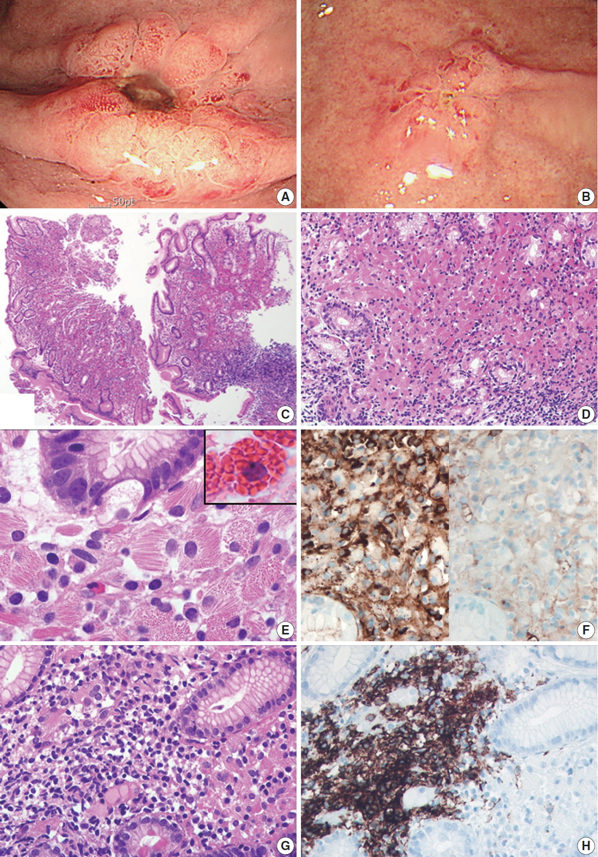
- Gastric crystal-storing histiocytosis with concomitant mucosa-associated lymphoid tissue lymphoma
- Mee Joo, Nam-Hoon Kim
- J Pathol Transl Med. 2020;54(4):332-335. Published online May 22, 2020
- DOI: https://doi.org/10.4132/jptm.2020.04.20
- 6,651 View
- 115 Download
- 5 Web of Science
- 5 Crossref
-
 Abstract
 PDF
PDF - Crystal-storing histiocytosis (CSH) is a rare entity that is characterized by intrahistiocytic accumulation of crystallized immunoglobulins. CSH is not a malignant process per se, but the majority of CSH cases are associated with underlying lymphoproliferative disorder. Although CSH can occur in a variety of organs, gastric CSH is very rare. We present a localized gastric CSH with concomitant mucosaassociated lymphoid tissue (MALT) lymphoma, manifesting as an ulcer bleeding in a 56-year-old man. Histologically, the biopsied gastric mucosa demonstrated expansion of the lamina propria by prominent collections of large eosinophilic mononuclear cells containing fibrillary crystalloid inclusions. Immunohistochemical studies revealed that the crystal-storing cells were histiocytes harboring kappa light chain-restricted immunoglobulin crystals. Within the lesion, atypical centrocyte-like cells forming lymphoepithelial lesions were seen, consistent with MALT lymphoma. Since this entity is rare and unfamiliar, difficulties in diagnosis may arise. Particularly, in this case, the lymphomatous area was obscured by florid CSH, making the diagnosis more challenging.
-
Citations
Citations to this article as recorded by
- Crystal-Storing Histiocytosis of the Stomach: An Unusual Clinical Context of a Rare Entity
Jenna Magri, Katsiaryna Khatskevich, Lauren Shealy, David Lewin, Chadi Hajar
International Journal of Surgical Pathology.2026; 34(1): 222. CrossRef - Sunny side up
João Pedro Pereira, Joanne Lopes, Fatima Carneiro, Francisco Baldaque-Silva
Frontline Gastroenterology.2025; 16(4): 344. CrossRef - Crystal-storing histiocytosis in the stomach: A case report and review of the literature
Linghong Kong, Liyan Xue, Yanfeng Zhong, Shenglan Wang, Danfeng Zheng, Lining Wang, Yang Jiao, Xinpeng Zhang, Huizhong Xue, Xiaogang Liu
Frontiers in Oncology.2022;[Epub] CrossRef - Lambda-Restricted Crystal-Storing Histiocytosis of Stomach: A Case Report and Review of Literature
Nalini Bansal, Pankaj Puri, Nishant Nagpal, Rahul Naithani, Rahul Gupta
Cureus.2021;[Epub] CrossRef - Immunoglobulin-Storing Histiocytosis: A Case Based Systemic Review
Hanne Wiese-Hansen, Friedemann Leh, Anette Lodvir Hemsing, Håkon Reikvam
Journal of Clinical Medicine.2021; 10(9): 1834. CrossRef
- Crystal-Storing Histiocytosis of the Stomach: An Unusual Clinical Context of a Rare Entity
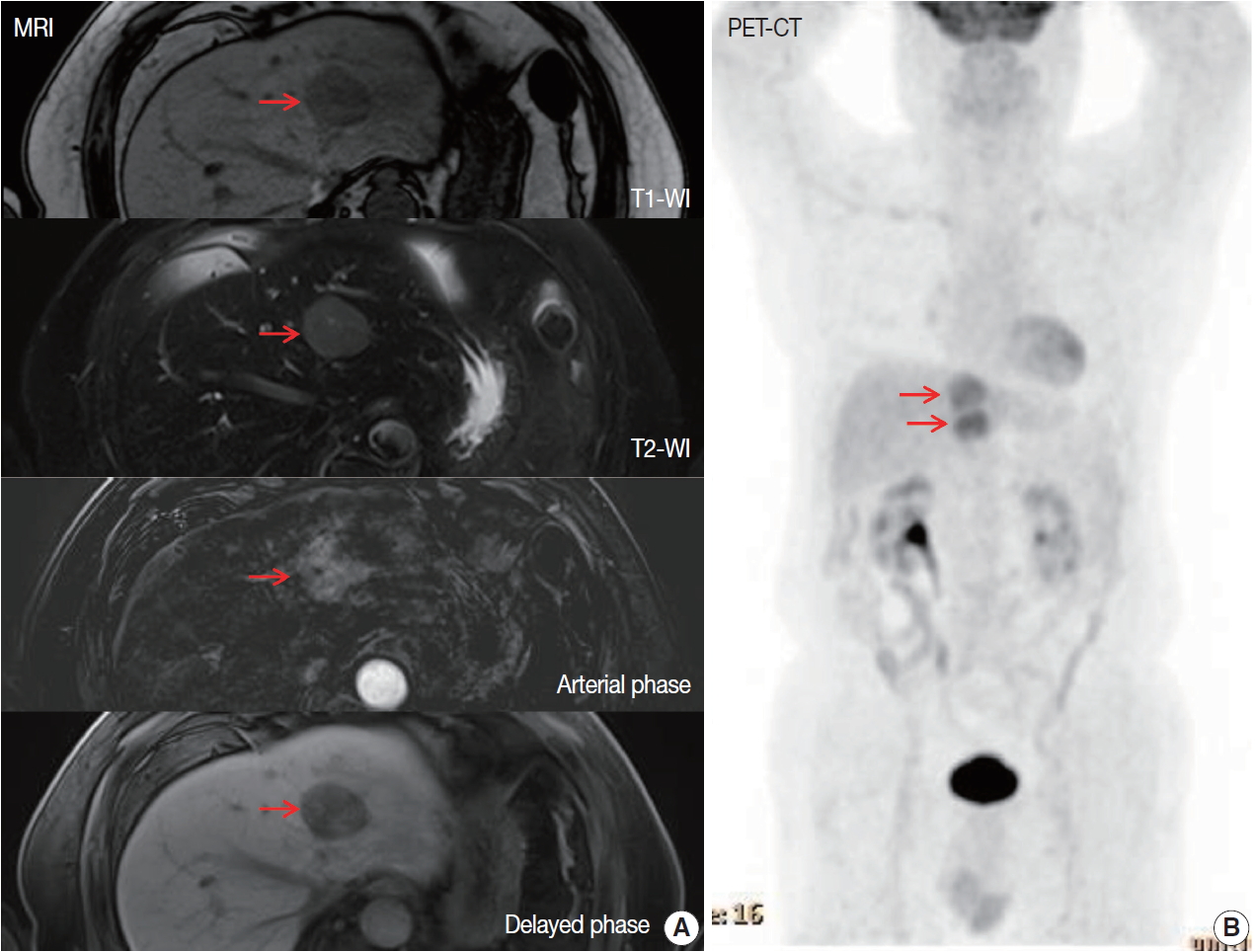
- Primary hepatic extranodal marginal zone lymphoma of mucosa-associated lymphoid tissue
- Soyeon Choi, Ji Hye Kim, Kyungbin Kim, Misung Kim, Hye Jeong Choi, Young Min Kim, Jae Hee Suh, Min Jung Seo, Hee Jeong Cha
- J Pathol Transl Med. 2020;54(4):340-345. Published online April 15, 2020
- DOI: https://doi.org/10.4132/jptm.2020.03.18
- 8,209 View
- 136 Download
- 14 Web of Science
- 14 Crossref
-
Abstract
PDF
- Extranodal marginal zone lymphoma of mucosa-associated lymphoid tissue (MALT lymphoma), is one of the specific type of low-grade B-cell lymphoma not infrequently found worldwide. It typically involves mucosal sites such as stomach and conjunctiva; however, primary hepatic MALT lymphoma has been extremely rarely reported. We describe a case of hepatic MALT lymphoma in a 70-year-old male patient who underwent left hepatectomy due to the incidentally detected liver masses at a medical checkup. The resected specimen revealed multinodular masses consisting of small-to-intermediate-sized lymphoid cells with serpentine pattern and focal lymphoepithelial lesions. The tumor cells were diffusely positive for CD20 and Bcl-2 but negative for CD3, CD10, CD5, CD23, CD43, and cyclinD1. The Ki-67 labeling index was 10% and immunoglobulin heavy chain gene rearrangement study confirmed monoclonal proliferation. In this paper, we discuss several unique clinicopathologic characteristics which will be helpful to the differential diagnosis of hepatic MALT lymphoma.
-
Citations
Citations to this article as recorded by- Oral administration of Limosilactobacillus reuteri VHProbi® M07 alleviates ovalbumin-induced allergic asthma in mice
Guoqing Meng, Hongchang Cui, Congrui Feng, Chaoqun Guo, Lei Song, Zhi Duan, Misbahuddin Rafeeq
PLOS ONE.2025; 20(1): e0317587. CrossRef - Response‑adapted involved site radiation therapy for hepatic marginal zone B‑cell lymphoma: A case report
Shin-Ting Chen, Yu-Guang Chen, Wen-Yen Huang, Cheng-Hsiang Lo
Oncology Letters.2025;[Epub] CrossRef - Management approaches for primary hepatic lymphoma: 10 year institutional experience with comprehensive literature review
Jennifer Ma, Remy Daou, Josiane Bou Eid, Beatrice Fregonese, Joe El-Khoury, N. Ari Wijetunga, Brandon S. Imber, Joachim Yahalom, Carla Hajj
Frontiers in Oncology.2025;[Epub] CrossRef - Primary Hepatic Mucosa-Associated B-Cell Lymphoma in a Patient with Primary Sclerosing Cholangitis—A Case Ultimately Requiring Liver Transplantation
Jerica Novak, Mihajlo Đokić, Miha Petrič, Diana Vozlič, Milanka Živanović, Branislava Ranković, Blaž Trotovšek
Diagnostics.2025; 15(16): 2082. CrossRef - Primary Hepatic Mucosa-Associated Lymphoid Tissue Lymphoma: A Case Report and Literature Review
Sook Hyun Shin, Jeong-Ju Yoo, Sang Gyune Kim, Young Seok Kim, Susie Chin
Clinical Ultrasound.2025; 10(2): 119. CrossRef - Primary hepatic mucosa-associated lymphoid tissue lymphoma: a case report and literature review
Tao He, Jieyu Zou
Frontiers in Oncology.2024;[Epub] CrossRef - “Speckled Enhancement” on Gd-EOB-DTPA Enhanced MR Imaging of Primary Hepatic Mucosa-associated Lymphoid Tissue Lymphoma
Ryota Hyodo, Yasuo Takehara, Ayumi Nishida, Masaya Matsushima, Shinji Naganawa
Magnetic Resonance in Medical Sciences.2023; 22(3): 273. CrossRef - Primary hepatic extranodal marginal zone B-cell mucosa-associated lymphoid tissue lymphoma treated by laparoscopic partial hepatectomy: a case report
Keisuke Okura, Satoru Seo, Hironori Shimizu, Hiroto Nishino, Tomoaki Yoh, Ken Fukumitsu, Takamichi Ishii, Koichiro Hata, Hironori Haga, Etsuro Hatano
Surgical Case Reports.2023;[Epub] CrossRef - Incidental Findings in Pediatric Patients: How to Manage Liver Incidentaloma in Pediatric Patients
Andrius Cekuolis, Dagmar Schreiber-Dietrich, Rasa Augustinienė, Heike Taut, Judy Squires, Edda L. Chaves, Yi Dong, Christoph F. Dietrich
Cancers.2023; 15(8): 2360. CrossRef - Primary hepatic mucosa‐associated lymphoid tissue lymphoma: Case report and literature review
Wing Yu Lau, Kit‐Man Ho, Fiona Ka‐Man Chan, Shi Lam, Kai‐Chi Cheng
Surgical Practice.2022; 26(1): 56. CrossRef - 18F-FDG Versus 68Ga-FAPI PET/CT in Visualizing Primary Hepatic Extranodal Marginal Zone Lymphoma of Mucosa-Associated Lymphoid Tissue
Yizhen Pang, Long Zhao, Qihang Shang, Tinghua Meng, Haojun Chen
Clinical Nuclear Medicine.2022; 47(4): 375. CrossRef - Primary hepatopancreatobiliary lymphoma: Pathogenesis, diagnosis, and management
Qianwen Wang, Kangze Wu, Xuzhao Zhang, Yang Liu, Zhouyi Sun, Shumei Wei, Bo Zhang
Frontiers in Oncology.2022;[Epub] CrossRef - Positive effect of Bifidobacterium animalis subsp. lactis VHProbi YB11 in improving gastrointestinal movement of mice having constipation
Hongchang Cui, Qian Wang, Congrui Feng, Chaoqun Guo, Jingyan Zhang, Xinping Bu, Zhi Duan
Frontiers in Microbiology.2022;[Epub] CrossRef - A case of primary hepatic extranodal marginal zone B-cell mucosa-associated lymphoid tissue (MALT) lymphoma treated by radiofrequency ablation (RFA), and a literature review
Zhe Xu, Chong Pang, Jidong Sui, Zhenming Gao
Journal of International Medical Research.2021;[Epub] CrossRef
- Oral administration of Limosilactobacillus reuteri VHProbi® M07 alleviates ovalbumin-induced allergic asthma in mice
Original Articles
- Methylotion Analysis of p16/INK4A in Gastric Low-Grade Mucosa-Associated Lymphoid Tissue Lymphomas after Helicobacter pylori Eradication Therapy.
- Young A Kim, Sung Shin Park, Bo Young Lee, You Sun Kim, In Sung Song, Chul Woo Kim
- Korean J Pathol. 2002;36(1):13-20.
- 1,997 View
- 16 Download
-
Abstract
PDF
- BACKGROUND
Inactivation of p16 has been associated with promoter region hypermethylation in different types of malignancies, including non-Hodgkin's lymphomas (NHLs). This loss of p16 was found frequently in cases of mucosa-associated lymphoid tissue (MALT) lymphomas. Recent studies indicate that promoter hypermethylation is often an early event in tumor progression in the follow-up of NHLs.
METHODS
To investigate the usefulness of p16 methylation in the diagnosis and follow-up of gastric low-grade MALT lymphomas, we analyzed methylation status of p16 using methylation-specific polymerase chain reaction methods in the sequential biopsy specimens of 13 patients with gastric low-grade MALT lymphomas undergoing Helicobacter pylori eradication therapy.
RESULTS
Five of thriteen cases showed p16 hypermethylation upon diagnosis. In four of five methylation positive cases, abnormal methylation was detected in the specimen even after the treatment, although there were no histologic evidence of disease. This methylation disappeared in the later samples of two of the cases, and they have remained in complete remission. Immunohistochemically, the loss of p16 protein expression was detected in one of three methylation-positive cases, and in none of the methylation-negative cases.
CONCLUSIONS
These results suggest that p16 methylation is relatively fequent in low-grade gastric MALT lymphomas, and it may have clinical applications in the management and follow-up of low-grade gastric MALT lymphomas.
- Expression of bcl-2 and p53 Protein in Primary Gastric Lymphomas.
- Young Rok Cho, Yu Na Kang, Sang Sook Lee, Hong Suk Song, Soo Sang Sohn, Dong Sug Kim
- Korean J Pathol. 1998;32(11):978-984.
- 2,570 View
- 10 Download
-
Abstract
- The bcl-2 gene is a proto-oncogene which extends cell survival by blocking apoptosis. Bcl-2 expression has been detected in many types of nodal and MALT lymphoma. The p53 gene is a tumor suppressor gene and p53 mutation is the most common genetic alteration in human malignancies. The relationship between the expression of bcl-2 and p53 protein in primary gastric lymphoma has been rarely reported. The authors investigated the expression of bcl-2 and p53 protein in 37 cases of primary gastric lymphoma by immunohistochemical method using bcl-2 and p53 monoclonal antibodies. There were five cases of low grade B-cell MALT lymphomas and thirty two cases of high grade B-cell lymphomas. Fifteen of 37 cases (41%) showed bcl-2 protein expression in the cytoplasm of tumor cells and 26 cases (70%) showed p53 protein expression in the nucleus of tumor cells. Bcl-2 protein was detected in 4 of 5 (80%) low grade MALT lymphomas, and in 11 of 32 (34%) high grade lymphomas. There was no significant correlation between bcl-2 expression and histologic grade of primary gastric lymphomas (p>0.05). p53 protein was positive in 25 of 32 (78%) high grade lymphomas, and in 1 of 5 (20%) low grade MALT lymphomas. The expression of p53 protein is significantly higher in high grade lymphoma than in low grade MALT lymphoma (p<0.05). The p53 expression in the bcl-2 negative cases (86%) was significantly higher than in the bcl-2 positive cases (47%). There was an inverse relationship between bcl-2 and p53 expression in primary gastric lymphoma. These results suggest that bcl-2 and p53 expression in primary gastric lymphoma may be involved in the transition from low grade MALT lymphoma to high grade lymphoma.
 First
First Prev
Prev


