 E-submission
Search
- Page Path
- HOME > Search
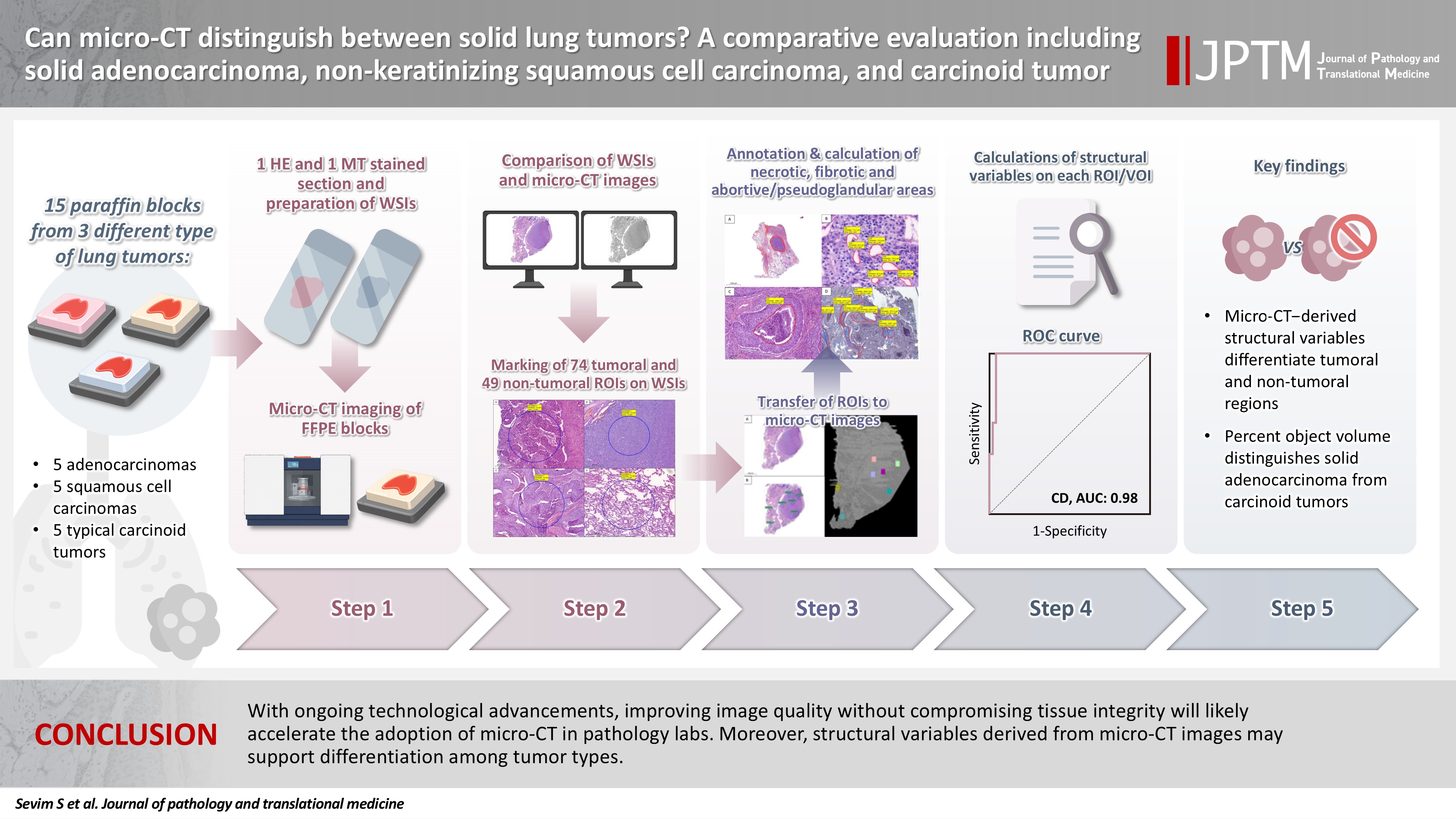
- Can micro-CT distinguish between solid lung tumors? A comparative evaluation including solid adenocarcinoma, non-keratinizing squamous cell carcinoma, and carcinoid tumor
- Selim Sevim, Serpil Dizbay Sak, Kaan Orhan, Arda Buyuksungur, Duru Karasoy, Hilal Ozakinci, Ayten Kayi Cangir
- J Pathol Transl Med. 2026;60(2):231-245. Published online March 10, 2026
- DOI: https://doi.org/10.4132/jptm.2025.12.16
- 1,823 View
- 121 Download
-
 Abstract
 PDF
PDF Supplementary Material
 - Background
Some pulmonary carcinomas display a solid pattern, and immunohistochemistry is commonly used for tumor differentiation. Micro–computed tomography (micro-CT), with its ability to produce detailed three-dimensional images using small voxel sizes, may offer additional insights. This study investigates whether three solid tumor types, solid adenocarcinoma (sAC), non-keratinizing squamous cell carcinoma, and carcinoid tumor (CaT), can be differentiated using micro-CT. Methods: Fifteen paraffin blocks, five for each type, were scanned with micro-CT (Skyscan 1275, Bruker). These images were compared to whole slide images (WSIs) of the same tumors. Consequently, tumoral (n = 74) and non-tumoral (n = 49) regions of interest (tumor ROIs [tROIs] and non-tumor ROIs [ntROIs]) were selected on the micro-CT images and evaluated in terms of certain structural variables (percent object volume, structure model index, structure thickness, structure linear density, connectivity, connectivity density, open porosity, closed porosity) to investigate whether tumors can be differentiated from normal parenchyma and from each other. Results: Although detailed images comparable to WSIs could not be obtained, it was considered an important advantage to be able to examine the entire depth of the paraffin blocks. tROIs and ntROIs could be distinguished based on all variables (p < .001). Additionally, sAC showed a notable difference from CaT in “percent object volume” (p = .011). Conclusions: With ongoing technological advancements, improving image quality without compromising tissue integrity will likely accelerate the adoption of micro-CT in pathology labs. Moreover, structural variables derived from micro-CT images may support differentiation among tumor types.
- Rhabdomyosarcoma of the skull with EWSR1 fusion and ALK and cytokeratin expression: a case report
- Hyeong Rok An, Kyung-Ja Cho, Sang Woo Song, Ji Eun Park, Joon Seon Song
- J Pathol Transl Med. 2024;58(5):255-260. Published online September 5, 2024
- DOI: https://doi.org/10.4132/jptm.2024.08.15
- 6,034 View
- 226 Download
- 1 Web of Science
- 3 Crossref
-
Abstract
PDF
- Rhabdomyosarcoma (RMS) comprises of heterogeneous group of neoplasms that occasionally express epithelial markers on immunohistochemistry (IHC). We herein report the case of a patient who developed RMS of the skull with EWSR1 fusion and anaplastic lymphoma kinase (ALK) and cytokeratin expression as cytomorphologic features. A 40-year-old man presented with a mass in his forehead. Surgical resection was performed, during which intraoperative frozen specimens were obtained. Squash cytology showed scattered or clustered spindle and epithelioid cells. IHC revealed that the resected tumor cells were positive for desmin, MyoD1, cytokeratin AE1/ AE3, and ALK. Although EWSR1 rearrangement was identified on fluorescence in situ hybridization, ALK, and TFCP2 rearrangement were not noted. Despite providing adjuvant chemoradiation therapy, the patient died of tumor progression 10 months after diagnosis. We emphasize that a subset of RMS can express cytokeratin and show characteristic histomorphology, implying the need for specific molecular examination.
-
Citations
Citations to this article as recorded by
- Rhabdomyosarcomas of Bone
Ahmed Shah, Andrew L. Folpe
Surgical Pathology Clinics.2025; 18(3): 503. CrossRef - Review of imaging modalities and radiological findings of calvarial lesions
Erkan Gökçe, Murat Beyhan
World Journal of Radiology.2025;[Epub] CrossRef - Molecular Morphology of Telangiectatic Osteosarcoma Associated With Сystic Content: A Case Report
David Makaridze, Armaz Mariamidze, Tamuna Gvianishvili, Giulia Ottaviani , Liana Gogiashvili
Cureus.2025;[Epub] CrossRef
- Rhabdomyosarcomas of Bone
- Peripheral type squamous cell carcinoma of the lung: clinicopathologic characteristics in comparison to the central type
- Yeoun Eun Sung, Uiju Cho, Kyo Young Lee
- J Pathol Transl Med. 2020;54(4):290-299. Published online June 17, 2020
- DOI: https://doi.org/10.4132/jptm.2020.05.04
- 12,747 View
- 213 Download
- 15 Web of Science
- 17 Crossref
-
Abstract
PDF
- Background
Squamous cell carcinomas (SqCCs) of the lung are known to arise more often in a central area but reports of peripheral SqCCs have increased, with a pathogenesis that is obscured. In this study, the clinicopathologic characteristics of peripheral lung SqCCs were studied and compared with those of the central type.
Methods
This study included 63 peripheral lung SqCCs and 48 randomly selected central cases; hematoxylin and eosin-stained slides of surgically resected specimens were reviewed in conjunction with radiologic images and clinical history. Cytokeratin-7 immunohistochemical staining of key slides and epidermal growth factor receptor (EGFR)/KRAS mutations tested by DNA sequencing were also included.
Results
Stages of peripheral SqCCs were significantly lower than central SqCCs (p=.016). Cystic change of the mass (p=.007), presence of interstitial fibrosis (p=0.007), and anthracosis (p=.049) in the background lung were significantly associated with the peripheral type. Cytokeratin-7 positivity was also higher in peripheral SqCCs with cutoffs of both 10% and 50% (p=.011). Pathogenic mutations in EGFR and KRAS were observed in only one case out of the 72 evaluated. The Cox proportional hazard model indicated a significantly better disease-free survival (p=.009) and the tendency of better overall survival (p=.106) in the peripheral type.
Conclusions
In peripheral type, lower stage is a favorable factor for survival but more frequent interstitial fibrosis and older age are unfavorable factors. Multivariate Cox analysis revealed that peripheral type is associated with better disease-free survival. The pathogenesis of peripheral lung SqCCs needs further investigation, together with consideration of the background lung conditions. -
Citations
Citations to this article as recorded by- Lepidic and alveolar subepithelial squamous cell carcinoma: expansion of the concept of peripheral squamous cell carcinoma with proposal for revised terminology based on morphologic, immunophenotypic, and clinical analysis of 22 cases
Federica Filipello, Francesca Ambrosi, Hans Blaauwgeers, Johanna Grefte, Wim Vos, Luisella Righi, Erik Thunnissen, Teodora Radonic
Virchows Archiv.2026; 489(1): 27. CrossRef - Adenosquamous carcinoma of the lung: Comparative CT and pathological features versus adenocarcinoma and squamous cell carcinoma
Qianyao Yuan, Dai Zhang, Rui Xu, Wenjun Yao, Hong Zhao, Kota V Ramana
PLOS One.2026; 21(6): e0352454. CrossRef - Assessing the performance of chest x‐ray screening in detecting early‐stage lung cancer in the general population
Choy‐Lye Chei, Sho Nakamura, Kaname Watanabe, Takashi Mizutani, Hiroto Narimatsu
International Journal of Cancer.2025; 156(11): 2127. CrossRef - Whole lung radiomic features are associated with overall survival in patients with locally advanced non-small cell lung cancer treated with definitive radiotherapy
Meng Yan, Zhen Zhang, Jia Tian, Jiaqi Yu, Andre Dekker, Dirk de Ruysscher, Leonard Wee, Lujun Zhao
Radiation Oncology.2025;[Epub] CrossRef - Imaging appearances, CT evolution patterns, and surgical prognosis of stage I lung squamous cell carcinoma
Wei-hua Zhao, Tian-you Luo, Fa-jin Lv, Qi Li
Cancer Imaging.2025;[Epub] CrossRef - Pulmonary squamous cell carcinoma and lymphoepithelial carcinoma – morphology, molecular characteristics and differential diagnosis
Sabina Berezowska, Marie Maillard, Mark Keyter, Bettina Bisig
Histopathology.2024; 84(1): 32. CrossRef - Assessment of seasonal variability of PM, BC and UFP levels at a highway toll stations and their associated health risks
Nazneen, Aditya Kumar Patra, Soma Sekhara Rao Kolluru, Abhishek Penchala, Sachidanand Kumar, Namrata Mishra, Naragam Bhanu Sree, Samrat Santra, Ravish Dubey
Environmental Research.2024; 245: 118028. CrossRef - Association between Airport Ultrafine Particles and Lung Cancer Risk: The Multiethnic Cohort Study
Arthur Bookstein, Justine Po, Chiuchen Tseng, Timothy V. Larson, Juan Yang, Sung-shim L. Park, Jun Wu, Salma Shariff-Marco, Pushkar P. Inamdar, Ugonna Ihenacho, Veronica W. Setiawan, Mindy C. DeRouen, Loïc Le Marchand, Daniel O. Stram, Jonathan Samet, Bea
Cancer Epidemiology, Biomarkers & Prevention.2024; 33(5): 703. CrossRef - Clinical and Bronchoscopy Assessment in Diagnosing the Histopathology Type of Primary Central Lung Tumors
Mia Elhidsi, Jamal Zaini, Lisnawati Rachmadi, Asmarinah Asmarinah, Aria Kekalih, Noni Soeroso, Menaldi Rasmin
The Open Respiratory Medicine Journal.2024;[Epub] CrossRef - Possible thoracic metastasis from squamous cell carcinoma of the external auditory canal: A case report
Hiroshi Takehara, Ken Kodama, Toru Momozane, Masashi Takeda, Kaichi Shigetsu, Hiroki Kishima
Clinical Case Reports.2024;[Epub] CrossRef - Radiological precursor lesions of lung squamous cell carcinoma: Early progression patterns and divergent volume doubling time between hilar and peripheral zones
Haruto Sugawara, Yasushi Yatabe, Hirokazu Watanabe, Hiroyuki Akai, Osamu Abe, Shun-ichi Watanabe, Masahiko Kusumoto
Lung Cancer.2023; 176: 31. CrossRef - Loss of GSTO2 contributes to cell growth and mitochondria function via the p38 signaling in lung squamous cell carcinoma
Ryusuke Sumiya, Masayoshi Terayama, Teruki Hagiwara, Kazuaki Nakata, Keigo Sekihara, Satoshi Nagasaka, Hideki Miyazaki, Toru Igari, Kazuhiko Yamada, Yuki I. Kawamura
Cancer Science.2022; 113(1): 195. CrossRef - Primary tumor location in lung cancer: the evaluation and administration
Xueqi Xie, Xiaolin Li, Wenjie Tang, Peng Xie, Xuefen Tan
Chinese Medical Journal.2022; 135(2): 127. CrossRef - Pulmonary squamous cell carcinoma with a lepidic-pagetoid growth pattern
Claudio Guerrieri, Mark Lindner, Joanna Sesti, Abhishek Chakraborti, Rachel Hudacko
Pathologica.2022; 114(4): 304. CrossRef - Deposition modeling of ambient particulate matter in the human respiratory tract
Salman Khan, Bhola Ram Gurjar, Veerendra Sahu
Atmospheric Pollution Research.2022; 13(10): 101565. CrossRef - Selection of the surgical approach for patients with cStage IA lung squamous cell carcinoma: A population-based propensity score matching analysis
Shengteng Shao, Guisong Song, Yuanyong Wang, Tengfei Yi, Shuo Li, Fuhui Chen, Yang Li, Xiaotong Liu, Bin Han, Yuhong Liu
Frontiers in Oncology.2022;[Epub] CrossRef - Virus Nanoparticles & Different Nanoparticles Affect Lung Cancer- A New Approach
Ranajit Nath, Ratna Roy, Soubhik bhattacharyya, Sourav Datta
International Journal of Scientific Research in Science and Technology.2021; : 867. CrossRef
- Lepidic and alveolar subepithelial squamous cell carcinoma: expansion of the concept of peripheral squamous cell carcinoma with proposal for revised terminology based on morphologic, immunophenotypic, and clinical analysis of 22 cases
- Double cocktail immunostains with high molecular weight cytokeratin and GATA-3: useful stain to discriminate in situ involvement of prostatic ducts or acini from stromal invasion by urothelial carcinoma in the prostate
- Junghye Lee, Youngeun Yoo, Sanghui Park, Min-Sun Cho, Sun Hee Sung, Jae Y. Ro
- J Pathol Transl Med. 2020;54(2):146-153. Published online February 10, 2020
- DOI: https://doi.org/10.4132/jptm.2019.11.12
- 9,713 View
- 138 Download
- 2 Web of Science
- 2 Crossref
-
Abstract
PDF
- Background
Distinguishing prostatic stromal invasion (PSI) by urothelial carcinoma (UC) from in situ UC involving prostatic ducts or acini with no stromal invasion (in situ involvement) may be challenging on hematoxylin and eosin stained sections. However, the distinction between them is important because cases with PSI show worse prognosis. This study was performed to assess the utility of double cocktail immunostains with high molecular weight cytokeratin (HMWCK) and GATA-3 to discriminate PSI by UC from in situ UC involvement of prostatic ducts or acini in the prostate.
Methods
Among 117 radical cystoprostatectomy specimens for bladder UCs, 25 cases showed secondary involvement of bladder UC in prostatic ducts/acini only or associated stromal invasion and of these 25 cases, seven cases revealed equivocal PSI. In these seven cases with equivocal PSI, HMWCK, and GATA-3 double immunohistochemical stains were performed to identify whether this cocktail stain is useful to identify the stromal invasion.
Results
In all cases, basal cells of prostate glands showed strong cytoplasmic staining for HMWCK and UC cells showed strong nuclear staining for GATA-3. In cases with stromal invasion of UC, GATA-3-positive tumor cells in the prostatic stroma without surrounding HMWCK-positive basal cells were highlighted and easily recognized. Among seven equivocal cases, two cases showed PSI and five in situ UC in the prostate. In two cases, the original diagnoses were revised.
Conclusions
Our study suggested that HMWCK and GATA-3 double stains could be utilized as an adjunct method in the distinction between PSI by UC from in situ UC involving prostatic ducts or acini. -
Citations
Citations to this article as recorded by- Aberrant expression of GATA3 in metastatic adenocarcinoma of the prostate: an important pitfall
João Lobo, Nazario P Tenace, Sofia Cañete‐Portillo, Isa Carneiro, Rui Henrique, Roberta Lucianò, Lara R Harik, Cristina Magi‐Galluzzi
Histopathology.2024; 84(3): 507. CrossRef - Utility of D2-40, Cytokeratin 5/6, and High–Molecular-weight Cytokeratin (Clone 34βE12) in Distinguishing Intraductal Spread of Urothelial Carcinoma From Prostatic Stromal Invasion
Oleksii A. Iakymenko, Laurence M. Briski, Katiana S. Delma, Merce Jorda, Oleksandr N. Kryvenko
American Journal of Surgical Pathology.2022; 46(4): 454. CrossRef
- Aberrant expression of GATA3 in metastatic adenocarcinoma of the prostate: an important pitfall
- Basaloid Squamous Cell Carcinoma of the Head and Neck: Subclassification into Basal, Ductal, and Mixed Subtypes Based on Comparison of Clinico-pathologic Features and Expression of p53, Cyclin D1, Epidermal Growth Factor Receptor, p16, and Human Papillomavirus
- Kyung-Ja Cho, Se Un Jeong, Sung Bae Kim, Sang-wook Lee, Seung-Ho Choi, Soon Yuhl Nam, Sang Yoon Kim
- J Pathol Transl Med. 2017;51(4):374-380. Published online June 8, 2017
- DOI: https://doi.org/10.4132/jptm.2017.03.03
- 24,555 View
- 506 Download
- 11 Web of Science
- 12 Crossref
-
Abstract
PDF
- Background
Basaloid squamous cell carcinoma (BSCC) is a rare variant of squamous cell carcinoma with distinct pathologic characteristics. The histogenesis of BSCC is not fully understood, and the cancer has been suggested to originate from a totipotent primitive cell in the basal cell layer of the surface epithelium or in the proximal duct of secretory glands.
Methods
Twenty-six cases of head and neck BSCC from Asan Medical Center, Seoul, Korea, reported during a 14-year-period were subclassified into basal, ductal, and mixed subtypes according to the expression of basal (cytokeratin [CK] 5/6, p63) or ductal markers (CK7, CK8/18). The cases were also subject to immunohistochemical study for CK19, p53, cyclin D1, epidermal growth factor receptor (EGFR), and p16 and to in situ hybridization for human papillomavirus (HPV), and the results were clinico-pathologically compared.
Results
Mixed subtype (12 cases) was the most common, and these cases showed hypopharyngeal predilection, older age, and higher expression of CK19, p53, and EGFR than other subtypes. The basal subtype (nine cases) showed frequent comedo-necrosis and high expression of cyclin D1. The ductal subtype (five cases) showed the lowest expression of p53, cyclin D1, and EGFR. A small number of p16- and/or HPV-positive cases were not restricted to one subtype. BSCC was the cause of death in 19 patients, and the average follow-up period for all patients was 79.5 months. Overall survival among the three subtypes was not significantly different.
Conclusions
The results of this study suggest a heterogeneous pathogenesis of head and neck BSCC. Each subtype showed variable histology and immunoprofiles, although the clinical implication of heterogeneity was not determined in this study. -
Citations
Citations to this article as recorded by- Histopathological variants of head and neck squamous cell carcinomas: A multicenter study in Latin America
Heitor Albergoni Silveira, Karina Helen Martins, Ana Lia Anbinder, Thais Aguiar Santos, Elton Fernandes Barros, Pollianna Muniz Alves, Cassiano Francisco Weege Nonaka, Ana Terezinha Marques Mesquita, Matheus Henrique Lopes Dominguete, Rafael Rodrigues Dia
Annals of Diagnostic Pathology.2026; 80: 152565. CrossRef - HPV-associated oropharyngeal cancer: epidemiology, molecular biology and clinical management
Matt Lechner, Jacklyn Liu, Liam Masterson, Tim R. Fenton
Nature Reviews Clinical Oncology.2022; 19(5): 306. CrossRef - Neoadjuvant treatment combined with planned endoscopic surgery in locally advanced sphenoid sinus basaloid squamous cell carcinoma
Yinghong Zhang, Suqing Tian, Yali Du, Qiang Zuo, Li Zhu, Furong Ma
Medicine: Case Reports and Study Protocols.2022; 3(6): e0044. CrossRef - Cetuximab and paclitaxel combination therapy for recurrent basaloid squamous cell carcinoma in the ethmoid sinus
Satoshi Koyama, Kazunori Fujiwara, Tsuyoshi Morisaki, Taihei Fujii, Yosuke Nakamura, Takahiro Fukuhara, Hiromi Takeuchi
Auris Nasus Larynx.2021; 48(6): 1189. CrossRef - Constitutive Hedgehog/GLI2 signaling drives extracutaneous basaloid squamous cell carcinoma development and bone remodeling
Marina Grachtchouk, Jianhong Liu, Mark E Hutchin, Paul W Harms, Dafydd Thomas, Lebing Wei, Aiqin Wang, Donelle Cummings, Lori Lowe, Jonathan Garlick, James Sciubba, Arul M Chinnaiyan, Monique E Verhaegen, Andrzej A Dlugosz
Carcinogenesis.2021; 42(8): 1100. CrossRef - Conjunctival ‘mucoepidermoid carcinoma’ revisited: a revision of terminology, based on morphologic, immunohistochemical and molecular findings of 14 cases, and the 2018 WHO Classification of Tumours of the Eye
Hardeep S. Mudhar, Tatyana Milman, Paul J.L. Zhang, Carol L. Shields, Ralph C. Eagle, Sara E. Lally, Jerry A. Shields, Sachin M. Salvi, Paul A. Rundle, Jennifer Tan, Ian G. Rennie
Modern Pathology.2020; 33(7): 1242. CrossRef - Basaloid squamous cell carcinoma with adenoid cystic‐like features of the head and neck region: A report of two cases
Kimihide Kusafuka, Haruna Yagi, Satoshi Baba, Hiroshi Inagaki, Chinatsu Tsuchiya, Kazuki Hirata, Aya Muramatsu, Makoto Suzuki, Kazumori Arai, Tadashi Terada
Pathology International.2020; 70(10): 767. CrossRef - Association study of cell cycle proteins and human papillomavirus in laryngeal cancer in Chinese population
Lifang Cui, Congling Qu, Honggang Liu
Clinical Otolaryngology.2019; 44(3): 323. CrossRef - Liver metastatic basaloid squamous cell carcinoma with negative expression of pancytokeratin: a case report and literature review
Linxiu Liu, Xuemin Xue, Liyan Xue
Diagnostic Pathology.2019;[Epub] CrossRef - Basaloid Squamous Cell Carcinoma at the Floor of the Mouth and Mandible: A Case Report
Jun-Sang Lee, Uk-Kyu Kim, Dae-Seok Hwang, Jun-Ho Lee, Hong-Seok Choi, Na-Rae Choi, Mi Heon Ryu, Gyoo Cheon Kim
The Korean Journal of Oral and Maxillofacial Pathology.2019; 43(5): 197. CrossRef - p53 and p16 expression in oral cavity squamous cell and basaloid squamous cell carcinoma
Allisson Filipe Lopes Martins, Carlos Henrique Pereira, Marília Oliveira Morais, Paulo Otávio Carmo Souza, Lucas Borges Fleury Fernandes, Aline Carvalho Batista, Elismauro Francisco Mendonça
Oral Cancer.2018; 2(1-2): 7. CrossRef - Expression and role of EGFR, cyclin�D1 and KRAS in laryngocarcinoma tissues
Xinsheng Lin, Guofeng Wen, Shuangle Wang, Hangui Lu, Chuangwei Li, Xin Wang
Experimental and Therapeutic Medicine.2018;[Epub] CrossRef
- Histopathological variants of head and neck squamous cell carcinomas: A multicenter study in Latin America
- A Rare Case of Primary Tubular Adenocarcinoma of the Thymus, Enteric Immunophenotype: A Case Study and Review of the Literature
- Hae Yoen Jung, Hyundeuk Cho, Jin-Haeng Chung, Sang Byoung Bae, Ji-Hye Lee, Hyun Ju Lee, Si-Hyong Jang, Mee-Hye Oh
- J Pathol Transl Med. 2015;49(4):331-334. Published online June 1, 2015
- DOI: https://doi.org/10.4132/jptm.2015.04.16
- 12,120 View
- 86 Download
- 14 Web of Science
- 15 Crossref
-
Abstract
PDF
- Thymic carcinomas are uncommon malignant tumors, and thymic adenocarcinomas are extremely rare. Here, we describe a case of primary thymic adenocarcinoma in a 59-year-old woman. Histological examination of the tumor revealed tubular morphology with expression of cytokeratin 20 and caudal-type homeobox 2 according to immunohistochemistry, suggesting enteric features. Extensive clinical and radiological studies excluded the possibility of an extrathymic primary tumor. A review of the literature revealed only two global cases of primary tubular adenocarcinomas of the thymus with enteric immunophenotype.
-
Citations
Citations to this article as recorded by- Non-mucinous, enteric-type thymic adenocarcinoma: genetic analysis of a case
Eiji Narusawa, Yoichi Ohtaki, Genichiro Ishii, Seshiru Nakazawa, Natsuko Kawatani, Tomohiro Yazawa, Kazuki Numajiri, Yuka Yoshida, Keisuke Nimura, Ken Shirabe
General Thoracic and Cardiovascular Surgery Cases.2026;[Epub] CrossRef - Unresectable Primary Enteric‐Type Thymic Adenocarcinoma Treated With FOLFOX Chemotherapy: A Case Report
Carl He, Georgia Bentick, Patrick Hosking, Andrew Mant
Cancer Reports.2025;[Epub] CrossRef - Enteric thymic adenocarcinoma: Understanding a unique pathology in the mediastinum
Raja Chhabra, Kartik Mittal, Anmol Tufchi, Sajjan Rajpurohit, Deepak Kumar Mittal, Aditya Vidushi, Swati Saxena, Md Ali Osama
Indian Journal of Pathology and Microbiology.2025; 68(4): 820. CrossRef - Clinical characteristics and prognosis of primary thymic adenocarcinoma: A single‐center retrospective analysis
Qian Hong, Rui Han, Chen Chen, Fuquan Wang, Sining Zhang, Chenguang Zhao, Fang Li, Juwei Mu, Jiagen Li
Thoracic Cancer.2024; 15(24): 1815. CrossRef - Case report: Primary adenocarcinoma NOS of the thymus and cytological features
Jonathan Willner, Osvaldo Hernandez, Lea Azour, Andre L. Moreira
Diagnostic Cytopathology.2023;[Epub] CrossRef - Enteric-type thymic adenocarcinoma: a case report and literature review focusing on prognosis based on histological subtypes
Rurika Hamanaka, Kei Nakano, Takaaki Tsuboi, Kazuhito Hatanaka, Mitsutomo Kohno, Ryota Masuda, Masayuki Iwazaki
General Thoracic and Cardiovascular Surgery.2022; 70(5): 501. CrossRef - Metastatic thymic-enteric adenocarcinoma responding to chemoradiation plus anti-angiogenic therapy: A case report
Man Li, Xiao-Yu Pu, Li-Hua Dong, Peng-Yu Chang
World Journal of Clinical Cases.2021; 9(7): 1676. CrossRef - A case report: primary thymic adenocarcinoma with enteric differentiation
Yuuki Kou, Hirokazu Tanaka, Nobuhisa Yamazaki, Hiroyoshi Watanabe, Makoto Sonobe
The Journal of the Japanese Association for Chest Surgery.2020; 34(2): 107. CrossRef - Thymic enteric type adenocarcinoma: A case report with cytological features
Marie Tamai, Mitsuaki Ishida, Yusuke Ebisu, Hisashi Okamoto, Chika Miyasaka, Chisato Ohe, Yoshiko Uemura, Tomohito Saito, Tomohiro Murakawa, Koji Tsuta
Diagnostic Cytopathology.2018; 46(1): 92. CrossRef - Histologic characteristics of thymic adenocarcinomas: Clinicopathologic study of a nine-case series and a review of the literature
Ah-Young Kwon, Joungho Han, Jinah Chu, Yong Soo Choi, Byeong-Ho Jeong, Myung-Ju Ahn, Yong Chan Ahn
Pathology - Research and Practice.2017; 213(2): 106. CrossRef - Characterization of genetic aberrations in a single case of metastatic thymic adenocarcinoma
Yeonghun Lee, Sehhoon Park, Se-Hoon Lee, Hyunju Lee
BMC Cancer.2017;[Epub] CrossRef - Cytologic Characteristics of Thymic Adenocarcinoma with Enteric Differentiation: A Study of Four Fine-Needle Aspiration Specimens
Ah-Young Kwon, Joungho Han, Hae-yon Cho, Seokhwi Kim, Heejin Bang, Jiyeon Hyeon
Journal of Pathology and Translational Medicine.2017; 51(5): 509. CrossRef - Mucinous cystic tumor with CK20 and CDX2 expression of the thymus: Is this a benign counterpart of adenocarcinoma of the thymus, enteric type?
Jun Akiba, Hiroshi Harada, Shintaro Yokoyama, Toshihiro Hashiguchi, Akihiko Kawahara, Masahiro Mitsuoka, Shinzo Takamori, Hirohisa Yano
Pathology International.2016; 66(1): 29. CrossRef - Colon cancer chemotherapy for a patient with CDX2-expressing metastatic thymic adenocarcinoma: a case report and literature review
Akihiko Sawaki, Mikiya Ishihara, Yuji Kozuka, Hiroyasu Oda, Satoshi Tamaru, Yumiko Sugawara, Yoshiki Yamashita, Toshiro Mizuno, Taizo Shiraishi, Naoyuki Katayama
International Cancer Conference Journal.2016; 5(2): 113. CrossRef - Metastatic Thymic Adenocarcinoma from Colorectal Cancer
Mina Lee, Suk Jin Choi, Yong Han Yoon, Joung-Taek Kim, Wan Ki Baek, Young Sam Kim
The Korean Journal of Thoracic and Cardiovascular Surgery.2015; 48(6): 447. CrossRef
- Non-mucinous, enteric-type thymic adenocarcinoma: genetic analysis of a case
- Clinical and Prognostic Significances of Cytokeratin 19 and KIT Expression in Surgically Resectable Pancreatic Neuroendocrine Tumors
- Eun-Mi Son, Joo Young Kim, Soyeon An, Ki-Byung Song, Song Cheol Kim, Eunsil Yu, Seung-Mo Hong
- J Pathol Transl Med. 2015;49(1):30-36. Published online January 15, 2015
- DOI: https://doi.org/10.4132/jptm.2014.10.23
- 14,386 View
- 103 Download
- 22 Web of Science
- 21 Crossref
-
Abstract
PDF
- Background
Pancreatic neuroendocrine tumors (PanNETs) are malignant endocrine neoplasms that present diverse clinical behaviors. Therefore, identification of biomarkers of PanNETs is important for stratification of the prognosis of PanNET patients. Recently, cytokeratin 19 (CK19) and KIT expression were reported to have prognostic significance in PanNET patients. Methods: To identify their prognostic significance, CK19 and KIT protein expression were assessed in 182 surgically resected PanNETs and compared with clinicopathologic factors. Results: Of 182 PanNETs cases, CK19 and KIT expression was noted in 97 (53.3%) and 16 (8.8%) cases, respectively. PanNET patients with CK19 expression had larger tumors (p=.006), higher World Health Organization (WHO) grade (p=.002) and pT classification (p<.001), increased distant metastasis (p=.004), and lymphovascular (p=.012) and perineural (p=.019) invasion. Similarly, those with KIT expression had larger tumors (p=.030), higher WHO grade (p=.001), advanced pT classification (p<.001), distant metastasis (p=.001), and lymphovascular invasion (p=.014). The 5-year survival rate for PanNET patients with KIT expression was significantly lower (62%) than that of patients without KIT expression (77%, p=.011), as determined by univariate but not by multivariate analyses. Conclusions: CK19 and KIT expression correlate with higher metastatic potential and advanced disease stage, and KIT expression is associated with worse survival in PanNET patients. -
Citations
Citations to this article as recorded by- Case Report: Extra-adrenal retroperitoneal paraganglioma in a young adult cat diagnosed by imaging, pathology, and immunohistochemistry
Sang-June Sohn, Sohee Lim, Junghoon Park, Ulsoo Choi, Yeon-Jung Hong
Frontiers in Veterinary Science.2025;[Epub] CrossRef - Appendiceal mucinous tumour resulting in autoamputation of the appendix: A case report and literature review
Chenao Wang, Yufeng Liu, Yaqing Liu, Baicheng Li, Xingdong Hou, Bowei Lu, Zhao Chen, Shili Ning
Experimental and Therapeutic Medicine.2025; 31(2): 1. CrossRef - Expression profiles of cadherin 17 and claudin 18.2 in comparison with peptide hormonal expression in pancreatic neuroendocrine tumours: Implications for targeted immunotherapy
Kahoko Maeda, Takeshi Uehara, Waki Hosoda, Yasuhiro Kuraishi, Hiroyoshi Ota
Pathology - Research and Practice.2024; 262: 155537. CrossRef - Glypican-3 and Cytokeratin-19 Expression in Pancreatic Cancer in a Canadian Population
Carley Bekkers, Ravi Ramjeesingh, Thomas Arnason
Journal of Clinical Medicine.2024; 13(22): 6893. CrossRef - Combined Infiltrative Macroscopic Growth Pattern and Infiltrative Microscopic Tumor Border Status Is a Novel Surrogate Marker of Poor Prognosis in Patients With Pancreatic Neuroendocrine Tumor
Bokyung Ahn, Joo Young Kim, Seung-Mo Hong
Archives of Pathology & Laboratory Medicine.2023; 147(1): 100. CrossRef -
Tumor-associated nonmyelinating Schwann cell–expressed
PVT1
promotes pancreatic cancer kynurenine pathway and tumor immune exclusion
Chengcao Sun, Youqiong Ye, Zhi Tan, Yuan Liu, Yajuan Li, Wei Hu, Ke Liang, Sergey D. Egranov, Lisa Angela Huang, Zhao Zhang, Yaohua Zhang, Jun Yao, Tina K. Nguyen, Zilong Zhao, Andrew Wu, Jeffrey R. Marks, Abigail S. Caudle, Aysegul A. Sahin, Jianjun Gao,
Science Advances.2023;[Epub] CrossRef - Diagnostic and prognostic impact of cytokeratin 19 expression analysis in human tumors: a tissue microarray study of 13,172 tumors
Anne Menz, Rifka Bauer, Martina Kluth, Clara Marie von Bargen, Natalia Gorbokon, Florian Viehweger, Maximilian Lennartz, Cosima Völkl, Christoph Fraune, Ria Uhlig, Claudia Hube-Magg, Noémi De Wispelaere, Sarah Minner, Guido Sauter, Simon Kind, Ronald Simo
Human Pathology.2021; 115: 19. CrossRef - The molecular biology of pancreatic neuroendocrine neoplasms: Challenges and translational opportunities
Kate Young, Naureen Starling, Anguraj Sadanandam
Seminars in Cancer Biology.2020; 61: 132. CrossRef - Pancreatic acinar cell carcinomas and mixed acinar-neuroendocrine carcinomas are more clinically aggressive than grade 1 pancreatic neuroendocrine tumours
Joo Young Kim, Jacqueline A. Brosnan-Cashman, Jiyoon Kim, Soyeon An, Kyoung-Bun Lee, Haeryoung Kim, Do Youn Park, Kee-Taek Jang, Young-Ha Oh, Ralph H. Hruban, Christopher M. Heaphy, Seung-Mo Hong
Pathology.2020; 52(3): 336. CrossRef - Morphologic Variants of Pancreatic Neuroendocrine Tumors: Clinicopathologic Analysis and Prognostic Stratification
Yue Xue, Michelle D. Reid, Burcin Pehlivanoglu, Rebecca C. Obeng, Hongmei Jiang, Bahar Memis, Shu K. Lui, Juan Sarmiento, David Kooby, Shishir K. Maithel, Bassel El-Rayes, Olca Basturk, Volkan Adsay
Endocrine Pathology.2020; 31(3): 239. CrossRef - Histological grades and prognostic markers of well-differentiated pancreatic neuroendocrine tumor (WDPNET)
Yongchao Li, Daniel Rowan, Claire P. Williamson, Meiyun Fan, Ali G. Saad, Lizhi Zhang
Journal of Pancreatology.2020; 3(4): 188. CrossRef - Clinical and histopathologic prognostic implications of the expression of cytokeratins 8, 10, 13, 14, 16, 18 and 19 in oral and oropharyngeal squamous cell carcinoma
Rima A. Safadi, Niveen I. Abdullah, Rolla F. Alaaraj, Dima H. Bader, Darshan D. Divakar, Abed A. Hamasha, Maher A. Sughayer
Archives of Oral Biology.2019; 99: 1. CrossRef - Prognostic and predictive factors on overall survival and surgical outcomes in pancreatic neuroendocrine tumors: recent advances and controversies
Lingaku Lee, Tetsuhide Ito, Robert T Jensen
Expert Review of Anticancer Therapy.2019; 19(12): 1029. CrossRef - Carbonic anhydrase 9 expression in well-differentiated pancreatic neuroendocrine neoplasms might be associated with aggressive behavior and poor survival
Joo Young Kim, Sang Hwa Lee, Soyeon An, Sung Joo Kim, You-Na Sung, Ki-Byung Song, Dae Wook Hwang, Song Cheol Kim, Seung-Mo Hong
Virchows Archiv.2018; 472(5): 739. CrossRef - CD133 expression in well-differentiated pancreatic neuroendocrine tumors: a potential predictor of progressive clinical courses
Yasuhiro Sakai, Seung-Mo Hong, Soyeon An, Joo Young Kim, Denis Corbeil, Jana Karbanová, Kyoko Otani, Kohei Fujikura, Ki-Byung Song, Song Cheol Kim, Masayuki Akita, Yoshihide Nanno, Hirochika Toyama, Takumi Fukumoto, Yonson Ku, Takanori Hirose, Tomoo Itoh,
Human Pathology.2017; 61: 148. CrossRef - Prognostic and predictive biomarkers in neuroendocrine tumours
David L. Chan, Stephen J. Clarke, Connie I. Diakos, Paul J. Roach, Dale L. Bailey, Simron Singh, Nick Pavlakis
Critical Reviews in Oncology/Hematology.2017; 113: 268. CrossRef - Loss of Progesterone Receptor Expression Is an Early Tumorigenesis Event Associated with Tumor Progression and Shorter Survival in Pancreatic Neuroendocrine Tumor Patients
Sung Joo Kim, Soyeon An, Jae Hoon Lee, Joo Young Kim, Ki-Byung Song, Dae Wook Hwang, Song Cheol Kim, Eunsil Yu, Seung-Mo Hong
Journal of Pathology and Translational Medicine.2017; 51(4): 388. CrossRef - Prognostic significance of cytokeratin 19 expression in pancreatic neuroendocrine tumor: A meta-analysis
Dong Cen, Jiang Chen, Zheyong Li, Jie Zhao, Xiujun Cai, Aamir Ahmad
PLOS ONE.2017; 12(11): e0187588. CrossRef - A retrospective cohort study of pancreatic neuroendocrine tumors at single institution over 15 years: New proposal for low- and high-grade groups, validation of a nomogram for prognosis, and novel follow-up strategy for liver metastases
Liangtao Ye, Huilin Ye, Quanbo Zhou, Zhihua Li, Qing Lin, Langping Tan, Wenchao Gao, Zhiqiang Fu, Shangyou Zheng, Rufu Chen
International Journal of Surgery.2016; 29: 108. CrossRef - Correlating and Combining Genomic and Proteomic Assessment withIn VivoMolecular Functional Imaging: Will This Be the Future Roadmap for Personalized Cancer Management?
Bhakti Basu, Sandip Basu
Cancer Biotherapy and Radiopharmaceuticals.2016; 31(3): 75. CrossRef - Recent Updates on Neuroendocrine Tumors From the Gastrointestinal and Pancreatobiliary Tracts
Joo Young Kim, Seung-Mo Hong
Archives of Pathology & Laboratory Medicine.2016; 140(5): 437. CrossRef
- Case Report: Extra-adrenal retroperitoneal paraganglioma in a young adult cat diagnosed by imaging, pathology, and immunohistochemistry
- Clear Cell Papillary Renal Cell Carcinoma: A Report of 15 Cases Including Three Cases of Concurrent Other-Type Renal Cell Carcinomas
- Jeong Hwan Park, Cheol Lee, Ja Hee Suh, Kyung Chul Moon
- Korean J Pathol. 2012;46(6):541-547. Published online December 26, 2012
- DOI: https://doi.org/10.4132/KoreanJPathol.2012.46.6.541
- 11,030 View
- 65 Download
- 23 Crossref
-
Abstract
PDF
Background Clear cell papillary renal cell carcinoma (CCPRCC) is a recently established subtype of renal epithelial tumor. The aim of this study was to identify the diagnostic criteria of CCPRCC with an emphasis on immunohistochemical studies, and to report three cases with concurrent other-type renal cell carcinoma (RCC).
Methods A total of 515 RCC patients that consecutively underwent surgical resection at Seoul National University Hospital from 1 January 2010 to 31 December 2011 were screened. Each case was reviewed based on the histologic features and was evaluated immunohistochemically.
Results A total of 15 CCPRCCs were identified, which composed 2.9% of the total RCCs. The mean age was 52 years, and the average tumor size was 1.65 cm. All 15 cases showed low nuclear grade, no lymph node metastasis and no distant metastasis. The CCPRCCs showed variable architectural patterns including cystic, trabecular, papillary, and acinar. All of the cases showed moderate to intense immunoreactivity for cytokeratin 7 (CK7). CD10 was negative or showed focal weak positivity. Three cases had concurrent other-type RCC, including a clear cell RCC and an acquired cystic disease-associated RCC.
Conclusions The strong CK7 and negative or focal weak CD10 expression will be useful for the diagnosis of CCPRCC.
-
Citations
Citations to this article as recorded by- Vascular, adipose tissue, and/or calyceal invasion in clear cell tubulopapillary renal cell tumour: potentially problematic diagnostic scenarios
Ankur R Sangoi, Harrison Tsai, Lara Harik, Jonathan Mahlow, Maria Tretiakova, Sean R Williamson, Michelle S Hirsch
Histopathology.2024; 84(7): 1167. CrossRef - Clinical features and Surgical Outcome of Clear Cell Papillary Renal Cell Tumor: result from a prospective cohort
Si Hyun Kim, Jang Hee Han, Seung-hwan Jeong, Hyeong Dong Yuk, Ja Hyeon Ku, Cheol Kwak, Hyeon Hoe Kim, Kyung Chul Moon, Chang Wook Jeong
BMC Urology.2023;[Epub] CrossRef - Coexistence of multiple clear cell papillary renal cell carcinoma with renal oncocytoma: a case report
Amine Hermi, Ahmed Saadi, Seif Mokadem, Ahlem Blel, Marouene Chakroun, Mohamed Riadh Ben Slama
Annals of Medicine & Surgery.2023; 85(5): 2017. CrossRef - Renal Cell Carcinoma in End-Stage Renal Disease: A Review and Update
Ziad M. El-Zaatari, Luan D. Truong
Biomedicines.2022; 10(3): 657. CrossRef - The Clinicopathologic and Molecular Landscape of Clear Cell Papillary Renal Cell Carcinoma: Implications in Diagnosis and Management
Stanley Weng, Renzo G. DiNatale, Andrew Silagy, Roy Mano, Kyrollis Attalla, Mahyar Kashani, Kate Weiss, Nicole E. Benfante, Andrew G. Winer, Jonathan A. Coleman, Victor E. Reuter, Paul Russo, Ed Reznik, Satish K. Tickoo, A. Ari Hakimi
European Urology.2021; 79(4): 468. CrossRef - Clear cell papillary renal cell carcinoma: Characteristics and survival outcomes from a large single institutional series
James E. Steward, Sean Q. Kern, Liang Cheng, Ronald S. Boris, Yan Tong, Clint D. Bahler, Timothy A. Masterson, K. Clint Cary, Hristos Kaimakliotis, Thomas Gardner, Chandru P. Sundaram
Urologic Oncology: Seminars and Original Investigations.2021; 39(6): 370.e21. CrossRef - Clear cell papillary renal cell carcinoma: an update after 15 years
Sean R. Williamson
Pathology.2021; 53(1): 109. CrossRef - Clear Cell Papillary Renal Cell Carcinoma
Jianping Zhao, Eduardo Eyzaguirre
Archives of Pathology & Laboratory Medicine.2019; 143(9): 1154. CrossRef - Clear cell papillary renal cell carcinoma – An indolent subtype of renal tumor
Wei-Jen Chen, Chin-Chen Pan, Shu-Huei Shen, Hsiao-Jen Chung, Chih-Chieh Lin, Alex T.L. Lin, Yen-Hwa Chang
Journal of the Chinese Medical Association.2018; 81(10): 878. CrossRef - Clear cell papillary renal cell carcinoma: A case report and review of the literature
Sung Han Kim, Whi-An Kwon, Jae Young Joung, Ho Kyung Seo, Kang Hyun Lee, Jinsoo Chung
World Journal of Nephrology.2018; 7(8): 155. CrossRef - Clinical features and survival analysis of clear cell papillary renal cell carcinoma: A 10‑year retrospective study from two institutions
Yiqiu Wang, Ying Ding, Jian Wang, Min Gu, Zengjun Wang, Chao Qin, Conghui Han, Hongxia Li, Xia Liu, Pengfei Wu, Guangchao Li
Oncology Letters.2018;[Epub] CrossRef - A contemporary series of renal masses with emphasis on recently recognized entities and tumors of low malignant potential: A report based on 624 consecutive tumors from a single tertiary center
Maria Rosaria Raspollini, Ilaria Montagnani, Rodolfo Montironi, Liang Cheng, Guido Martignoni, Andrea Minervini, Sergio Serni, Giulio Nicita, Marco Carini, Antonio Lopez-Beltran
Pathology - Research and Practice.2017; 213(7): 804. CrossRef - Renal Neoplasms With Overlapping Features of Clear Cell Renal Cell Carcinoma and Clear Cell Papillary Renal Cell Carcinoma
Hari P. Dhakal, Jesse K. McKenney, Li Yan Khor, Jordan P. Reynolds, Cristina Magi-Galluzzi, Christopher G. Przybycin
American Journal of Surgical Pathology.2016; 40(2): 141. CrossRef - New and emerging renal tumour entities
Naoto Kuroda, Ondřej Hess, Ming Zhou
Diagnostic Histopathology.2016; 22(2): 47. CrossRef - Immunohistochemical Panel for Differentiating Renal Cell Carcinoma with Clear and Papillary Features
Hanan AlSaeid Alshenawy
Pathology & Oncology Research.2015; 21(4): 893. CrossRef - Immunohistochemical panel for differentiating renal cell carcinoma with clear and papillary features
Hanan AlSaeid Alshenawy
Journal of Microscopy and Ultrastructure.2015; 3(2): 68. CrossRef - Clear Cell-Papillary Renal Cell Carcinoma of the Kidney Not Associated With End-stage Renal Disease
Manju Aron, Elena Chang, Loren Herrera, Ondrej Hes, Michelle S. Hirsch, Eva Comperat, Philippe Camparo, Priya Rao, Maria Picken, Michal Michal, Rodolfo Montironi, Pheroze Tamboli, Federico Monzon, Mahul B. Amin
American Journal of Surgical Pathology.2015; 39(7): 873. CrossRef - Papillary or pseudopapillary tumors of the kidney
Fang-Ming Deng, Max X. Kong, Ming Zhou
Seminars in Diagnostic Pathology.2015; 32(2): 124. CrossRef - Do Clear Cell Papillary Renal Cell Carcinomas Have Malignant Potential?
Mairo L. Diolombi, Liang Cheng, Pedram Argani, Jonathan I. Epstein
American Journal of Surgical Pathology.2015; 39(12): 1621. CrossRef - Targeted next‐generation sequencing and non‐coding RNA expression analysis of clear cell papillary renal cell carcinoma suggests distinct pathological mechanisms from other renal tumour subtypes
Charles H Lawrie, Erika Larrea, Gorka Larrinaga, Ibai Goicoechea, María Arestin, Marta Fernandez‐Mercado, Ondrej Hes, Francisco Cáceres, Lorea Manterola, José I López
The Journal of Pathology.2014; 232(1): 32. CrossRef - Clear cell papillary renal cell carcinoma is the fourth most common histologic type of renal cell carcinoma in 290 consecutive nephrectomies for renal cell carcinoma
Haijun Zhou, Shaojiang Zheng, Luan D. Truong, Jae Y. Ro, Alberto G. Ayala, Steven S. Shen
Human Pathology.2014; 45(1): 59. CrossRef - Clear cell papillary renal cell carcinoma: Incidence, morphological features, immunohistochemical profile, and biologic behavior: A single institution study
Borislav A. Alexiev, Cinthia B. Drachenberg
Pathology - Research and Practice.2014; 210(4): 234. CrossRef - MRI Phenotype in Renal Cancer
Naomi Campbell, Andrew B. Rosenkrantz, Ivan Pedrosa
Topics in Magnetic Resonance Imaging.2014; 23(2): 95. CrossRef
- Vascular, adipose tissue, and/or calyceal invasion in clear cell tubulopapillary renal cell tumour: potentially problematic diagnostic scenarios
- CD56 and High Molecular Weight Cytokeratin as Diagnostic Markers of Papillary Thyroid Carcinoma.
- Mi Kyung Shin, Jeong Won Kim, Young Su Ju
- Korean J Pathol. 2011;45(5):477-484.
- DOI: https://doi.org/10.4132/KoreanJPathol.2011.45.5.477
- 6,763 View
- 38 Download
- 10 Crossref
-
Abstract
PDF
- BACKGROUND
The incidence of papillary thyroid carcinoma (PTC) has been increasing recently and a precise diagnosis is essential for optimal treatment. Ancillary immunohistochemical stains are important for diagnosing some difficult cases.
METHODS
The dignostic value of CD56, high molecular weight cytokeratin (HMCK), galectin-3 (GAL3), and cytokeratin 19 (CK19) were evaluated to distinguish PTC from other benign thyroid lesions (BTL). We studied 23 cases of papillary thyroid overt carcinomas, 57 papillary thyroid microcarcinomas, five follicular adenomas, five cases of Hashimoto's thyroiditis, and 12 nodular hyperplasias.
RESULTS
The statistical analysis showed significantly different expressions of CD56, HMCK, GAL3, and CK19 in PTC vs other BTL. The diagnostic specificity of HMCK and CD56 (90.9% and 72.7%, respectively) was higher than that of GAL3 and CK19 (50.0% and 36.4%, respectively). However, the sensitivity of HMCK and CD56 detection (92.5% and 95.0%, respectively) was lower than that of GAL3 and CK19 (98.8% and 100.0%, respectively). The combined use of CD56, HMCK, GAL3, and CK19 showed 87.5% sensitivity, 100.0% specificity, and 100.0% positive predictive value in differentiating PTC from other BTL.
CONCLUSIONS
Although the differential diagnosis of thyroid follicular lesions are based on histological and cytomorphological criteria, CD56 and HMCK might be useful markers for diagnosing PTC. -
Citations
Citations to this article as recorded by- Diagnostic role of immunohistochemical markers CK19 and CD56 in thyroid neoplasms
Pallavi Priyadarshini, Manoj Kumar Patro, Prasanta Kumar Das
MGM Journal of Medical Sciences.2023; 10(2): 176. CrossRef - CD56 Expression in Papillary Thyroid Carcinoma Is Highly Dependent on the Histologic Subtype: A Potential Diagnostic Pitfall
Uiju Cho, Yourha Kim, Sora Jeon, Chan Kwon Jung
Applied Immunohistochemistry & Molecular Morphology.2022; 30(5): 389. CrossRef - CD-56 IMMUNOREACTIVITY IN FOLLICULAR CELL DERIVED LESIONS OF THYROID
Elvin Merin Cherian, Priya P. V, Sankar S
Journal of Evolution of Medical and Dental Sciences.2018; 7(17): 2066. CrossRef - Diagnostic utility of CK19 and CD56 in the differentiation of thyroid papillary carcinoma from its mimics
Nehal S. Abouhashem, Suzan M. Talaat
Pathology - Research and Practice.2017; 213(5): 509. CrossRef - Use of CD56 and cyclin D1 in differentiating thyroid hyperplasia from papillary thyroid carcinoma
Maha E. Salama, Wael S. Ibrahim
Egyptian Journal of Pathology.2016; 36(1): 39. CrossRef - CD56, HBME-1 and cytokeratin 19 expressions in papillary thyroid carcinoma and nodular thyroid lesions
Senay Erdogan-Durmus, Deniz Ozcan, Enver Yarikkaya, Ali Kurt, Aynur Arslan
Journal of Research in Medical Sciences.2016;[Epub] CrossRef - Defining the value of CD56, CK19, Galectin 3 and HBME-1 in diagnosis of follicular cell derived lesions of thyroid with systematic review of literature
Duško Dunđerović, Jasmina Marković Lipkovski, Ivan Boričic, Ivan Soldatović, Vesna Božic, Dubravka Cvejić, Svetislav Tatić
Diagnostic Pathology.2015;[Epub] CrossRef - Utility of immunohistochemical markers in differential diagnosis of follicular cell-derived thyroid lesions
HananAlSaeid Alshenawy
Journal of Microscopy and Ultrastructure.2014; 2(3): 127. CrossRef - Utility of Immunohistochemical Markers in Diagnosis of Follicular Cell Derived Thyroid Lesions
Hanan AlSaeid Alshenawy
Pathology & Oncology Research.2014; 20(4): 819. CrossRef - Potential diagnostic utility of CD56 and claudin-1 in papillary thyroid carcinoma and solitary follicular thyroid nodules
Rasha M. Abd El Atti, Lobna S. Shash
Journal of the Egyptian National Cancer Institute.2012; 24(4): 175. CrossRef
- Diagnostic role of immunohistochemical markers CK19 and CD56 in thyroid neoplasms
- The Cytologic Features of Desmoplastic Small Round Cell Tumor with Intranuclear Inclusions : A Case Report .
- Ho Chang Lee, Hye Suk Han, Ok Jun Lee
- Korean J Pathol. 2009;43(3):279-284.
- DOI: https://doi.org/10.4132/KoreanJPathol.2009.43.3.279
- 3,432 View
- 30 Download
-
Abstract
PDF
- Desmoplastic small round cell tumor (DSRCT) is a rare neoplasm of young adults and it is characterized by polyphenotypic differentiation. We experienced a case of abdominal DSRCT that occurred in a 19-year-old female who presented with painful swelling of her right forearm. The tumor was cytokeratin-negative and it exhibited some tumor cells with intranuclear inclusions. Molecular demonstration of EWS-WT1 fusion transcripts is particularly useful to confirm the diagnosis of DSRCT without epithelial differentiation. We report here on a case of cytokeratin-negative DSRCT that showed an unusual feature of intranuclear inclusions.
- Odontogenic Gingival Epithelial Hamartoma; with Reference to the Expression of Ameloblastin Gene by in situ Hybridization and Immunohistochemistry.
- Na Rae Kim, Yeon Lim Suh, Je G Chi, Young Joon Lee, Suk Keun Lee, Jae Il Lee, Chang Yun Lim, Ji Young Park
- Korean J Pathol. 2004;38(2):116-120.
- 2,549 View
- 33 Download
-
Abstract
PDF
- Odontogenic gingival epithelial hamartoma (OGEH) is an extremely rare lesion characterized by an abnormal proliferation of odontogenic epithelium. This lesion is thought to arise from the rest of the dental lamina lying dormant in the gingival tissue after odontogenesis. Distinguishing OGEH from the granular cell variant of ameloblastoma and central odontogenic fibroma is important. To date, only eleven cases have been reported, and its pathogenesis remains unclear. We report here on a case of OGEH, where the epithelial strands in the lesion were conspicuously positive for the antisera of cytokeratin 19 and ameloblastin. Tumor cells intensely expressed ameloblastin mRNA by in situ hybridization. To the best of our knowledge, this is the first case of OGEH to which ameloblastin immunohistochemical stain and in situ hybridization were applied. Although our study is limited to a single case, the coexpression of cytokeratin 19 and ameloblastin might indicate the origin and specific cytodifferentiation of OGEH is quite different and unique, when contrasted to other odontogenic tumors.
- A Cystic Mesothelioma in the Inguinal Area.
- Im Joong Yoon, Nam Bok Cho, Tae Jin Lee, Mee Kyung Kim, Se Chul Kim, Kye Yong Song
- Korean J Pathol. 1997;31(3):284-287.
- 2,240 View
- 18 Download
-
Abstract
PDF
- The cystic mesothelioma is a very rare tumor which has a clinically and histologically benign nature. Here in reported is the case of a cystic mesothelioma presented as a palpable mass of the inguinal area in a 28-year-old male. Ultrasound showed a cystic tumor at the inguinal canal, and the other physical and laboratory examinations were within normal limits. Grossly, the tumor consisted of cysts containing clear serous fluid and focally solid areas. Microscopically, the tumor was encapsulated with fibrocollagenous wall, and the tumor cells were cuboidal or polygonal epithelial cells with single or multiple layers and had clear cytoplasm. Some areas showed thyroid follicle-like structures. The content of follicle-like structures showed eosinophilia in the H&E section, but positive in mucin stain. Neither cytologic atypia nor mitoses were present. Immunohistochemical staining revealed positive reaction for keratins of low molecular weight, while negative for the thyroglobulin and CEA. These findings suggested mesothelial in origin. We concluded that this tumor was primary rather than metastatic, because he had no evidence of a tumor in gastrointestinal, genitourinary tracts and scrotum.
- Expression of Cytokeratin 1, 10 and 14 in Fetal Skin.
- Kye Yong Song, Sun Lee, Dong Hye Suh, Mi Kyung Kim, Hye Jung Min, Je G Chi
- Korean J Pathol. 2001;35(3):226-231.
- 2,691 View
- 38 Download
-
Abstract
PDF
- BACKGROUND
During the fetal stage, the epidermis and adnexal epithelium might express different types of cytokeratin (CK) by developmental stages. The objective of this study is to observe the expressions of CK1, CK10 and CK14 in the skin of human fetuses.
METHODS
Immunohistochemical stains were applied to the skin of 42 fetuses ranging from 10 to 36 gestation weeks. Three different portions of the body (i.e., scalp, chest and sole) were sampled. Immunohistochemical staining with monoclonal antibodies against CK1, 10, 14 were done.
RESULTS
We found that CK14 was expressed in the basal layer of the epidermis and adnexae of fetuses beween 10 to 36 gestation weeks. However, stronger expression in the middle than the basal layer was noted in the soles of 15-week fetuses followed by exclusive basal expression. The sebaceous gland, the outer root sheath of the hair follicle and the eccrine duct epithelium also showed CK14 expressions, while CK14 was negative in hair germ and acini. Both CK1 and CK10 were expressed in the epidermis of fetuses ranging between 10 to 36 gestation weeks at the suprabasal layer of the scalp, chest and sole; while they were negative in the basal layer and skin adnexae including sebaceous, hair and eccrine gland.
CONCLUSIONS
Expression of cytokeratins in the fetal skin were noted at 10 weeks throughout the entire gestation period and were similar in the three different sites, except in the early stage of the sole. The main expression sites of K14 were the basal layer of the epidermis, the eccrine ducts and the outer root sheath cells of hair, suggesting the same origin, while those of K1 and K10 were in the suprabasal layer of epidermis.
- Morphological Observations on the Epidermal Development of Human Fetal Skin.
- Joong Seok Seo, Kye Yong Song, Je G Chi
- Korean J Pathol. 1990;24(1):27-38.
- 2,413 View
- 24 Download
-
Abstract
PDF
- To observe developing process of human fetal skin during intrauterine life, morphological studies in light microscopic level were made based on 27 human embryos and 76 fetuses ranging from 4 to 40 gestation weeks. The fetuses were the products of induced abortion and were found to have no associated diseases of congenital anomalies at the autopsy. Ten different portions of the body were sampled and examined. They were scalp, forehead, face, chest, abdomen, back, palm, sole, finger and toe. In embryos two different portions; cephalic and caudal portions were examined: The following results were obtained: 1) A single layer of undifferentiated cell was the primitive epidermis at the 4th week and it was followed by two layered epidermis consisting of periderm and primitive basal cell layer. Epidermal ridges started to develop along with primitive eccrine and hair germs as clustering of basal cells at the llth week. Stratum inter-medium was formed at the 12th week, and primitive granular cell layers and keratin formation in association with hair follicles at the 19th week forming earliest adult type epidermis, followed by progressive maturation. 2) The thickness of the fetal epidermis and keratin layer increased as the fetal age approached to the term with its slightly different developmental pattern by the site of body. Cephalic protions developed slightly earlier than the other parts. 3) The developmental pattern of various portions of epidermis could be categorized into three groups; (1) scalp, forehead and face; (2) chest, abdomen and back; (3) palm, sole, finger and toe.
- Pseudometastasis in Sentinel Lymph Nodes with Cytokeratin Debris-containing Histiocytes in Breast Cancer Patient: A Case Report.
- Keum Ha Choi, Eun Jung Cha, Ha Na Choi, Woo Sung Moon
- Korean J Pathol. 2007;41(6):427-429.
- 2,877 View
- 49 Download
-
Abstract
PDF
- Immunohistochemical staining for cytokeratins can detect false negative nodes in patients with breast carcinoma. We report on a patient with breast carcinoma and pseudometastasis detected by immunohistochemical staining within a negative sentinel lymph node. A 66-year-old woman underwent a simple mastectomy and sentinel lymph node biopsy. Immunohistochemical staining of the sentinel nodes for cytokeratin in permanent sections showed cells with intense cytoplasmic staining in the subcapsular sinus. The cells were negative for epithelial membrane antigen staining, but positive for CD68. In combination with morphologic findings and immunohistochemistry, cytokeratin-positive cells were confirmed as histiocytes with phagocytized cytokeratin debris. Careful correlation with histology and additional IHC could help avoid a misinterpretation of this type of pseudometastasis.
- The Utility of HMW-CK and CK5/6 Immunohistochemical Stains for Differentiating Ductal Proliferative Lesions and Ducal Carcinoma of the Breast.
- Sung Hee Son, Ju Yeon Song, Hye Kyoung Yoon
- Korean J Pathol. 2008;42(1):21-26.
- 4,679 View
- 122 Download
-
Abstract
PDF
- BACKGROUND
Basal-type cytokeratins may help to distinguish benign from malignant intraductal proliferative lesions. The basal-type cytokeratins expression is markedly decreased or absent in atypical ductal hyperplasia (ADH), ductal carcinoma in situ (DCIS) and invasive ductal carcinomas (IDC). However, the expression patterns vary according to the antibodies that are used for staining.
METHODS
HMW-CK (clone 34 E12) was applied to 175 lesions, and CK5/6 (clone D5/16B4) was applied to 145 lesions. The specimens were IDC (n=165), DCIS (n=35), ADH (n=37), florid ductal hyperplasia (FDH) (n=38) and columnar cell lesion (CCL) (n=45). The expression patterns of HMW-CK and CK5/6 were categorized as negative, focal positive and positive.
RESULTS
Loss of the HMW-CK expression was noted in 76% (66/87) of the IDC, 78% (21/27) of the DCIS, 78% (21/28) of the ADH, and 55% (10/18) of the FDH. Loss of the CK5/6 expression was found in 96% (75/78) of the IDC, in all the DCIS (n=8) and ADH (n=9), and in none of the FDH (n=20). Loss of the CK5/6 expression is more reliable than that of the HMW-CK expression for differentiating FDH, ADH and malignant intraductal proliferatve lesions. Eleven (73%) of 15 CCLs revealed the loss of the HMW-CK expression, but all the CCLs (n=30) were negative for CK5/6 (p=0.0161).
CONCLUSION
CK5/6 antibody is more reliable than HMW-CK antibody for differentiating FDH from ADH or DCIS, and for discriminating CCL.
- Proliferating Trichilemmal Tumor: Report of four cases.
- Yeong Jin Choi, Mi Kyung Jee, Seok Jin Gang, Byoung Kee Kim, Sun Moo Kim, Soo Il Chung
- Korean J Pathol. 1990;24(2):176-182.
- 2,343 View
- 22 Download
-
Abstract
PDF
- Proliferating trichilemmal tumor is relatively rare, and is generally considered to be a benign tumor that can be histologically mistaken for well-differentiated squamous cell carcinoma. The proliferating trichilemmal tumor is thought to be a tumor with differentiation toward the hair structure because the characteristic trichilemmal keratinization in this tumor is analogous to that of the outer root sheath of anagen hair or the trichilemmal sac surrounding catagen hair. We report four cases of proliferating trichilemmal tumor removed by surgical excision.
- Expression of ICAM-1 on Short-Term Cultured Human Keratinocytes: Modulation by IFN-gamma, UVB and retinoic acid.
- Bang Hur, Duck Ha Kim, Man Ha Hur
- Korean J Pathol. 1995;29(6):746-755.
- 2,141 View
- 14 Download
-
Abstract
PDF
- Intercellular adhesion molecule I(ICAM-1; CD 54), a 90 kD glycoprotein, counter-receptor for lymphocyte function-associated antigen-I(LFA-1) on T-cells, is critically important to a wide variety of adhesion-dependent leukocyte functions, including antigen presentation and target cell lysis. Induction of ICAM-1 on the keratinocytes(KCs) is an important regulator in initiation, maintenance, and resolution of cutaneous inflammation, which is modulated with cytokines produced by activated T-lymphocytes. This study was designed to further our understanding on modulation effects of ultraviolet B(UVB), gamma interferon(IFN-;v), and retinoic acid(all trans) upon expression of ICAM-1 on cultured human KCs, with emphasis on their correlation. Cell surface expression of ICAM-1 in cultured human KCs was analyzed with the use of indirect immunofluorescence and fluorescence activating cell sorting(FACS) by flow cytometry. The results of this study were as follows: 1) Expression of ICAM-1 was significantly induced with IFN-,-(20 U/ml)(p<0.005). 2) UVB irradiation of 30mJ/cm2 significantly suppressed ICAM-1 expression of KCs 24 hours after irradiation(p<0.05). However, at 72 hours after irradiation, ICAM-1 expression of KCs was considerably increased in comparison to that of initial phase (24 hours after irradiation). 3) High concentrations(10(-5)M) of retinoic acid reduced UVB-induced expression of ICAM-1 in late phase(72 hours after irradiation), although retinoic acid showed induction effect of ICAM- I expression of KCs. In summary, these results indicate that ICAM- I may contribute to the biphasic effect of UVB on delayed hypersensitivity in vivo. Also, retinoic acid, a vitamin A derivative, may have a cutaneous photoprotective effect through a regulation of UVB-induced ICAM-1 expression on the KCs.
- A Positive Hybrid (HMW-CK and E-Cadherin) Carcinoma in situ Arising in a Phyllodes Tumor of the Breast: A Case Report.
- Yun Kyung Kang, Young Hyeh Ko
- Korean J Pathol. 2008;42(2):113-117.
- 2,288 View
- 22 Download
-
Abstract
PDF
- Malignant transformation in phyllodes tumor (PT) is uncommon and almost always confined to the stromal component. Epithelial changes like hyperplasia, metaplasia, and varying degrees of atypia are not uncommon in PT, whereas carcinomatous change is extremely rare. We report a 37-year-old woman with carcinoma in situ (CIS) arising in a benign PT. Grossly, it was a well circumscribed, 4.5 cm-sized mass. The CIS component was confined to the PT and showed overlapping ductal and lobular features with coexpression of E-cadherin and high molecular weight cytokeratin (HMW-CK). The present case emphasizes that careful investigation of multiple microscopic sections is mandatory to find a small carcinomatous lesion within PT. Expression of E-cadherin and HWM-CK in this hybrid CIS suggests that intraepithelial neoplasia of the breast arising in PT may be derived from a common progenitor of the terminal duct-lobular unit.
- Histopathologic and Immunohistochemical observation on Malignant Schwannoma.
- Tae Sook Hwang, Seong Hoe Park, Eui Keun Ham
- Korean J Pathol. 1990;24(4):446-455.
- 2,301 View
- 12 Download
-
Abstract
PDF
- Histopathologic and immunohistochemical analysis using antibodies for S-100 protein and keratin has been conducted on 21 cases of malignant schwannomas. The 21 cases were divided into the following three groups Group A: tumors originating from the nerve trunk or neurofibroma; Group B: tumors related to von Recklinghausen's disease; and Group C: other tumors not belonging to the above groups but histologically diagnosed as malignant schwannoma. The commonest histological pattern consisted of either closely packed or loosely arranged interlacing fascicles of slender spindle cells with wavy fibrillar cytoplasm, followed by myxoid change, perithelial pattern, hyaline change of the blood vessels, and hyalinlzed cords or nodules. Nine out of 12 cases of malignant schwannomas in group A and B, and 7 out of 9 cases of group C were positive for S-100 protein. None of the above cases showed positive staining reaction for keratin. Since 7 of 9 malignant schwannomas in Group C stained with S-100 protein, we can conclude that careful histological analysis supplemented by immunohistichemical study can make a conclusive diagnosis in most of the cases of malignant schwannomas even in cases that do not fulfil the traditional strict criteria.
- Immunohistochemical Staining of Ovarian Tumors.
- Young Seak Kim, Yang Seok Chae, In Sun Kim, Seung Yong Paik
- Korean J Pathol. 1991;25(1):11-20.
- 3,176 View
- 58 Download
-
Abstract
PDF
- Forty-four ovairan tumors were immunohistochemically studied for the presence of broad-spectrum keratin, vimentin, desin, carcinoembryonic antigen (CEA), alpha-fetoprotein (AFP), and alpha 1-antitrypsin (AAT) in formalin-fixed, paraffin-embedded tissues. 1) Among the common epithelial tumors, all the serous carcinomas (4) expressed keratin and AAT, and one additionally CEA. Six mucinous carcinomas exhibited keratin-positivity in two. One endometrioid carcinoma coexpressed keratin and vimentin as well as AAT, but one clear cell carcinoma expressed only keratin. Keratin-and CEA-positivity in epithelial cell nests and vimentin-positivity in stromal cells were observed in two Brenner tumors. Two undifferentiated carcinomas showed keratin-positivity in one and focal CEA positivity in the other. 2) In sex cord-stromal tumors, four out of six granulsa cell tumors, all four thecomas and three fibromas expressed vimentin, and two granulosa cell tumors and two thecomas showed AAT-positivity. The others were negative. 3) Among germ cell tumors, four dysgerminomas showed focal vimentin-positive cells in two and diffuse staining for AAT. Seven endodermal sinus tumors expressed AAT in all. Additionally, AFP were positive in two and CEA in three out of them. One embryonal carcinoma expressed CEA, AAT and AFP. 4) In four metastatic carcinomas, three exhibited keratin-and CEA-positivity, whereas one exhibited keartin-and vimentin-positivity. All showed AAT-positivity. 5) There was no positive case for desmin among ovarian tumors.
- Expression of Cytokeratin 7 and 20 According to The Anatomical Location of Colon Cancer and The Differential Diagnosis with Cholangiocarcinoma.
- Yoon Kyung Jeon, Sun Lee, Byoung Kwon Kim, Woo Ho Kim, Gyeong Hoon Kang
- Korean J Pathol. 2002;36(3):146-153.
- 3,559 View
- 59 Download
-
Abstract
PDF
- BACKGROUND
Colonic adenocarcinoma usually shows CK7 negativity and CK20 positivity, which helps to differentiate it from cholangiocarcinoma usually showing a reverse immunohistochemical profile. We immunohistochemically investigated the pattern of CK7 and 20 expressions according to the anatomical location of colon cancer to refine the usefulness of CK expression in differential diagnosis.
METHODS
Immunohistochemical staining was done on 90 cases of surgically resected colon cancers and 84 cases of cholangiocarcinomas.
RESULTS
When the cases of colon cancer were divided into CATD (from the cecum to the descending colon) (32), sigmoid (26), and rectum (32), the positivity of CK7 was 41%, 15% and 28%, respectively, and the negativity of CK20 was 25%, 0 and 9% (p=0.013), respectively. In sigmoid colon cancers, 22 cases (85%) exhibited CK7-/CK20+ immunophenotype. However, the percentage decreased to 63% in the rectum and 47% in CATD. The CK7+/CK20- immunophenotype was found only in cancers in the cecum and ascending colon. The expression of CK7 was related to histologic differentiation (p=0.017).
CONCLUSIONS
The aberrant expressions of CKs were frequent in cancers of the rectum and ascending colon which are located in the transition site from the anus and small bowel, respectively. If adenocarcinoma in the liver were CK7+/CK20+ or CK7-/CK20-, the possibility of metastatic adenocarcinoma from CATD and rectum should be considered.
- Merkel Cell Carcinoma: A case report associated with squamous cell carcinoma.
- Chang Hun Lee, Gyeong Yeob Gong, Kang Suek Suh, Sun Kyung Lee
- Korean J Pathol. 1991;25(2):164-171.
- 2,399 View
- 20 Download
-
Abstract
PDF
- Merkel cell carcinoma is a relatively uncommon, cutaneous, neuroendocrine neoplasm that was first recognized by Toker in 1972. Occasionally it is found concurrent with squamous cell carcinoma or basal cell carcinoma and in them cases, the coexistence of them is suggesive of presenting the effect of a common carcinogenic influence on two distinct precursor cells. Now the authors report a case of Merkel cell carcinoma associated with squamous cell carcinoma arising in the overlying epidermis, and a brief review of literatures is introduced. The patient was a 75-year-old female, who had noticed a reddish brown, ulcerated mass on the right buttock. It had progressively enlarged to become lemon-size during last 4 months. The right buttock mass excised measured 10x8x3 cm and was gray white, solid, with an ill-defined marigin. Histologically the tumor was located in the dermis and was lacking in connection with the epidermis in which invasive squamous cell carcinoma developed. The neoplastic cells were arranged in a diffuse, lymphoma-like pattern or trabecular arrangement and their cytologic details were reminiscent of small cell carcinoma of the lung, On electron microscopy the cells displayed many neurosecretory granules averaging about 100nm in diameter, intermediate filaments and desmosomes. Immunohistochemically a ball-like immunostaining for keratin, resembling an inclusion body, was seen, but other markers, including neuron-specific enolase, vimentin, S-100 protein and leukocyte common antigen, were unrewarded.
- Mucinous Tumors of the Appendix Associated with Mucinous Tumors of the Ovary and Pseudomyxoma Peritonei: A Clinicopathologic Analysis of 5 Cases Supporting an Appendiceal Origin.
- Eung Seok Lee, Han Kyeom Kim, In Sun Kim
- Korean J Pathol. 1998;32(2):131-137.
- 3,057 View
- 53 Download
-
Abstract
PDF
- Pseudomyxoma peritonei often have synchronous appendiceal and ovarian mucinous tumors. There has been considerable debate as to whether the ovarian tumors are secondary to the appendiceal tumor or they are independent primary ovarian tumors. It is important to reveal the primary site for treatment and prognosis of a patient. Five cases of synchronous mucinous tumors of the ovary and appendix were studied. Four cases had pseudomyxoma peritonei and pseudomyxoma ovarii. The ovarian tumors were bilateral in two cases, right in two, and left in one. The ovarian tumors were four mucinous cystadenoma of borderine malignancy and one mucinous cystadenocarcinoma, and the appendiceal tumors consisted of four mucinous tumors of borderline malignancy and one mucinous adenocarcinoma. The histology of the ovarian and appendiceal tumors was similar. Rupture of the tumor was seen in all appendiceal tumors and two ovarian tumors. It has been reported that cytokeratin 7 is a useful marker for distinguishing primary ovarian neoplasms from metastases of intestinal origin. All ovarian and appendiceal tumors showed positive reaction for broad-spectrum cytokeratin, but negative for cytokeratin 7. Based on the clinicopathologic and immunohistochemical features, it should be considered that the appendiceal tumors are primary and ovarian tumors are secondary in the synchronous presentation of the ovarian and appendiceal mucinous tumors.
- Expression of Cytokeratin 7 and 20 in Periampullary Carcinomas.
- Jong Sun Choi, Na Rae Kim, Geung Hwan Ahn, Cheol Keun Park
- Korean J Pathol. 2000;34(1):34-38.
- 2,285 View
- 29 Download
-
Abstract
PDF
- The distinction of carcinomas involving periampullary region is often difficult, even in the surgically resected specimens. To examine the differences in the expressions of cytokeratin (CK) 7 and 20 in the periampullary carcinomas, we performed immunohistochemical studies on surgically resected 20 pancreatic duct adenocarcinomas (PDA), 13 distal bile duct adenocarcinomas (DBA), 10 duodenal adenocarcinomas (DA), and 18 ampulla of Vater adenocarcinomas (AVA). We analyzed the relationships between CK 7/CK 20 immunoprofile, and tumor cell differentiation and tumor size. We interpreted diffuse cytoplasmic reactivity found in > or =5% of tumor cells as positive. In the majority of cases, PDA were CK 7 /20 (95%), DBA CK 7 /20 (92.3%), DA either CK 7 /20 (40%) or CK 7 /20 (30%), AVA either CK 7 /20 (50%) or CK 7 /20 (44.4%). In DA, there was an increased CK 20 negativity in less differentiated (moderately or poorly differentiated) cases (p<0.05) and in larger (> or =5 cm) tumor size (p=0.049). In AVA, there was a tendency of increased CK 20 positivity in less differentiated cases (p=0.10). In conclusion, the CK 7/CK 20 immunophenotype is useful in the differentiation of periampullary carcinomas: the CK 7 /CK 20 immunophenotype strongly suggests DA or AVA, whereas the CK 7 /CK 20 immunophenotype suggests PDA or DBA.
- Fibrolamellar Hepatocellular Carcinoma with Cytokeratin 7 Expression: A Case Report.
- Mi Jung Kim, Eun Yoon Cho, Mi Sun Choe, Eun Sil Yu
- Korean J Pathol. 2002;36(5):344-347.
- 2,496 View
- 24 Download
-
Abstract
PDF
- Fibrolamellar carcinoma (FLC) is a rare variant of hepatocellular carcinoma (HCC). A 26-year-old female presented a hepatic mass and mild elevation of liver enzymes. Viral markers were negative, and levels of tumor markers were normal. Radiologically, the mass was well demarcated with central dot-like calcification and hypervascularity. Under the diagnosis of hepatocellular carcinoma, right lobectomy was performed. The tumor was grayish yellow with central fibrosis and focal hemorrhage and invaded a septal bile duct. Non-neoplastic liver was unremarkable. Microscopically, the tumor consisted of large polygonal cells in sheets, cords, and pseudoglands that were interwound by dense collagenous stroma. Tumor cells had abundant deeply eosinophilic cytoplasm and large nuclei with prominent nucleoli. Intracellular bile pigments and pale bodies were present. Tumor cells were diffusely immunostained for cytokeratin 7 (CK7), but not for cytokeratin 20 (CK20). Strong expression of CK7 in the present case suggests dual differentiation of FLC.
- The Usefulness of Cytokeratin 7 and Colon Ovarian Tumor Antigen in the Differential Diagnosis of Primary and Metastatic Ovarian Tumors.
- Eung Seok Lee, Hyun Deuk Cho, In Sun Kim
- Korean J Pathol. 1998;32(3):201-207.
- 2,301 View
- 13 Download
-
Abstract
PDF
- Cytokeratin 7 has been known to be present in various types of human epithelial cells including the ovarian neoplasms, but not in colon cancers. The antibody to colon ovarian tumor antigen (COTA) has been introduced as a marker of colon and ovarian tumors. The aim of this study was to evaluate the usefulness of cytokeratin 7 and COTA in the differential diagnosis between ovarian primary and metastatic tumors. Nineteen primary ovarian epithelial tumors, seven metastatic carcinomas of the ovary from the stomach, three metastatic carcinomas of the ovary from the colon, one mucinous tumor of the ovary associated with a mucinous tumor of the appendix and pseudomyxoma peritonei, and nineteen colonic and twenty gastric adenocarcinomas were stained with monoclonal antibodies to cytokeratin 7 and COTA. The results are summerized as follows; In the primary ovarian tumors, 94.4% were positive for cytokeratin 7 and 50% were positive for COTA. In the primary colonic adenocarcinomas, 94.7% were negative for cytokeratin 7 and 68% were positive for COTA. In the metastatic ovarian tumor from the colonic adenocarcinomas, 100% were negative for cytokeratin 7 and positive for COTA. In the primary gastric adenocarcinomas, 40% were negative for cytokeratin 7 and 85% were negative for COTA. In the metastatic ovarian tumor from the gastric adenocarcinomas, 43% were negative for cytokeratin 7 and 14% were negative for COTA. From the results of this study, it could be concluded that in the differential diagnosis of primary ovarian tumors from metastatic colonic carcinomas, positive reaction for cytokeratin 7 suggests a primary ovarian tumor but a negative reaction for cytokeratin 7 and positive reaction for COTA suggest metastatic colonic carcinomas. The results of this study also reveal that cytokeratin 7 and COTA are not useful in the differential diagnosis of primary ovarian tumors from metastatic gastric carcinomas.
- Clinicopathologic Significance of Lymph Node Micrometastasis in Advanced Gastric Carcinoma.
- Youngmee Kwon, Jae Y Ro, Gyeong Hoon Kang
- Korean J Pathol. 2000;34(2):125-131.
- 2,092 View
- 12 Download
-
Abstract
PDF
- There have been some controversies on prognostic significance of lymph node (LN) micrometastasis (MM) in advanced gastric carcinomas (AGCs). The present study aimed at 1) determination of prognostic significance of MM, 2) evaluation of the relationship between MM and clinicopathological parameters, and 3) determination of LN group where MMs were frequently found. We studied 70 cases of AGC without LN metastasis on initial examination. The tumors were examined for location, size, depth of invasion, differentiation, histologic type, lymphatic invasion, and c-erbB-2 expression. To evaluate MM, pancytokeratin immunohistochemistry was performed in all LNs from 70 cases of AGCs. Among 2,203 dissected LNs from 70 patients, 37 (1.6%) LNs from 19 (27.1%) patients revealed MM. Micrometastases were seen in only group 1 and 2 LNs: none had group 3 and 4 LN involvement. The gender, age, tumor size, location of tumor, histologic type, differentiation, depth of invasion, lymphatic invasion, and c-erbB-2 expression were not significantly associated with MM status. The survival time of the MM-positive group (mean: 62 months) was significantly shorter than that of the MM-negative group (mean: 72 months) (p=0.046). The findings of this study indicate that the presence of MM in LNs is an important prognostic factor in AGC patients.
- An Application of Immunohistochemical Study of Cytokeratin in Tumor Diagnosis.
- Hye Rim Park, In Sun Kim, Seung Yong Paik
- Korean J Pathol. 1988;22(1):1-12.
- 3,219 View
- 53 Download
-
Abstract
PDF
- Cytokeratins are a family of polypeptides of intermediate filaments which in diverse epithelia are expressed in diffeent, yet specific combinations. To evaluate the diagnostic value of keratin, immunohistochemical staining was done in formalin-fixed, paraffin-embedded normal and neoplastic tissues by PAP and StreptABC methods. The antiserum for cytokeratin in monoclonal antibody which gives the specificity for 40, 46, 50, 52, 56, 58, and 65-67 Kd keratin classes. The results are as follows: 1) The staining was positive for cytokeratin in all of the squamous epithelium, ductular epithelial cells of various glands, respiratory and urinary tract epithelium, and mesothelial cells. 2) No staining for cytokeratin was ovserved in respiratory alveolar epithelium, acinar cells of various glands, renal glomeruli, hepatocytes, and many mesoderm-derived tissues such as muscle, hematopoieitc and lymphoid tissues, nerve, bone, cartilage, and fibroblasts. 3) Squamous cell carcinomas, transitional cell carcinomas, mesotheliomas, and some of the adenocarcinomas (stomach, colon, uterine cervix, biliary tract and breast) exhibited positive staining for cytokeratin. Epithelial cells of thymoma, adenomatoid tumor, plemorphic adenoma of salivary gland, papillary carcinoma of thyroid, lymphoepithelioma, and craniopharyngioma were also positive. 4) Some of the adenocarcinomas (prostate and pancreas), renal cell carcinoma, ovarian stromal and germ cell tumors, hepatocellular carcinoma, malignant melanoma, and mesoderm-derived tumors including malignant lymphoma were uniformly negative for staining. 5) From the above results, the immunohistochemical study in paraffin-embedded tissues using monoclonal antibody for cyto keratin may be useful to differentiate various tumors, especially in differential of hepatocellular carcinoma from bile duct adenocarcinoma, lymphoepithelioma and other undifferentiated carcinomas from lymphoma, thymoma from lymphoma, and squamous cell carcinoma from melanoma. It will be helpful in the diagnosis of transitional cell carcinoma in which the differentiation from renal cell carcinoma and prostatic adenocarcinoma be difficult.
- Three Dimensional Reconstitution of Oral Mucosal Keratinocytes and Its Biologic Characteristics.
- In Ho Cha, Jong In Yook, Young Sook Son, Eun Ha Lee, So Young Jeong, Kyung Joo Kim, Jin Kim
- Korean J Pathol. 2000;34(3):181-189.
- 2,186 View
- 19 Download
-
Abstract
PDF
- The purposes of this study were to develop an in vitro co-culture model of epithelial tissue with dermal equivalent, cultured at an air-liquid interface, and to evaluate the effects of extracellular matrix and concentration of calcium and fetal bovine serum in medium to find optimized culture condition. Oral keratinizing epithelial cells in monolayer culture were grown in Mitomycin-treated 3T3 feeder. Primary cultured oral epithelial cells were reconstituted onto the dermal equivalents consisting of 3T3 fibroblast and type I collagen, and co-culture was grown at the air-liquid interface. The histomorphological development of reconstituted oral epithelium in vitro for 21 days revealed 10~12 layered statified epithelium, closely similar to the parakeratinized gingival epithelium. Neither laminin nor type IV collagen was able to induce keratinocyte differentiation. But a mixture of laminin and type IV collagen induced well-polarized keratinizing tissue with anchoring structure of basal cells. When the reconstituted oral epithelium was incubated in 1.0% and 0.5% serum-containing medium, the granular cell layers with orthokeratinization developed. The reconstituted epidermis generated in serum-free keratinocyte growth medium (KGM)-containing pituitary extract showed features of incomplete differentiation. The present study shows that the dermal equivalents containing fibroblasts will support epidermal morphogenesis and differentiation. And these results suggest that extracellular matrix and calcium concentration are important factors during the reconstitution of keratinizing epithelium in vitro.
- Usefulness of Galectin-3, Cytokeratin 19, p53, and Ki-67 for the Differential Diagnosis of Thyroid Tumors.
- Moon Il Park, Dae Young Kang
- Korean J Pathol. 2006;40(2):86-92.
- 2,444 View
- 16 Download
-
Abstract
PDF
- BACKGROUND
The expressions of galectin-3, cytokeratin 19, p53, and Ki-67 in papillary carcinoma (PC), follicular carcinoma (FC), follicular adenoma (FA), and nodular hyperplasia (NH) are characteristic for the differential diagnosis between benign and malignant thyroid tumors.
METHODS
The expressions of the four markers were evaluated in PC (n=37), FC (n=12), FA (n=22), and NH (n=23) by immunohistochemical staining.
RESULTS
Statistical analyses revealed that galectin-3 was significantly expressed in the malignant tumor cells of PC and FC, while CK19 was expressed only in PC.
CONCLUSION
These results show that galectin-3 is useful in differential diagnosis between malignant and benign thyroid lesions, especially between FC and FA in the patients over 20 years old, and indicate that CK19 is valuable in differentiating between follicular variant of PC and FC and between PC and papillary area of nodular hyperplasia.
- Sinonasal Low-Grade Adenocarcinoma: Report of Three Cases with the Clinicopathologic and Immunohistochemical Findings.
- Joon Seon Song, Shin Kwang Khang, Jooryung Huh, Bong Jae Lee, Kyung Ja Cho
- Korean J Pathol. 2006;40(3):235-240.
- 2,467 View
- 23 Download
-
Abstract
PDF
- Low-grade adenocarcinomas that primarily arise within the sinonasal tract are uncommon tumors. We report here on three cases of primary sinonasal low-grade adenocarcinomas. The patients were 2 females and 1 male with ages of 48, 57 and 64, respectively. Microscopically, the tumors had a well developed tubulopapillary growth pattern that consisted of columnar or pseudostratified cells with eosinophilic cytoplasm, round to oval nuclei and rare mitotic activity. On immunohistochemistry, the tumor cells were strongly positive for cytokeratin 7, but they were negative for cytokeratin 20, CDX-2 and p53. The Ki-67 labeling index was very low (mean: 1.9%). Two patients developed recurrent tumors at the primary site after the initial surgery, but all the patients are presently alive without metastasis 6 years 8 months, 8 years 8 months, and 11 months after the initial diagnosis. When considering the progress of these tumors, we think that it's important to understand the pathology of this entity to avoid underdiagnosis because a complete excision is required for effective treatment.
- Immunohistochemical Sdtudy of Cytokeratin and Epithelial Membrane Antigen Expression in Osteosarcoma.
- Jong Yup Bae, Mee Yon Cho, Soon Hee Jung
- Korean J Pathol. 1996;30(10):920-927.
- 3,067 View
- 42 Download
-
Abstract
PDF
- Immunohistochemical analysis of 24 paraffin-embedded osteosarcomas was studied to evaluate the expression of simple cytokeratin, basal cytokeratin and epithelial membrane antigen(EMA) according to the histologic subtypes and anatomical locations. Mean age of the patients was 18 years. Anatomical locations of the tumors were femur(8), tibia(10), humerus(4), lumbar spine(1), and zygomatic arch(1). Histologic subtypes included osteoblastic(14), fibroblastic(4), chondroblastic(4), epithelioid(1), and mixed osteoblastic and fibroblastic(1). All were positive in the immunohistochemical stain for vimentin. The expression of cytokeratin and/or EMA was found in 10 cases(41.7%) regardless of anatomical locations and histologic subtypes. Positive immunoreaction for EMA was demonstrated in osteoblastic(5), chondroblastic(2), epithelioid(1), and mixed osteoblastic and fibroblastic(1) types. Osteoblastic (2), chondroblastic(2), and epithelioid(1) types among them also showed immunoreactivity with anti-simple cytokeratin monoclonal antibody, NCL-5D3. The expression of basal cytokeratin (NCL-LL002) was found in two osteoblastic, one chondroblastic, one epithelioid, and one mixed osteoblastic and fibroblastic types. These findings indicate that cytokeratin and EMA immunoreactivity can not be regarded as an absolute specific marker of the epithelial origin of tumor and may also occur in osteosarcoma.
- Immunohistochemical Profile of Sclerosing Hepatic Carcinoma.
- Chan Il Park, Young Nyun Park
- Korean J Pathol. 1994;28(6):636-642.
- 2,175 View
- 22 Download
-
Abstract
PDF
- Sclerosing hepatic carcinoma (SHC) is composed of slender cords or small nests of tumor cells with peripheral palisading, and abundant intervening sclerosis. The tumor seems to have the histologic features of both hepatocellular carcinoma (HCC) and cholangiocarcinoma. To evaluate the phenotypic expression of SHC and to investigate its cellular origin, immunohistochemical studies on three cases of SHC were performed. In all cases, the tumor cells showed positive staining for cytokeratins AE1, AE3 and 19, carcinoembryonic antigen (CEA) and epithelial membrane antigen (EMA). The expressions of cytokeratins AE1 and 19 were stronger in the palisading cells than the interior of the cords and nests. Conversely, CEA and EMA were expressed mainly in the inner portion. Alpha-fetoprotein was expressed in only one case, mainly in the palisading cells. In summary, SHC has the histological as well as the immunohistochemical profiles intermediate between HCC and cholangiocarcinoma, and the immunohistochemical profile suggests that SHC arises from primitive hepatoblast with a tendency of differentiation to the bile duct epithelium.
- Histological and Immunohistochemical Findings of the Endometrium in Ectopic and Intrauterine Pregnancy.
- Yee Jeong Kim, Soon Won Hong, Kyu Rae Kim, Chanil Park
- Korean J Pathol. 1995;29(1):33-39.
- 2,828 View
- 28 Download
-
Abstract
PDF
- We reviewed histological and immunohistochemical findings of the endometrium in 28 cases of ectopic pregnancy and 11 cases of intrauterine pregnancy without chorionic villi or syncytiotrophoblast. 1) Twenty cases(71.41/o) of ectopic pregnancy revealed gestational patterns and 8 cases(28.6%) showed non-gestational patterns, which were menstrual phase in 3 cases, proliferative phase in I case, early secretary phase in 3 cases and mid secretary phase in 3 cases, respectively. Implantation sites were present in 36.40/o of intrauterine pregnancy. 2) Endometrial spiral arterioles tend to be more prominent with frequent intimal proliferation and thickening of the wall in intrauterine pregnancy than in ectopic pregnancy although it was not statistically significant(p=0.271). 3) Deposition of fibrinoid material in the endometrium was present in 72.7% of intrauterine pregnancy and 25% of ectopic pregnancy. Thrombosis was present in 72.7% of intrauterine pregnancy and 5% of ectopic pregnancy. Hyalinized vessels were also present in 90.9% of intrauterine pregnancy and 200/o of ectopic pregnancy. These were statistically significant(p=0.0002, 0.0209 and 0.0004), but not diagnostic. 4) On immunohistochemical study for intrauterine pregnancy, the rates of positive reaction to human placental lactogen, cytokeratin and human chorionic gonadotropin were 45.5%, 45.5% and 9%, respectively. We concluded that HFIL and cytokeratin are reliable and sensitive markers for implantation site.
- Immunohistochemical Study of Cytokeratin in Human Trophoblastic Tissue.
- Jeong Hee Kang, Kang Suek Suh
- Korean J Pathol. 1992;26(5):459-465.
- 2,337 View
- 31 Download
-
Abstract
PDF
- The use of human chorionic gonadotropin(hCG), human placental lactogen(hPL) as markers for trophoblastic tissue has been well documented in the literature. However, it is not widely recognized that cytokeratin is a very sensitive and reliable marker for various types of trophoblastic tissue. The authors have studied 15 cases of human placental tissue ranging in age from first to third trimesters. Unlike hCG and hPL, which stain only the syncytiotrophoblast and intermediate trophoblast, cytokeratin(low molecular weight) stains all three types trophoblastic tissue. The staining of placental tissue for cytokeratin is marked and very consistent throughout pregnancy. Because of its high sensitivity and ability to stain cytotrophoblast, it is believed that it could be very useful in detecting trophoblasts of early pregnancy and in the study of the pathologic process of trophoblastic diseases.
- Expression of Cytokeratins 7 and 20 in Cholangiocarcinoma and Metastatic Colonic Adenocarcinoma of the Liver.
- Cheol Keun Park, Mi Kyung Kim
- Korean J Pathol. 1999;33(1):42-47.
- 3,483 View
- 38 Download
-
Abstract
PDF
- The distinction between cholangiocarcinoma (CC) and metastatic colonic adenocarcinoma of the liver (MCA) is often difficult, particularly in needle biopsy and fine-needle aspiration specimens, if histologic features alone are used. To examine the differences in the expressions of the cytokeratin (CK) 7 and 20 in the CCs and MCAs, we performed immunohistochemical studies on surgically resected 19 CCs and 23 MCAs. We used monoclonal antibodies against CK 7 and CK 20, and applied microwave antigen retrieval technique on formalin-fixed, paraffin-embedded tissue. We interpreted diffuse cytoplasmic reactivity found in > or =5% of tumor cells as positive. CCs showed CK 7+/CK 20- immunophenotype in 63%, CK 7+/CK 20+ in 32%, CK 7-/CK 20+ in 5%, and CK 7-/CK 20- in 0%. MCAs exhibited CK 7-/CK 20+ immunophenotype in 87%, CK 7+/CK 20+ in 9%, CK 7-/CK 20- in 4%, and CK 7+/CK 20- in 0%. CK 20-reactive cells in CCs were frequently columnar in shape (p<0.05). In conclusion, the CK 7/CK 20 immunophenotype was useful in the differentiation of CCs from MCAs: the CK 7+/CK 20- immunophenotype strongly suggested CCs, whereas the CK 7-/CK 20+ immunophenotype strongly suggested MCAs.
 First
First Prev
Prev


