 E-submission
Search
- Page Path
- HOME > Search
Review
- Professional biobanking education in Korea based on ISO 20387
- Jong Ok Kim, Chungyeul Kim, Sangyong Song, Eunah Shin, Ji-Sun Song, Mee Sook Roh, Dong-chul Kim, Han-Kyeom Kim, Joon Mee Kim, Yeong Jin Choi
- J Pathol Transl Med. 2025;59(1):11-25. Published online January 15, 2025
- DOI: https://doi.org/10.4132/jptm.2024.11.04
- 8,299 View
- 201 Download
- 4 Web of Science
- 4 Crossref
-
 Abstract
 PDF
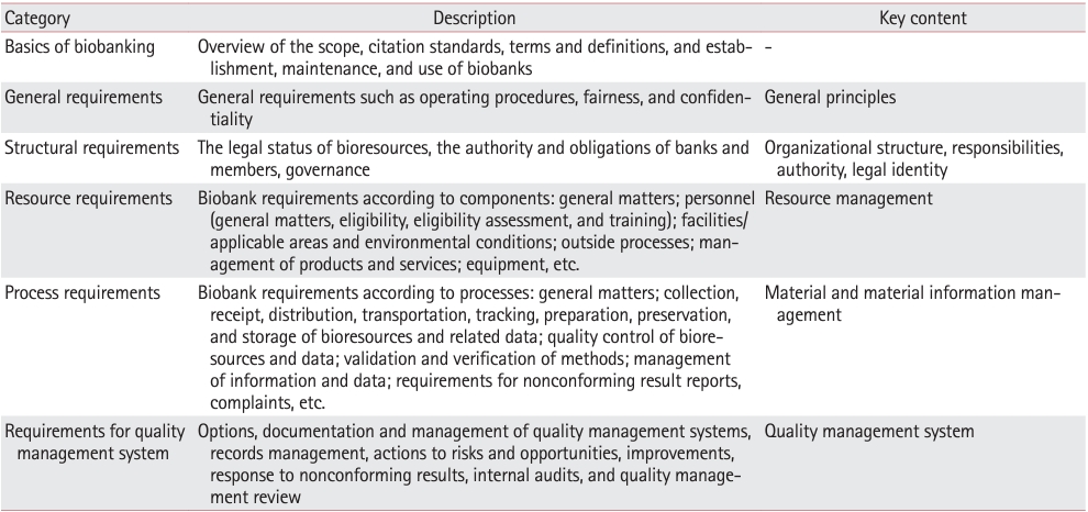
PDF - To ensure high-quality bioresources and standardize biobanks, there is an urgent need to develop and disseminate educational training programs in accordance with ISO 20387, which was developed in 2018. The standardization of biobank education programs is also required to train biobank experts. The subdivision of categories and levels of education is necessary for jobs such as operations manager (bank president), quality manager, practitioner, and administrator. Essential training includes programs tailored for beginner, intermediate, and advanced practitioners, along with customized training for operations managers. We reviewed and studied ways to develop an appropriate range of education and training opportunities for standard biobanking education and the training of experts based on KS J ISO 20387. We propose more systematic and professional biobanking training programs in accordance with ISO 20387, in addition to the certification programs of the National Biobank and the Korean Laboratory Accreditation System. We suggest various training programs appropriate to a student’s affiliation or work, such as university biobanking specialized education, short-term job training at unit biobanks, biobank research institute symposiums by the Korean Society of Pathologists, and education programs for biobankers and researchers. Through these various education programs, we expect that Korean biobanks will satisfy global standards, meet the needs of users and researchers, and contribute to the advancement of science.
-
Citations
Citations to this article as recorded by
- Establishing and Managing a Biobank at an Academic Institution in a Resource-Limited Setting: A Case Study from Ecuador
Alexander Maldonado, Andrés Herrera-Yela, Evaluna Chicango, Micaela Gómez, Gabriela Naranjo, Camila Maldonado, Paula Echeverría
Biopreservation and Biobanking.2026;[Epub] CrossRef - Biobanking for intelligent medicine: assessment and evaluation with the SHARE principle
Yin Yang, Amin Ullah, Yingbo Zhang, Hui Zong, Xingyun Liu, Chi Zhang, Shanshan Hu, Jiakun Li, Bairong Shen
Journal of the American Medical Informatics Association.2026; 33(7): 1333. CrossRef - Development of a big data platform for collecting and utilizing clinical information from the Korea Biobank Network
Yun Seon Im, Seol Whan Oh, Ki Hoon Kim, Wona Choi, In Young Choi
BMC Medical Informatics and Decision Making.2025;[Epub] CrossRef - Frozen section histopathology and preanalytical factors affecting nucleic acid integrity in biobanked fresh-frozen human cancer tissues
Soungeun Kim, Jaewon Kang, Boyeon Kim, Yoonjin Kwak, Hye Seung Lee
Journal of Pathology and Translational Medicine.2025; 59(6): 398. CrossRef
- Establishing and Managing a Biobank at an Academic Institution in a Resource-Limited Setting: A Case Study from Ecuador
Case Report
- Strongyloidiasis of Gastric and Colonic Mucosa in a Patient with Monoclonal Gammopathy of Undetermined Significance: A Case Report.
- Jung Uee Lee, Sang Bum Kang, Hae Joung Sul, Jong Ok Kim
- Korean J Pathol. 2011;45:S75-S78.
- DOI: https://doi.org/10.4132/KoreanJPathol.2011.45.S1.S75
- 4,304 View
- 21 Download
- 1 Crossref
-
Abstract
PDF
- Here we report a case of Strongyloides stercoralis infection of the gastric and pancolonic mucosa in a 79-year-old female with a monoclonal gammopathy of undetermined significance. Endoscopic biopsies were performed in gastric antrum, cecum, distal ascending colon, and hepatic flexure of the colon. On microscopic examination, there were many adult worms, larvae and eggs in the gastric and colonic mucosa. Worms, larvae, and eggs were located in the crypts and within the lumen of the crypts. The body wall of the adult worm was composed of cuticle and a weak muscle layer. A routine stool examination failed to detect larvae or ova. Based on the histopathologic examination, these parasites were confirmed as S. stercoralis.
-
Citations
Citations to this article as recorded by- Is Gastric Involvement by Strongyloides stercoralis in an Immunocompetent Patient a Common Finding? A Case Report and Review of the Literature
Irene Pecorella, Tom Richard Okello, Gaia Ciardi, David Martin Ogwang
Acta Parasitologica.2022; 67(1): 94. CrossRef
- Is Gastric Involvement by Strongyloides stercoralis in an Immunocompetent Patient a Common Finding? A Case Report and Review of the Literature
Original Article
- Evaluation of the HPV ISH Assay in Cervical Cancer.
- Jung Uee Lee, Jung Ha Shin, Jong Ok Kim, Yeong Jin Choi, Kyo Young Lee, Jong Sup Park, Won Chul Lee, Ahwon Lee
- Korean J Pathol. 2010;44(5):513-520.
- DOI: https://doi.org/10.4132/KoreanJPathol.2010.44.5.513
- 5,998 View
- 121 Download
- 3 Crossref
-
Abstract
PDF
- BACKGROUND
Human papillomavirus (HPV) infection can be detected by in situ hybridization (ISH), in which a punctate signal pattern indicates integrated HPV DNA and a diffuse pattern denotes the presence of episomal viral DNA. This study was conducted to evaluate the usefulness of an HPV ISH assay for invasive cervical cancer.
METHODS
The HPV ISH assay for high-risk HPV and immunohistochemical staining for p16(INK4a), p53, bcl-2, and Ki-67 were performed in a tissue microarray of 279 cervical cancers.
RESULTS
High-risk HPV ISH was positive in 194 (69.5%) of the samples. Punctate, diffuse, and mixed signal patterns were observed in 157 (56.3%), one (0.4%), and 36 cases (12.9%), respectively. Positive results in high-risk HPV ISH were associated with p16 and bcl-2 expression (p = 0.01 and p < 0.01, respectively). According to a Cox regression analysis, HPV infection and its surrogate immunohistochemical markers such as p16, bcl-2, and Ki-67 were not independent prognostic factors, but stage and grade were independent prognostic factors.
CONCLUSIONS
Our results confirm that an HPV ISH assay is reasonably sensitive for HPV infection and that it might be useful to identify integrated HPV DNA in formalin-fixed and paraffin-embedded specimens. Further study encompassing HPV type, E2/E6 ratio, and therapeutic modality is necessary to understand the clinical meaning of HPV status in cervical cancer. -
Citations
Citations to this article as recorded by- Prevalence of human papillomavirus in eyelid carcinoma among Koreans: a clinicopathological study
Min Kyu Yang, Namju Kim, Hokyung Choung, Ji Eun Kim, Sang In Khwarg
BMC Ophthalmology.2023;[Epub] CrossRef - Cervical cancer screening by molecular Pap‐transformation of gynecologic cytology
Shaikhali M Barodawala, Kirti Chadha, Vikas Kavishwar, Anuradha Murthy, Shamma Shetye
Diagnostic Cytopathology.2019; 47(5): 374. CrossRef - Prognostic Significance of Amplification of thec-MYCGene in Surgically Treated Stage IB-IIB Cervical Cancer
Tae-Jung Kim, Ahwon Lee, Sung-Jong Lee, Won-Chul Lee, Yeong-Jin Choi, Kyo-Young Lee, Chang Suk Kang
The Korean Journal of Pathology.2011; 45(6): 596. CrossRef
- Prevalence of human papillomavirus in eyelid carcinoma among Koreans: a clinicopathological study
Case Reports
- Completely Isolated Enteric Duplication Cyst Presenting as an Inguinal Hernia.
- Jung Uee Lee, Jong Ok Kim, Say June Kim, Hye Jung Sul
- Korean J Pathol. 2010;44(2):204-206.
- DOI: https://doi.org/10.4132/KoreanJPathol.2010.44.2.204
- 4,400 View
- 35 Download
- 4 Crossref
-
Abstract
PDF
- Enteric duplication cysts are uncommon congenital anomalies whose embryogenesis remains unknown. We report here on an isolated enteric duplication cyst, that presents as an inguinal hernia. A 21-year-old woman was admitted with a month-long history of a palpable mass in the left groin. Radiologically, a computed tomography scan revealed a 3.5 x 2.5 cm sized cystic mass in subcutaneous layers of the left suprapubic area. Microscopically, the cystic wall resembled gut wall. The wall was composed of two distinct muscle layers with the presence of Auerbach's plexus. On examining the entire sections of the cyst wall very carefully, no epithelial lining was found on the inner surface. The submucosa was slightly fibrotic. The diagnosis was a completely isolated enteric duplication cyst.
-
Citations
Citations to this article as recorded by- A case of completely isolated advanced enteric duplication cyst cancer performed partial pancreatectomy
Shinsuke Nakashima, Terumasa Yamada, Go Sato, Takaaki Sakai, Yoshinao Chinen, Hiroaki Itakura, Ryo Kato, Masami Ueda, Yujiro Tsuda, Katsuya Ohta, Jin Matsuyama, Masakazu Ikenaga
International Journal of Surgery Case Reports.2019; 54: 83. CrossRef - A huge completely isolated duplication cyst complicated by torsion and lined by 3 different mucosal epithelial components in an adult
Ai Xiao-Ming, Lu Jin-Jing, Ho Li-Chen, Han Lu-Lu, Yue Xiong, Zhang Hong-Hai, Yang Nian-Yin
Medicine.2018; 97(44): e13005. CrossRef - An isolated intestinal duplication cyst masquerading as a mucinous cystic neoplasm of the pancreas
Evan Weitman, Sameer Al Diffalha, Barbara Centeno, Pamela Hodul
International Journal of Surgery Case Reports.2017; 39: 208. CrossRef - Isolated omental duplication cyst with respiratory epithelium & pancreatic glands: Case report & review of literature
Phuoc T. Nguyen, Novae B. Simper, Charles K. Childers
Journal of Pediatric Surgery Case Reports.2016; 11: 17. CrossRef
- A case of completely isolated advanced enteric duplication cyst cancer performed partial pancreatectomy
- Mediastinal Hemangioma: Report of a case.
- Jong Ok Kim, Bum Kyeong Kim, Kyoung Hee Kim, Dae Young Kang, Kwang Sun Suh
- Korean J Pathol. 1997;31(9):891-894.
- 2,136 View
- 24 Download
-
Abstract
PDF
- Benign hemangioma of the mediastinum is rare. This slowly growing tumor is described as well circumscribed, cystic, hemorrhagic tumor. Histologically it can be differentiated into capillary or cavernous form. We present a case of mediastinal hemangioma. A 20-year-old-man was presented with a slowly growing posterior mediastinal mass of 6 years duration, 8x6 cm in size. The mass was relatively well defined but focally invasive. Microscopically, it was differentiated into vessels of capillary, cavernous, and venous patterns. A solid cellular proliferation with inconspicuous capillary lumens was focally seen. The stroma between variable-sized vessels showed marked myxoid change associated with some smooth muscle bundles and adipose tissue. Ultrastructurally, areas of solid cellular proliferation showed formation of lumens. These lumens were lined by active endothelial cells showing plasmalemmal vesicles and Weibel-Palade bodies on the abluminal surface.
Original Articles
- Morphometric Analysis for Cytological Diagnosis of Thyroid Papillary Carcinoma.
- Jong Ok Kim, Bo Seong Yang, Hye Soo Kim, Jong Min Lee, Dong Ho Lee, So Young Shin, Chang Suk Kang, Hye Kyung Lee
- J Pathol Transl Med. 2006;17(2):116-119.
- 2,651 View
- 27 Download
-
Abstract
PDF
- The diagnosis of papillary thyroid cancer is generally based on the findings of intranuclear cytoplasmic inclusions and nuclear grooves. Although anisokaryosis and poikilokaryosis, in papillary thyroid cancer, are not distinct when compared to other cancers, cytological examination can provide useful preoperative information. Our study evaluated the diagnostic role of computer-assisted image analysis for the pre-surgical assessment of papillary thyroid carcinoma. Thyroid aspirates from twenty female patients who were histologically confirmed to have both papillary carcinoma and benign nodules were studied. Different populations of 50 benign cells and 50 malignant cells were analyzed. Five morphometric parameters were selected for analysis: nuclear area, perimeter, maximum length, maximum width and intensity standard variation. The values obtained for papillary carcinomas were higher than the surrounding benign nodules as follows: nuclear area 63.5 vs. 36.1 (p=0.000), nuclear perimeter were 29.4 vs. 22.0 (p=0.000), maximum length 9.6 vs. 7.1 (p=0.000), maximum width 8.2 vs. 6.3 (p=0.000), the ratio between maximal length and maximal width 1.16 vs. 1.13 (p=0.000), the standard variation of intensity 14.9 vs. 15.9 (p=0.101) respectively. Therefore, morphometric information can be helpful for the differential cytological diagnosis of papillary thyroid carcinoma.
- Renal Dysplasia: A Clinicopathologic Review of Six Cases.
- Gil Hyun Kang, Jong Ok Kim, Bum Kyung Kim, Kwang Sun Suh
- Korean J Pathol. 1997;31(1):34-39.
- 2,229 View
- 16 Download
-
Abstract
PDF
- Renal dysplasia results from aberrant histogenesis in metanephric differentiation. It is characterized morphologically by abnormal organization and a persistence of primitive structures, such as cartilage, undifferentiated mesenchyme, and immature tubules. Six cases of renal dysplasia from five children and one adult are reviewed. Five patients were female and one patient was male. The chief complaint was urinary incontinence in four patients, dysuria in one patient, and the sixth patient suffered from vesicoureteral reflux. No evidence of family history of renal dysplasia in any patient was seen. According to Risdon's classification, three cases were hypoplastic dysplasia, one case was dysplasia in a duplex system, one case was dysplasia in a triplex system, and one case was dysplasia with vesicoureteral reflux. The ipsilateral ectopic ureteral orifice was identified in four patients, two of which drained into a Gartner's duct cyst, and the orifice was suggested in one patient. On histologic examination, all cases showed primitive ducts surrounded by concentrically arranged primitive mesenchyme. Nests of metaplastic cartilage were observed within the stroma in three of the six cases.
 First
First Prev
Prev


