 E-submission
Search
- Page Path
- HOME > Search
- Clinicopathological profile of high-grade differentiated thyroid carcinoma in an Indonesian tertiary hospital
- Novita , Agnes Stephanie Harahap, Maria Francisca Ham, Alfianto Widiono, Chan Kwon Jung
- Received October 20, 2025 Accepted January 15, 2026 Published online April 23, 2026
- DOI: https://doi.org/10.4132/jptm.2026.01.15 [Epub ahead of print]
- 436 View
- 19 Download
-
 Abstract
 PDF
PDF Supplementary Material
 - Background
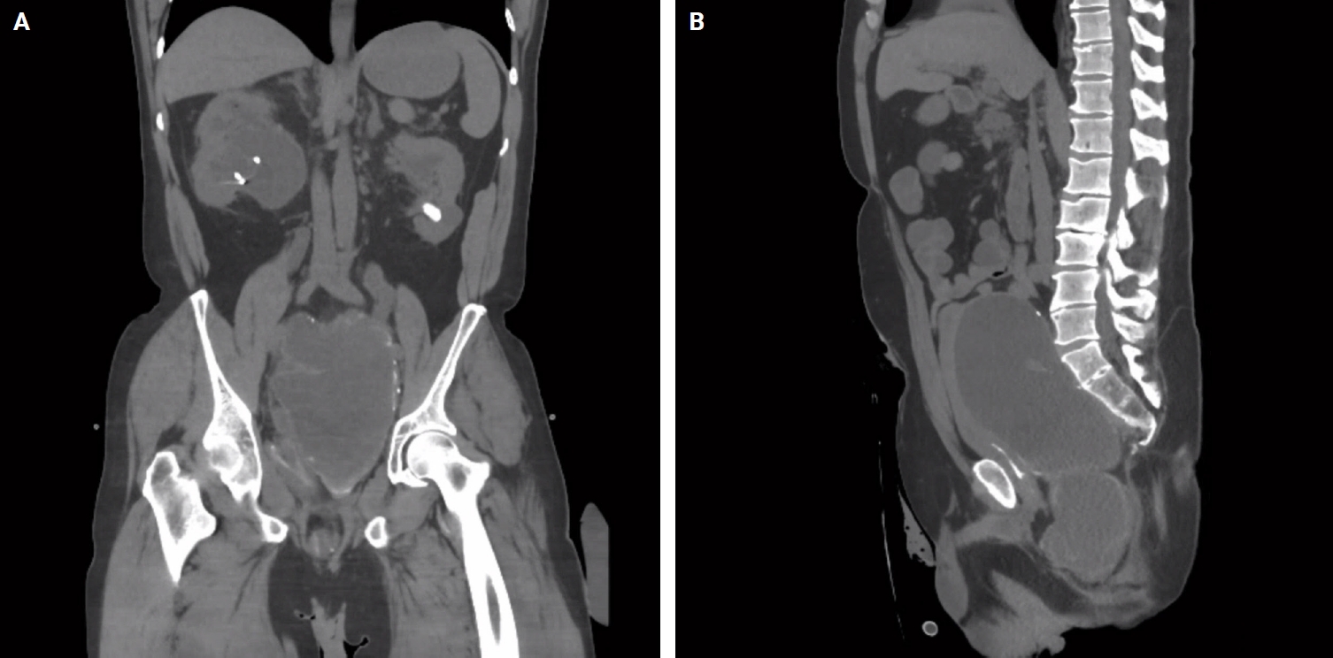
High-grade differentiated thyroid carcinoma (HGDTC) is a recently recognized entity in the 2022 World Health Organization classification, representing a more aggressive subtype of differentiated thyroid carcinoma. Previously, high-grade features such as increased mitotic activity and tumor necrosis were often overlooked, despite being important independent prognostic factors. Although rare, HGDTC carries significant diagnostic, prognostic, and therapeutic implications. Data remain limited in Indonesia. Methods: This retrospective descriptive study reviewed 565 thyroid carcinoma cases diagnosed at Cipto Mangunkusumo Hospital from 2019 to 2024. Eleven cases (1.9%) met HGDTC criteria. Clinicopathological characteristics, histologic subtypes, Ki-67 proliferation index, molecular alterations, treatment modalities, and clinical outcomes were analyzed. Results: Patients had a mean age of 54.6 years, with a female-to-male ratio of 2.7:1. Papillary thyroid carcinoma was the main type (90.9%), with the tall cell subtype predominating. Mean tumor size was 6.4 cm. Lymphatic invasion, vascular invasion, and extrathyroidal extension were present in 54.5%, 18.2%, and 45.5% of cases, respectively. All tumors showed necrosis. Mean mitotic count was 3 per 2 mm². The Ki-67 index ranged from 5% to 45% (median, 14%). BRAFV600E and TERT promoter mutations were detected in 18.2% and 36.4% of cases, respectively, with co-mutations in 18.2%. Six cases (54.5%) had metastases at time of diagnosis. During a mean follow-up of 20.5 months, one patient (9.1%) developed new vertebral metastases and all patients (100%) remained alive. Conclusions: HGDTC presents with more aggressive characteristics and a worse prognosis. Accurate diagnosis, molecular profiling, and long-term monitoring are essential for optimal management.
- Mucocele of the rectal stump: mucinous cystic neoplasm with low-grade dysplasia simulating low-grade appendiceal mucinous neoplasm
- Hasan Basri Aydin, Maria Faraz, A. David Chismark, Haiyan Qiu, Hwajeong Lee
- J Pathol Transl Med. 2025;59(2):139-146. Published online February 26, 2025
- DOI: https://doi.org/10.4132/jptm.2024.12.27
- 3,654 View
- 174 Download
-
Abstract
PDF
- Mucoceles, commonly observed in the appendix, are mucin-filled, dilated structures arising from a range of etiologies. Cases associated with dysplastic or neoplastic epithelium can rupture and disseminate within the abdominopelvic cavity. Similar lesions in other parts of the colon are exceedingly rare, with only 16 colonic mucoceles having been reported. The first case of a colonic mucinous neoplasm with dysplasia resembling a low-grade appendiceal mucinous neoplasm involving rectal stump was described in 2016. Here, we present the second such case arising in the rectal stump, identified in a 44-year-old male with extensive surgical history. Microscopic examination revealed low-grade dysplastic epithelium lining the cyst and mucin dissecting into the stroma, without evidence of rupture or extramural mucin. The patient was followed for 16 months without recurrence or peritoneal disease. The exact etiology and outcome of these rare lesions remain unknown, requiring close follow-up.
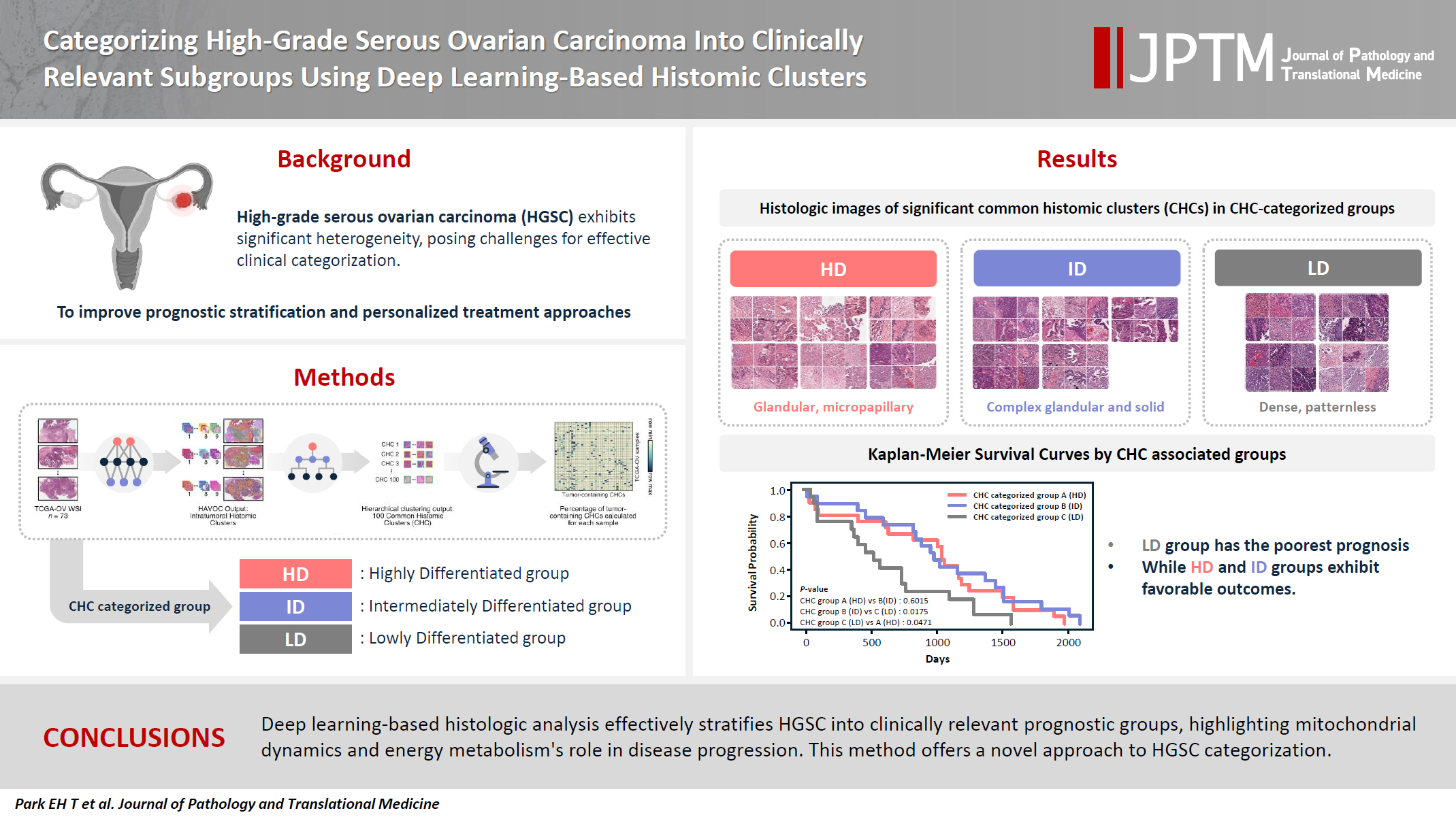
- Categorizing high-grade serous ovarian carcinoma into clinically relevant subgroups using deep learning–based histomic clusters
- Byungsoo Ahn, Eunhyang Park
- J Pathol Transl Med. 2025;59(2):91-104. Published online February 18, 2025
- DOI: https://doi.org/10.4132/jptm.2024.10.23
- 6,719 View
- 265 Download
- 1 Web of Science
- 2 Crossref
-
Abstract
PDFSupplementary Material
- Background
High-grade serous ovarian carcinoma (HGSC) exhibits significant heterogeneity, posing challenges for effective clinical categorization. Understanding the histomorphological diversity within HGSC could lead to improved prognostic stratification and personalized treatment approaches. Methods: We applied the Histomic Atlases of Variation Of Cancers model to whole slide images from The Cancer Genome Atlas dataset for ovarian cancer. Histologically distinct tumor clones were grouped into common histomic clusters. Principal component analysis and K-means clustering classified HGSC samples into three groups: highly differentiated (HD), intermediately differentiated (ID), and lowly differentiated (LD). Results: HD tumors showed diverse patterns, lower densities, and stronger eosin staining. ID tumors had intermediate densities and balanced staining, while LD tumors were dense, patternless, and strongly hematoxylin-stained. RNA sequencing revealed distinct patterns in mitochondrial oxidative phosphorylation and energy metabolism, with upregulation in the HD, downregulation in the LD, and the ID positioned in between. Survival analysis showed significantly lower overall survival for the LD compared to the HD and ID, underscoring the critical role of mitochondrial dynamics and energy metabolism in HGSC progression. Conclusions: Deep learning-based histologic analysis effectively stratifies HGSC into clinically relevant prognostic groups, highlighting the role of mitochondrial dynamics and energy metabolism in disease progression. This method offers a novel approach to HGSC categorization. -
Citations
Citations to this article as recorded by
- Ovarian Cancer: Epidemiology, Disease Mechanisms, New Diagnosis and Treatment Strategies, and Research Directions
Zunera Khalid, Weirong Fan, Farah Nazir, Yixiang Xing, Tengchuan Jin
iNew Medicine.2026;[Epub] CrossRef - Learning Disabilities in the 21st Century: Integrating Neuroscience, Education, and Technology for Better Outcomes
Syed Mohammed Basheeruddin Asdaq, Ahmad H. Alhowail, Syed Imam Rabbani, Naira Nayeem, Syed Mohammed Emaduddin Asdaq, Faiqa Nausheen
SAGE Open.2025;[Epub] CrossRef
- Ovarian Cancer: Epidemiology, Disease Mechanisms, New Diagnosis and Treatment Strategies, and Research Directions
- Malignant potential of neuroendocrine microtumor of the pancreas harboring high-grade transformation: lesson learned from a patient with von Hippel-Lindau syndrome
- Jongwon Lee, Kyung Jin Lee, Dae Wook Hwang, Seung-Mo Hong
- J Pathol Transl Med. 2024;58(2):91-97. Published online March 13, 2024
- DOI: https://doi.org/10.4132/jptm.2024.02.13
- 5,487 View
- 216 Download
- 3 Web of Science
- 4 Crossref
-
Abstract
PDF
- Pancreatic neuroendocrine microtumor (PNEMT) is a neuroendocrine tumor (NET) < 0.5 cm in diameter, and it is considered benign. We report a PNEMT with high-grade transformation (HGT). A man in his 60s with von Hippel-Lindau syndrome underwent surgical resection of a NET. A second sub-centimeter nodule with a nodule-in-nodule pattern was discovered. The 0.4 cm outer nodule contained clear columnar cells with round nuclei and indistinct nucleoli, while the 0.1 cm inner nodule had eosinophilic cells with an increased nuclear to cytoplasmic ratio, vesicular nuclei, and prominent nucleoli. Tumor cells in the outer and inner nodules were synaptophysin and chromogranin positive. Only the inner nodule was p53 positive, while the outer nodule was exclusively positive for carbonic anhydrase 9 and vimentin. The Ki-67 labeling indices for the outer and inner nodules were 2.1% (grade 1) and 44.3% (grade 3), respectively. This nodule was determined to be a PNEMT with HGT. Our findings suggest that a PNEMT may not always be benign and can undergo HGT.
-
Citations
Citations to this article as recorded by- Intraductal papillary mucinous neoplasm unveiling incidental multifocal pancreatic neuroendocrine tumors: a challenging case report
Faten Limaiem, Mohamed Hajri, Nafaa Arfa
International Journal of Surgery Case Reports.2026;[Epub] CrossRef - Decoding Pancreatic Neuroendocrine Tumors: Molecular Profiles, Biomarkers, and Pathways to Personalized Therapy
Linda Galasso, Federica Vitale, Gabriele Giansanti, Giorgio Esposto, Raffaele Borriello, Irene Mignini, Alberto Nicoletti, Lorenzo Zileri Dal Verme, Antonio Gasbarrini, Maria Elena Ainora, Maria Assunta Zocco
International Journal of Molecular Sciences.2025; 26(16): 7814. CrossRef - Pancreatic neuroendocrine microtumors in the elderly: A retrospective study using cadaveric pancreatic tissue
Ting Yang, Ke Ren, Xiang-Quan Chen, Taku Toriumi, Yutaro Natsuyama, Jun Li, Aoi Sukeda, Toshitaka Nagao, Shuang-Qin Yi
World Journal of Gastrointestinal Oncology.2025;[Epub] CrossRef - Molecular Basis of Pancreatic Neuroendocrine Tumors
Alesia Maluchenko, Denis Maksimov, Zoia Antysheva, Julia Krupinova, Ekaterina Avsievich, Olga Glazova, Natalia Bodunova, Nikolay Karnaukhov, Ilia Feidorov, Diana Salimgereeva, Mark Voloshin, Pavel Volchkov
International Journal of Molecular Sciences.2024; 25(20): 11017. CrossRef
- Intraductal papillary mucinous neoplasm unveiling incidental multifocal pancreatic neuroendocrine tumors: a challenging case report
- Trouble-makers in cytologic interpretation of the uterine cervix
- Eunah Shin, Jaeeun Yu, Soon Won Hong
- J Pathol Transl Med. 2023;57(3):139-146. Published online May 15, 2023
- DOI: https://doi.org/10.4132/jptm.2023.04.25
- 12,577 View
- 483 Download
- 4 Web of Science
- 5 Crossref
-
Abstract
PDF
- The development and standardization of cytologic screening of the uterine cervix has dramatically decreased the prevalence of squamous cell carcinoma of the uterine cervix. Advances in the understanding of biology of human papillomavirus have contributed to upgrading the histologic diagnosis of the uterine cervix; however, cytologic screening that should triage those that need further management still poses several difficulties in interpretation. Cytologic features of high grade intraepithelial squamous lesion (HSIL) mimics including atrophy, immature metaplasia, and transitional metaplasia, and glandular lesion masquerades including tubal metaplasia and HSIL with glandular involvement are described with accentuation mainly on the differential points. When the cytologic features lie in a gray zone between the differentials, the most important key to the more accurate interpretation is sticking to the very basics of cytology; screening the background and cellular architecture, and then scrutinizing the nuclear and cytoplasmic details.
-
Citations
Citations to this article as recorded by- Cytology–Biopsy Concordance in High-Risk Human Papillomavirus–Positive Women with Abnormal Cytology Findings: Menopause-Stratified Analysis
Isik Sozen, Gozde Sahin, Yuksel Ulu, Dilara Yitiz, Basak Ozge Kayan, Ilkbal Temel Yuksel
Medicina.2026; 62(4): 631. CrossRef - Pathologists Recommend Repeat Pap Testing: In Clinical Practice What Do Gylecologist Do?
Gizem Ay Haldız
Muğla Sıtkı Koçman Üniversitesi Tıp Dergisi.2026; 13(1): 1. CrossRef - Risk of cervical stenosis after cervical excision in postmenopausal patients
Eva Hauge, Line Winther Gustafson, Mette Tranberg, Pinar Bor
European Journal of Obstetrics & Gynecology and Reproductive Biology.2025; 308: 208. CrossRef - Pitfalls in Gynecological Cytology: Review of the Common and Less Frequent Entities in Pap Test
Danijela Vrdoljak-Mozetič, Snježana Štemberger-Papić, Damjana Verša Ostojić, Roberta Rubeša, Marko Klarić, Senija Eminović
Acta Cytologica.2024; 68(3): 281. CrossRef - Cytological features of human papillomavirus‐infected immature squamous metaplastic cells from cervical intraepithelial neoplasia grade 2
Mitsuaki Okodo, Kaori Okayama, Koji Teruya, Ruku Shinohara, Shuichi Mizuno, Rei Settsu, Yasuyoshi Ishii, Masahiko Fujii, Hirokazu Kimura, Mizue Oda
Journal of Medical Virology.2023;[Epub] CrossRef
- Cytology–Biopsy Concordance in High-Risk Human Papillomavirus–Positive Women with Abnormal Cytology Findings: Menopause-Stratified Analysis
- Myoferlin Expression and Its Correlation with FIGO Histologic Grading in Early-Stage Endometrioid Carcinoma
- Min Hye Kim, Dae Hyun Song, Gyung Hyuck Ko, Jeong Hee Lee, Dong Chul Kim, Jung Wook Yang, Hyang Im Lee, Hyo Jung An, Jong Sil Lee
- J Pathol Transl Med. 2018;52(2):93-97. Published online March 14, 2018
- DOI: https://doi.org/10.4132/jptm.2017.11.29
- 8,604 View
- 118 Download
- 11 Web of Science
- 9 Crossref
-
Abstract
PDF
- Background
For endometrioid carcinoma patients, International Federation of Gynecologists and Obstetricians (FIGO) histologic grading is very important for identifying the appropriate treatment method. However, the interobserver discrepancy with this three-tiered grading system is a serious potential problem. In this study, we used immunohistochemistry to analyze the relationship between FIGO histologic grading score and myoferlin expression.
Methods
We studied the endometrioid carcinoma tissues of 60 patients from Gyeongsang National University Hospital between January 2002 and December 2009. Immunohistochemical analysis of myoferlin was performed on tissue microarray blocks from surgical specimens.
Results
Myoferlin expression was observed in 58 of 60 patients. Moderate and strong myoferlin expression was observed in low-grade endometrioid carcinoma, while there was a tendency toward loss of myoferlin expression in high-grade endometrioid carcinoma (p<.001).
Conclusions
Our study revealed that myoferlin loss is significantly correlated with high FIGO grade of endometrioid carcinoma. -
Citations
Citations to this article as recorded by- Myoferlin: A Potential Marker of Response to Radiation Therapy and Survival in Locally Advanced Rectal Cancer
Hayley Fowler, Rachael E. Clifford, David Bowden, Paul A. Sutton, Naren Govindarajah, Matthew Fok, Mark Glenn, Michael Wall, Carlos Rubbi, Simon J.A. Buczacki, Amit Mandal, Hayley Francies, Jonathan Hughes, Jason L. Parsons, Dale Vimalachandran
International Journal of Radiation Oncology*Biology*Physics.2024; 120(4): 1111. CrossRef - Neoexpression of JUNO in Oral Tumors Is Accompanied with the Complete Suppression of Four Other Genes and Suggests the Application of New Biomarker Tools
Dominik Kraus, Simone Weider, Rainer Probstmeier, Jochen Winter
Journal of Personalized Medicine.2022; 12(3): 494. CrossRef - Correlation between myoferlin expression and lymph node metastasis in papillary thyroid carcinoma
Ji Min Na, Dong Chul Kim, Dae Hyun Song, Hyo Jung An, Hyun Min Koh, Jeong-Hee Lee, Jong Sil Lee, Jung Wook Yang, Min Hye Kim
Journal of Pathology and Translational Medicine.2022; 56(4): 199. CrossRef - PINCH-1 interacts with myoferlin to promote breast cancer progression and metastasis
Tao Qian, Chengmin Liu, Yanyan Ding, Chen Guo, Renwei Cai, Xiaoxia Wang, Rong Wang, Kuo Zhang, Li Zhou, Yi Deng, Chuanyue Wu, Ying Sun
Oncogene.2020; 39(10): 2069. CrossRef - Human colon cancer cells highly express myoferlin to maintain a fit mitochondrial network and escape p53-driven apoptosis
Gilles Rademaker, Brunella Costanza, Justine Bellier, Michael Herfs, Raphaël Peiffer, Ferman Agirman, Naïma Maloujahmoum, Yvette Habraken, Philippe Delvenne, Akeila Bellahcène, Vincent Castronovo, Olivier Peulen
Oncogenesis.2019;[Epub] CrossRef - Prognostic significance of immunohistochemical staining for myoferlin in clear cell renal cell carcinoma and its association with epidermal growth factor receptor expression
Minsun Jung, Cheol Lee, Jeong Hwan Park, Kyung Chul Moon
Urologic Oncology: Seminars and Original Investigations.2019; 37(11): 812.e9. CrossRef - Ferlin Overview: From Membrane to Cancer Biology
Olivier Peulen, Gilles Rademaker, Sandy Anania, Andrei Turtoi, Akeila Bellahcène, Vincent Castronovo
Cells.2019; 8(9): 954. CrossRef - Myoferlin, a multifunctional protein in normal cells, has novel and key roles in various cancers
Wei Zhu, Bolun Zhou, Chenxuan Zhao, Zhengqing Ba, Hongjuan Xu, Xuejun Yan, Weidong Liu, Bin Zhu, Lei Wang, Caiping Ren
Journal of Cellular and Molecular Medicine.2019; 23(11): 7180. CrossRef - Myoferlin, a Membrane Protein with Emerging Oncogenic Roles
Yimin Dong, Honglei Kang, Huiyong Liu, Jia Wang, Qian Guo, Chao Song, Yunlong Sun, Ya Zhang, Honghua Zhang, Zheng Zhang, Hanfeng Guan, Zhong Fang, Feng Li
BioMed Research International.2019; 2019: 1. CrossRef
- Myoferlin: A Potential Marker of Response to Radiation Therapy and Survival in Locally Advanced Rectal Cancer
- Cytomorphological Findings and Histological Correlation of Low-Grade Cribriform Cystadenocarcinoma of Salivary Gland in Fine-Needle Aspiration: A Case Study
- Young Sin Ko, Ja Seung Koo
- Korean J Pathol. 2013;47(6):592-595. Published online December 24, 2013
- DOI: https://doi.org/10.4132/KoreanJPathol.2013.47.6.592
- 9,580 View
- 70 Download
- 13 Crossref
-
Abstract
PDF
Low-grade cribriform cystadenocarcinoma (LGCCC) of the salivary gland is a rare tumor. We report the cytologic features and histologic correlation of a patient with LGCCC. A 57-year-old man had a hardly palpable, nontender mass in the right cheek area followed over nine months. Radiologic analysis revealed a 1.2 cm multiseptated, cystic, solid nodule in an anterior superficial lobe of the right parotid gland. Fine-needle aspiration cytology revealed many irregular overlapping sheets or clusters of ductal epithelial cells forming solid, pseudopapillary, and cribriform architectures. Nuclei of the tumor cells revealed inconspicuous atypia with minimal size variation. On the basis of these findings, we confirmed a diagnosis of ductal epithelial proliferative lesion, favoring neoplasm, with uncertain malignant potential. Tumor excision was performed, revealing a tiny multicystic nodule (0.7 cm). Histopathologically, this tumor showed the characteristic morphology of LGCCC. This is the first report of cytomorphological findings of LGCCC in Korea.
-
Citations
Citations to this article as recorded by- Duct tales of a parotid gland swelling
Swati Raj, Monika Singh, Mamta Gupta, Naveen Thapliyal
Cytojournal.2023; 20: 22. CrossRef - Salivary Gland Intraductal Carcinoma: How Do 183 Reported Cases Fit Into a Developing Classification
Lester D.R. Thompson, Justin A. Bishop
Advances in Anatomic Pathology.2023; 30(2): 112. CrossRef - Intraductal carcinoma of the parotid gland
Yukiya HIRATA, Kayoko HIGUCHI, Toshitaka NAGAO, Yoko ZUKERAN, Takao KINJO, Naoki WADA
The Journal of the Japanese Society of Clinical Cytology.2022; 61(6): 431. CrossRef - Intraductal carcinomas of the salivary glands: systematic review and classification of 93 published cases
Andrea Palicelli
APMIS.2020; 128(3): 191. CrossRef - What do we know about the cytological features of pure intraductal carcinomas of the salivary glands?
Andrea Palicelli
Cytopathology.2020; 31(3): 185. CrossRef - Diagnosing Recently Defined and Uncommon Salivary Gland Lesions in Limited Cellularity Specimens: Cytomorphology and Ancillary Studies
Esther Diana Rossi, Zubair Baloch, William Faquin, Liron Pantanowitz
AJSP: Reviews and Reports.2020; 25(5): 210. CrossRef - Low-grade intraductal carcinoma of salivary glands: A systematic review of this rare entity
Francesco Giovacchini, Caterina Bensi, Stefano Belli, Maria Elena Laurenti, Martina Mandarano, Daniele Paradiso, Michele Giansanti, Antonio Tullio
Journal of Oral Biology and Craniofacial Research.2019; 9(1): 96. CrossRef - The rare entity of cystadenocarcinoma (CAC) in parotid gland: A single-center experience
Bing Guo, Yu-an Cao, Xingjun Qin, Chunyue Ma
Journal of Cranio-Maxillofacial Surgery.2019; 47(5): 826. CrossRef - Cytopathology approach to rare salivary gland lesions with oncocytic features
Siba El Hussein, Samer N. Khader
Diagnostic Cytopathology.2019; 47(10): 1090. CrossRef - Unicystic high‐grade intraductal carcinoma of the parotid gland: cytological and histological description with clinic–pathologic review of the literature
Andrea Palicelli, Paola Barbieri, Narciso Mariani, Paola Re, Stefania Galla, Raffaele Sorrentino, Francesca Locatelli, Nunzio Salfi, Guido Valente
APMIS.2018; 126(9): 771. CrossRef - Low-grade cribriform cystadenocarcinoma arising from a minor salivary gland: a case report
Masashi Kimura, Shinji Mii, Shinichi Sugimoto, Kosuke Saida, Shojiroh Morinaga, Masahiro Umemura
Journal of Oral Science.2016; 58(1): 145. CrossRef - A Case of Cystadenocarcinoma Arising from Parotid Gland
Jong Chul Hong, Tae Kyoung Koh, Min Gyoung Pak, Heon Soo Park
Korean Journal of Otorhinolaryngology-Head and Neck Surgery.2016; 59(4): 300. CrossRef - Mammary analogue secretory carcinoma of parotid gland
Atsuko NASU, Sakae HATA, Masaru FUJITA, Toyoko YAMAUCHI, Satoko NAKAMURA, Takehiro TANAKA, Kouichi ICHIMURA, Hiroyuki YANAI
The Journal of the Japanese Society of Clinical Cytology.2016; 55(2): 112. CrossRef
- Duct tales of a parotid gland swelling
- Fine-Needle Aspiration Cytology of Low-Grade Cribriform Cystadenocarcinoma with Many Psammoma Bodies of the Salivary Gland
- Ji Yun Jeong, Dongbin Ahn, Ji Young Park
- Korean J Pathol. 2013;47(5):481-485. Published online October 25, 2013
- DOI: https://doi.org/10.4132/KoreanJPathol.2013.47.5.481
- 9,000 View
- 49 Download
- 12 Crossref
-
Abstract
PDF
Low-grade cribriform cystadenocarcinoma (LGCCC) is a rare salivary gland tumor that was recently defined as a variant of cystadenocarcinoma by the 2005 World Health Orgazniation (WHO) classification system. We report cytologic findings of an unusual case of LGCCC with many psammoma bodies. A 90-year-old man presented a palpable mass on his left parotid gland. Fine-needle aspiration (FNA) cytology showed tumor cells that were arranged in clusters and dispersed individually. The tumor cells showed mild atypia and had clear or dense cytoplasm with some vacuoles. Numerous psammoma bodies were noted. After surgical resection, the histologic examination revealed a mixed solid and cystic mass showing intraductal growth with focal stromal invasion. The S-100 protein expressed in the tumor cells, but smooth muscle actin and p63 were positive only in myoepithelial cells. Although LGCCCs resemble other salivary gland tumors, differentiating LGCCC during preoperative FNA is important to avoid unnecessary overtreatment.
-
Citations
Citations to this article as recorded by- Salivary Gland Intraductal Carcinoma: How Do 183 Reported Cases Fit Into a Developing Classification
Lester D.R. Thompson, Justin A. Bishop
Advances in Anatomic Pathology.2023; 30(2): 112. CrossRef - Duct tales of a parotid gland swelling
Swati Raj, Monika Singh, Mamta Gupta, Naveen Thapliyal
Cytojournal.2023; 20: 22. CrossRef - Intraductal carcinoma of the parotid gland
Yukiya HIRATA, Kayoko HIGUCHI, Toshitaka NAGAO, Yoko ZUKERAN, Takao KINJO, Naoki WADA
The Journal of the Japanese Society of Clinical Cytology.2022; 61(6): 431. CrossRef - Intraductal carcinoma of the retromolar trigone found with elevated serum CEA and CA19-9 levels: a case report
Mao KAWAKAMI, Nobuhiro UEDA, Yuka TAKAHASHI, Sho ARIKAWA, Nobuhiro YAMAKAWA, Tadaaki KIRITA
Japanese Journal of Oral and Maxillofacial Surgery.2021; 67(5): 292. CrossRef - Endoscopic trans‐pterygoid resection of a low‐grade cribriform cystadenocarcinoma of the infratemporal fossa
Vikram G. Ramjee, Landon J. Massoth, John P. Richards, Kibwei A. McKinney
World Journal of Otorhinolaryngology - Head and Neck Surgery.2020; 6(2): 115. CrossRef - Psammoma Bodies in a Large Myoepithelioma
Marcela Pessoa de Melo, Diego Filipe Bezerra Silva, Rodrigo Alves Ribeiro, Tony Santos Peixoto, Daliana Queiroga de Castro Gomes, Pollianna Muniz Alves, Cassiano Francisco Weege Nonaka, Bárbara Vanessa de Brito Monteiro
Journal of Craniofacial Surgery.2020; 31(4): e326. CrossRef - Low-grade intraductal carcinoma of salivary glands: A systematic review of this rare entity
Francesco Giovacchini, Caterina Bensi, Stefano Belli, Maria Elena Laurenti, Martina Mandarano, Daniele Paradiso, Michele Giansanti, Antonio Tullio
Journal of Oral Biology and Craniofacial Research.2019; 9(1): 96. CrossRef - What is your diagnosis? Submandibular mass in a dog
Julie Allen, Ashley M. Talley, Carol B. Grindem, Jennifer A. Neel
Veterinary Clinical Pathology.2018; 47(4): 676. CrossRef - Primary acinic cell carcinoma of the lung with psammoma bodies: A case report and review of literature
Xiu-Peng Zhang, Gui-Yang Jiang, Qing-Fu Zhang, Hong-Tao Xu, Qing-Chang Li, En-Hua Wang
Pathology - Research and Practice.2017; 213(4): 405. CrossRef - Cytology of low‐grade cribriform cystadenocarcinoma in salivary glands: Cytological and immunohistochemical distinctions from other salivary gland neoplasms
Yoshiki Ohta, Yuko Hirota, Yohko Kohno, Koji Kishimoto, Tomoko Norose, Nobuyuki Ohike, Masafumi Takimoto, Akira Shiokawa, Hidekazu Ota
Diagnostic Cytopathology.2016; 44(3): 241. CrossRef - Low-grade cribriform cystadenocarcinoma arising from a minor salivary gland: a case report
Masashi Kimura, Shinji Mii, Shinichi Sugimoto, Kosuke Saida, Shojiroh Morinaga, Masahiro Umemura
Journal of Oral Science.2016; 58(1): 145. CrossRef - A Case of Cystadenocarcinoma Arising from Parotid Gland
Jong Chul Hong, Tae Kyoung Koh, Min Gyoung Pak, Heon Soo Park
Korean Journal of Otorhinolaryngology-Head and Neck Surgery.2016; 59(4): 300. CrossRef
- Salivary Gland Intraductal Carcinoma: How Do 183 Reported Cases Fit Into a Developing Classification
- Intraosseous Well Differentiated Osteosarcoma: A case report.
- Mee Hye Oh, So Young Park, Yeon Lim Suh, Shin Khang Kang
- Korean J Pathol. 1992;26(6):627-631.
- 2,154 View
- 17 Download
-
Abstract
PDF
- Well differentiated osteosarcomas are variants of osteosarcoma composed mainly of fibrous and osseous tissue with minimal cystologic atypia. This tumor may be misinterpretated as a benign lesion if the radiologic and clinical features are not taken into account. We report a typical case of intraosseous well differentiated osteosarcoma occuring in the left distal femur of a 58-year-old woman. Radiologically, it appered as an ill-defined lesion with a mixture of sclerotic and osteolytic ares. But there was a lack of highly destructive appearance of conventional osteosarcoma. Grossly, the mass occupied a metaphysis of the distal femur with extension into the diaphysis and epiphysis. Multifocal cortical destruction and sclerosis were also associated. Histologically, the mass showed typical features of intraosseous well differentiated osteosarcoma. There were various patterns of osteoid deposits and bone formation mimicking those of fibrous dysplasia, nonossifying fibroma or parosteal osteosarcoma.
- A Pathological Study of Renal Cell Carcinoma.
- Kwang Hwa Park, Dong Hwan Shin, In Joon Choi
- Korean J Pathol. 1989;23(3):322-330.
- 2,287 View
- 16 Download
-
Abstract
PDF
- The most common malignant renal neoplasm is renal cell carcinoma. It is estimated that renal cell carcinoma accounts for 1% of all primary malignancies in Korea. Rell cell carcinoma presents diverse clinical courses with gross, histopathologic features. It has been known to be very difficult tumor to predict its clinical prognosis. In Korea, many studies have been reported concerning the clinical aspects of renal cell carcinoma. However, pathological studies of renal cell carcinoma are very few even though studies of nuclear grade have been attempted recently. We reviewed 93 cases of renal cell carcinoma examined in the period from 1978 to 1987 in the department of pathology, Yonsei university college of medicine, Yongdong Severance hospital, Wonju college of medicine and analyzed the histopathologic classification, including nuclear grade according to the Fuhrman's method. We abtained the following results by studying the relationship of the factors which had been known as correlated with the prognosis. 1) The ages of patients ranged from 9 to 74 years with a peak in the 6th decade. 2) The most common symptoms of the patients were hematuria, mass and pain, in that oder, and 7 patients complained to specific symptoms. The incidentally found cases characterized stage I, nuclear grade 2 small tumor size (not more than 4 cm) and clear cell type. 3) The renal cell carcinoma was more frequently located in the left kidney than the right by a ratio of 1.25 : 1. The incidence of intrarenal location was divided to the upper pole, 40% : mid portion, 29% : lower pole, 23% : diffuse involvement, 8%. The tumor shoing diffuse growth pattern had a large size, high nuclear grade and mixed cells. 4) The tumor size averaged 8 cm and there was no significant relationship between the size and stage. Seven cases of neoplasms not more than 3 cm were seen, of which 2 cases revealed an outcome of distant metastasis. 5) The histological pattern showed major solid, 53% : tubular, 11% : mixed, 18% : papillary, 9% and sarcomatoid type 9%. The sarcomatoid type was characterized by grade 4, a larger size(more than 10 cm), advanced stage. 6) There was no special relationship between the stage and grade but mostly grade 2 occupied the stage I. 7) The clear cell type was predominantly noted at grade 2 (65%), at the stage I (63%), granular or mixed cell type at grade 3 (87%), 4 (70%). According to these results, the tumors showing a sarcomatoid histologic pattern, diffuse growth pattern had unfavorable prognostic factors, and are thus estimated to have a poor prognosis. But the case which were incidentally found have favorable prognostic factors and probably a better prognosis. The tumor size alone can not exactly predict the metastasis and is not correlated with the stage. Small renal cell neoplasm (not more than 3 cm) generally has unfavorable prognostic factors and should be considered potentially malignant. The high grade frequently has granular cytoplasm. This represents the relationship between grade and cytoplasm, poor prognosis in the granular cell than the clear. The renal cell carcinoma shows variable prognosis and thus the prognosis should be estimated by all the factors. Nuclear grade can be used as one of the useful prognostic factors.
- Flow Cytometric DNA Analysis of Prostate Adenocarcinoma :Correlation with histologic grade and DNA ploidy.
- Hong Ki Lee, Kwang Sun Suh, Dae Young Kang, Jong Woo Park
- Korean J Pathol. 1993;27(1):40-49.
- 2,047 View
- 11 Download
-
Abstract
PDF
- Nuclear DNA content of 32 cases of prostate adenocarcinoma diagnosed 1986-1991 was determined by flow cytometry, with the use of paraffin-embedded archival tissue. The present study was done to define the relationship between clinical stage, histopathological grade, and DNA ploidy. Aneuploidy was found in 10(31.3%) cases including 7 cases of near-tetraploidy. Among diploid tumors, 36.4% were localized disease(stage A and B), 13.6% were characterized by invasion outside the prostate(stage C), and 50.0% showed distant metastasis(stage D). Among aneuploid tumors, 10.0% were stage B, 50.0% stage C, and 40.0% stage D. The degree of glandular differentiation was characterized by the Gleason score and the percentage of sampled tissue involved by carcinoma was graded by Dhom's method. Apparent correlation was found between Gleason grade and Dhom grade(P<0.05). All 13 tumors with a Gleason grade I(score of 2 to 5) were diploid. Four of 9 tumors with a Gleason grade II(score of 6 to 7) were aneuploid(near-tetraploidy 33.3%, aneuploidy 11.1%) and 60.0%, of tumors with a Gleason grade III(score of 8 to 10) were aneuploid(near-tetraploidy 40.0%, aneuploidy 20%). The percentage of aneuploid cases increased with advanced clinical stage, but the relationship between aneuploidy versus clinical stage was not significant. However, it can be concluded that DNA ploidy correlates well with Gleason grade(p<0.05), which may have predictive prognostic value for prostate adeno-carcinomas.
- Histological Grading and Staging of Chronic Hepatitis Standardized Guideline Proposed by the Korean Study Group for the Pathology of Digestive Diseases .
- Young Nyun Park, Ho guen Kim, Chae Yoon Chon, Jae Bok Park, Jin Hee Sohn, Seung Ha Yang, Eun Sil Yu, Mi Seon Lee, Ja June Jang, Hee Kyung Chang, Jong Jae Jeong, Dae Young Kang, Yong Il Kim, Chan Il Park
- Korean J Pathol. 1999;33(5):337-346.
- 3,672 View
- 219 Download
-
Abstract
PDF
- The terms chronic active hepatitis (CAH), chronic persistent hepatitis (CPH), and chronic lobular hepatitis (CLH) should be discontinued in favor of etiologic terminology. The activity of necro-inflammation and the degree of fibrosis should be evaluated for grading the severity and for the stage of disease. Members of the Korean Study Group for the Pathology of Digestive Diseases reviewed 30 cases of chronic hepatitis and reached the following consensus: 1) The pathology report of the biopsy samples with features of chronic hepatitis should include the etiology, grade and stage. 2) Grade and stage should be semiquantitatively evaluated as none, minimal, mild, moderate and severe. 3) For grading, lobular activity and periportal activity should be evaluated, separately. 4) To avoid confusion with other grading systems, simple report using descriptive terms rather than numerical records is recommended in daily practice. Criteria for each grade and stage should be presented and discussed. Histologic grading and staging of chronic hepatitis by new standardized guidelines will give more information about the prognosis as well as the present status of hepatitis. The terms CAH, CPH and CLH may be used in parentheses to facilitate relearning.
- Flow Cytometric DNA Analysis of Gastrointestinal Stromal Tumors .
- Mee Yon Cho, Soon Won Hong, Soon Hee Jung, Hogeun Kim, Chanil Park
- Korean J Pathol. 1997;31(7):608-616.
- 2,191 View
- 23 Download
-
Abstract
PDF
- To evaluate the correlation between the histologic grade and DNA ploidy or proliferation index/S phase fraction (SPF) of gastrointestinal stromal tumors, we performed the DNA analysis using the flow cytometry. Paraffin embedded tissue samples of 57 gastrointestinal stromal tumors were used. The sites of the tumors were: stomach (28), small intestine (23), and large intestine(6). DNA index, proliferative index, and SPF by the flow cytomery were compared with histologic grade. The histologic grade of the gastric tumors were benign (12), borderline (10), and malignant (6). Those of the small intestinal timors were benign (2), borderline (13), and malignant(8). The large intestine were borderline (2), and malignant (4). In stomach, aneuploidy was found in 25.0% of benign, 40.0% of borderline, and 100% of malignant. And there was statistically significant correlation between the histologic grade and ploidy (p < 0.05). By contrast, small and large intestinal tumors showed more frequent aneuploidy in benign than in malignant. The proliferative index was correlated with the histologic grade in gastric tumors (p<0.05), but the SPF was not. In conclusion, the ploidy and proliferative index of gastric tumors are closely correlated to the histologic grade. However, aneuploidy in tumors of the small and large intestine were difficult to predict the malignancy.
- Histological Grade of Prostatic Adenocarcinoma.
- Kyong Ho Kim, Soon Hee Jung, Chan Il Park
- Korean J Pathol. 1990;24(3):236-242.
- 2,376 View
- 12 Download
-
Abstract
PDF
- The authors attempted to choose what has the best reproducibility and predictability for prognosis of the prostatic adenocarcinoma among four most widely used gradings methods; the Gleason's Mostofi's, Bocking and MD Anderson hospital systems. According to these gradings systems, each of two pathologists made histologic gradings of 40 consecutive prostatic adenocarcinomas which had been diagnosed with the surgically resected specimens. Correlation between the histological grades and the clinical stages was studied and a comparison was made among each system. For the comparison, the Gleason's and MDAH systems were revised as 3 grades and adjusted to the other gradings systems. In this study, MDAH grading system yielded the highest reproducibility as represented by 90% agreement, as compared with the other systems which showed 82.5~87.5% agreement. By the Gleason's, Mostofi's and Bocking's systems, 46.2%, 23.1% and 46.2% of grade 3 tumors respectively fell under the clinical stage A. On the contrary, there were no cases of grade 3 in stage A and no cases of grade 1 in stage D, by MDAH gradings system. These results suggest that MDAH gradings system is superior to the other systems in reproducibility and for predicting the biological behavior.
- Qualification of Atypical Squamous Cells of Undetermined Significance - "ASCUS, R/O HSIL": Cytologic Features and Histologic Correlation.
- Hye Sun Kim, Bock Man Kim, Yee Jung Kim, Hy Sook Kim
- J Pathol Transl Med. 2002;13(1):14-20.
- 2,727 View
- 26 Download
-
Abstract
PDF
- Cytologic and histopathologic features and human papillomavirus (HPV) DNA detection associated with 101 cervicovaginal smears which are classified as 'atypical squamous cells of undetermined significance, rule out high grade squamous intraepithelial lesion(ASCUS, R/O HSIL)' were reviewed and compared to 89 smears of 'ASCUS, not otherwise specified(NOS)' . Cytologic fieatures of ASCUS, R/O HSIL included atypical single small cells(36.6%), hyperchromatic tissue fragments(35.6%), atypical metaplastic cells(18.8%), endometrial cell-like clusters(5.9%), and atypical parakeratotic cells(3.0%). A final diagnosis of HSIL on biopsy was assigned to 47(54.0%) of 87 women with ASCUS, R/O HSIL and to 13(14.6%) of 89 women with ASCUS, NOS ( p=0.000). There was no difference in HPV DNA detection rate between ASCUS, R/O HSIL and ASCUS, NOS smears. These data suggest that subclassification of ASCUS is helpful to manage patients because ASCUS, R/O HSIL is more often associated with an underlying HSIL on biopsy. Therefore, women with ASCUS, rule out HSIL should be actively managed with colposcopic examination.
- Cytologic Features of Papillary Immature Metaplasia of Uterine Cervix.
- Hye Sun Kim, Mee Im Seon, Yee Jung Kim, Hy Sook Kim
- J Pathol Transl Med. 2002;13(1):21-27.
- 3,443 View
- 64 Download
-
Abstract
PDF
- Papillary immature metaplasia (PIM) of the uterine cervix (immature condyloma) is a subset of low grade squamous intraepithelial lesion (LSIL) which is frequently associated with human papilloma virus (HPV) types 6 and 11. The histologic features of PIM include filiform papillae lined by evenly spaced immature metaplastic-type cells with frequent nucleoli, mild anisokaryosis, and a low mitotic index. To characterize the cytologic changes associated with PIM, we analyzed 14 cases of PIM from our file. We reviewed biopsy slides and the cervicovaginal smears taken proximate to the time of biopsy. Histologically, nine cases had either flat condyloma (7 cases) or high grade squamous intraepithelial lesion (HSIL) (2 cases). Cytologic changes included cells in various stages of maturation with karyomegaly (14 cases), cells with irregularities in the nuclear membrane (13 cases), intermediate cells with karyomegaly(13 cases), cells with binucleation (13 cases), and aborted koilocytes (11 cases). Cervicovaginal smears from all cases were interpreted as atypical squamous cells of undetermined significance (ASCUS), NOS or ASCUS, rule out squamous intraepithelial lesion (SIL) or LSIL in two cases with flat condyloma or HSIL in a case with severe dysplasia. PIM is a distinct histologic entity that can present with a spectrum of cytologic findings, but cytologic findings may resemble variable reactive conditions and immature HSIL. Therefore, it is difficult to diagnose PIM by cytology alone. However, the meticulous efforts for making the cytologic diagnoses which can induce active management of patients are recommended because PIM is a variant of LSIL and frequently has a flat condyloma or HSIL.
- p53 Protein Expression in Infiltrating Ductal Carcinoma of the Breast.
- Soon Hee Jung, Mee Yon Cho, Soo Yong Kim
- Korean J Pathol. 1996;30(1):7-14.
- 2,266 View
- 23 Download
-
Abstract
PDF
- Overexpression of the nuclear phosphoprotein p53 is the most common genetic anomaly found in primary human cancer and mutation of the tumor suppressor gene p53 has been identified in breast cancer cell lines. In this study, we evaluated the prognostic significance of p53 protein expression in patients with mammary infiltrating ductal carcinoma and its correlation with histopathologic grade, lymph node status, tumor size, p53 protein expression and survival. Among 53 cases, p53 protein expression was detected in 26(49.1%) cases by immunohistochemistry. There was no correlation between p53 protein overexpression and histopathologic grade(p=0.09) or lymph node status(p=0.38) and between survival and histopathologic grade (p=0.68) or lymph node status(p=0.52). However, p53 protein expression was significantly correlated with survival(p=0.01) and patients with p53 protein-positive tumors showed poorer survival times. But Cox multivariate analysis showed the lymph node status is significant(p=0.01). The authors conclude that the presence of mutant p53 protein and lymph node status may serve a prognostic role, in a subset of mammary infiltrating ductal carcinoma cases.
- Correlation between Nuclear Grades and the Numbers of AgNORs and PCNA Labeling Indices in Renal Cell Carcinoma.
- Hye Jin Lee, Young Im Han, Kang Suek Suh, Sun Kyung Lee
- Korean J Pathol. 1996;30(2):132-139.
- 1,899 View
- 12 Download
-
Abstract
PDF
- The author examined the number of AgNORs and PCNA labeling indices by histochemical and immunohistochemical studies in 20 cases of renal cell carcinoma, composed of 5 cases according to the nuclear grades. The results obtained are summarized as follows; 1) Mean number of AgNORs according to the nuclear grades of renal cell carcinoma were 1.38+/-0.40 (mean+/-standard deviation) for Grade I, 2.53+/-0.33 for Grade II, 5.43+/-0.66 for Grade III, and 7.88+/-0.72 for Grade IV. The mean numbers of AgNORs according to the nuclear grades were significantly increased(p=0.0005). 2) PCNA labeling indices (positive nuclear ratio) according to the nuclear grades of renal cell carcinoma were 5.90+/-2.36 for Grade I, 19.30+/-6.71 for Grade II, 45.73+/-8.62 for Grade III, and 61.83+/-6.34 for Grade IV. Also, the PCNA labeling indices according to the nuclear grades were significantly increased(p=0.0008). 3) The mean numbers of AgNORs directly correlated with the PCNA labeling indices (r=0.9861, p<0.001). On the basis of the above results, it was considered that the numbers of AgNORs and PCNA labeling indices as markers of proliferative activity of tumor cells correlate well with the nuclear grades of renal cell carcinoma.
- Papillary Neoplasm of the Endolymphatic Sac: A report of two cases.
- Jai Hyang Go, Yoon Jung Choi, Tae Seung Kim, Chan Il Park
- Korean J Pathol. 1996;30(2):150-154.
- 2,128 View
- 12 Download
-
Abstract
PDF
- Papillary tumor of the temporal bone or middle ear has been recognized as an aggressive neoplasm because of its invasive growth pattern. The site of origin is controversial so that most cases have been reported under various diagnostic terms. Recently, Heffner(1989) suggested that the endolymphatic sac is a possible site of origin, because the tumor resembles the endolymphatic sac in several aspects. We report two such cases. One patient was a 34-year-old female presenting with tinnitus and hearing difficulty for 1 year. Temporal bone CT revealed extensive bone destruction by the tumor which was located in the posterolateral aspect of temporal bone. The other patient was a 56-year-old female who complained of tinnitus, dizziness and otalgia for 2 years. Cranial MR imaging showed an irregularly marginated mass in the left jugular fossa with extension to the petrous bone. Histologically, both cases showed a papillary pattern and locally destructive growth that are typical of papillary tumor of the endolymphatic sac. The papillae were lined by a single layer of bland-looking cuboidal to low columnar cells. Immunohistochemically the lining cells expressed cytokeratin, epithelial membrane antigen, neuron specific antigen and in one case, S-100 protein, supporting the thesis that these neoplasms might be of endolymphatic sac origin.
- E-Cadherin Expression in Breast Carcinoma: Correlation with Tumor Grade and Hormone Receptor.
- Haeng Ji Kang, Chan Pil Park, Chan Kum Park
- Korean J Pathol. 1997;31(11):1172-1179.
- 2,418 View
- 10 Download
-
Abstract
- E-cadherin (E-CD), a Ca2+ -dependent adhesion molecule, plays a major role in the maintenance of intercellular junctions in normal epithelial cells in most organs. Recently, a correlation has been observed between a loss of E-CD and increased invasiveness of neoplastic cells. In this study, E-CD expression in the breast carcinoma was investigated using monoclonal antibody, anti-E-CD by immunohistochemical method. Expression of E-CD were evaluated in 57 breast carcinomas and correlated with their tumor grade, lymph node involvement, and hormonal receptor status. Histological types included in this study were 54 invasive ductal carcinomas (IDCs) of otherwise not specified and 3 invasive lobular carcinomas. Cases of histologic grade I IDC were 6, grade II 30, and grade III 18. Of 54 IDCs 39 (72.2%) showed moderate to strong linear staining at the cell borders regardless of their histologic grade, status of lymph node metastasis, and status of hormone receptor. Staining intensity of E-CD was reduced in 54 cases (83%) of IDC when compared with that of normal or benign breast lesions (P<0.01). All seven cases of intraductal carcinoma, which were included in 54 IDCs showed one or two grade reduced expression of E-CD than that of infiltrative lesions. Three invasive lobular carcinomas showed strong (1 case), moderate (1 case), and negative reactivity (1 case). The data indicated that loss of E-CD expression is a crucial event in the development of breast carcinoma.
- Urinary Cytologic Findings of Urothelial Lesions.
- Yoon Jung Choi, Kwang Gil Lee
- J Pathol Transl Med. 1994;5(2):130-136.
- 2,158 View
- 17 Download
-
Abstract
PDF
- Urinary cytology is increasingly accepted as a diagnostic tool in the detection and follow-up of patients with bladder cancer. However, its value is reduced by several limitations, especially by the tack of cytologic criteria specifically reflecting the morphology of low-grade urothelial neoplasm. We reviewed histologically proven 50 cases of urine cytology with emphasis on cytologic findings of benign atypia and differential findings of urothelial neoplasm according to the grade. The diagnoses included 17 benign lesions (including 5 cases of urine calculi) and 33 malignant lesions(including 28 transitional cell carcinomas. 3 squamous cell carcinomas, 1 adenocarcinoma and 1 prostate adenocarcinoma). Diagnostic accuracy was 92%. Important cytodiagnostic criteria for benign atypia and low grade malignancy were cellularity, number of cell clusters, and morphology and arrangement of urothelial cells. The cytologic findings of urothelial neoplasms according to histologic grade were relatively well correlated with the histologic findings. However, the cytologic criteria were not sufficient to readily distinguish grade I from grade II. In view of this, we think that cytologic nomenclature "low-grade" and "high-grade" is a more reliable criterion. Recognition of subtle cellular morphologic features specific for urothelial lesions(including benign or malignancy) and proper fixation, processing and staining of specimen can expand the role of urinary cytology in detection and follow-up of patients.
- Correlation of Clinical Stage and Presumptive Prognostic Factors in Renal Cell Carcinoma.
- Jin Ye Yoo, Hye Jae Cho
- Korean J Pathol. 1999;33(11):1061-1066.
- 2,072 View
- 16 Download
-
Abstract
PDF
- Renal cell carcinoma is the most common primary cancer of the kidney. The tumor stage is a reliable prognostic marker in renal cell carcinoma which is significantly associated with patient survival. But assessment of other prognostic factors has produced varying and often conflicting results. We reevaluated the significance of varied prognostic parameters in 33 cases of renal cell carcinoma; clinical stage, cell type, histologic pattern, DNA ploidy, Ki-67 labeling index, and bcl-2 oncoprotein expression. We could not statistically prove that DNA ploidy and bcl-2 expression were related to any examined parameters. Cell type was not related to clinical stage nor nuclear grade but there was a significant correlation (p=0.002) between cell type and histologic pattern. Nuclear grade (p=0.007) and Ki-67 labeling index (p=0.036) were significantly related to clinical stage, suggesting their value as complementary prognostic markers for renal cell carcinoma.
- p27Kip1 Expression and Apoptotic Index in Prostatic Adenocarcinoma.
- Eun Sook Nam, Duck Hwan Kim, Hyung Sik Shin, Young Euy Park, Dae Yul Yang
- Korean J Pathol. 1999;33(12):1139-1145.
- 1,971 View
- 14 Download
-
Abstract
PDF
- p27kip1, a cyclin dependent kinase inhibitor, has been recognized as a negative regulator of cell cycle. To investigate the role of p27kip1 on progression of cancer and apoptotic pathway, we analyzed p27kip1 expression using immunohistochemical stain in 40 cases of prostatic adenocarcinoma and apoptotic index by TUNEL method in 30 cases of prostatic adenocarinoma. Both were correlated with Gleason grade and Gleason score. Loss of p27kip1 expression was more frequent in prostatic adenocarcinomas of higher score (Gleason score 7 to 10) (60.7%) than in those of lower score (Gleason score 4 to 6) (33.3%) (p<0.05). The value of mean apoptotic index of carcinoma was 1.13+/-0.26, 1.80+/-0.91, 2.06+/-0.79, and 2.12+/-0.82 in grade 2, 3, 4, and 5, respectively, and was positively correlated with grade of carcinoma (p<0.05). Mean apoptotic index of higher Gleason score (score 7 to 10; 2.05+/-0.63) was also significantly increased than in lower Gleason score (score 4 to 6; 1.34+/-0.39) (p<0.05). Mean apoptotic index in cases with and without p27kip1 expression was 1.92+/-0.86 and 1.89+/-0.81, respectively (p>0.05). These results suggest that loss of p27kip1 expression and increased apoptotic index may be the morphologic markers to predict the behavior of prostatic adenocaricnoma. The role of p27kip1 on apoptotic pathway seems to be meager in this study and needs further study.
- p53 Expression and Ki-67 Labeling Index in Brain Tumor with Special Reference to Tumor and Histologic Grade.
- Duck Hwan Kim, Yeon Lim Suh, Dong Ik Shin, Hyung Jin Shin, Jong Hyun Kim
- Korean J Pathol. 1998;32(2):81-87.
- 3,772 View
- 87 Download
-
Abstract
PDF
- Mutation in the p53 suppressor gene is the most common genetic alteration found in human cancers including primary brain tumors. Ki-67 labeling index(LI) is known to be a marker of proliferating activity. The purpose of this study was to verify whether an immunohistochemical expression of p53 antibody and Ki-67 LI could be related to different clinicopathologic parameters including histologic grade, size, invasiveness and recurrence of the brain tumors. Materials were based on the 147 surgically resected brain tumors during the last two years. Of the 147 brain tumors, there were 35 astrocytic tumors, 35 meningiomas, 10 oligodendrogliomas, 7 craniopharyngiomas, 5 dysembryoplastic neuroepithelial tumors, 4 medulloblastomas, 5 ependymomas, 23 pituitary adenomas, 9 schwannomas, and 14 other brain tumors. The p53 expression and Ki-67 LI were higher in malignant brain tumors including astrocytic tumors, medulloblastoma, PNET and gliosarcoma. The p53 positivity was correlated with histologic grades and tumor recurrence. The brain tumors with a high Ki-67 LI(>6%) also showed a close relationship to a higher histologic grading, radiological invasiveness and recurrence. There was no evident correlation with the age and tumor size with p53 expression and Ki-67 LI. These results suggest that p53 overexpression and high proliferation potential of the tumor cells are associated with the higher histologic grade and aggressive clinical course in the central nervous system tumors.
- Non-Salivary Type Adenocarcinoma of the Sinonasal Tract A case report with low and high grade histologies.
- Jai Hyang Go, Min Chul Lee
- Korean J Pathol. 2000;34(1):85-87.
- 1,907 View
- 10 Download
-
Abstract
PDF
- Non-salivary type adenocarcinoma of the sinonasal tract is a rare entity and includes low grade and high grade adenocarcinomas, which show somewhat different clinical and histological features. We report a case of non-salivary type adenocarcinoma occurring in a 55-year-old man. Computed tomography showed a soft tissue mass in right nasal cavity and ethmoid sinus, which extended to the nasopharynx. Removed mass showed both high grade and low grade adenocarcinomatous areas, which have different histology from usual salivary type tumor. The high grade area mimicked intestinal adenocarcinoma and low grade area was similer to adenoma because of very well differentiated tumor glands.
- Urinary Cytologic Findings of Transitional Cell Carcinoma: Analysis of 83 Cases.
- Yeon Mee Kim, Hye Je Cho
- J Pathol Transl Med. 1995;6(2):148-155.
- 2,801 View
- 46 Download
-
Abstract
PDF
- Urinary cytology has become an essential element in the diagnosis and management of transitional cell carcinoma(TCC) of the urinary tract. It has the advantage of being noninvasive, inexpensive, and easily accessible. Besides that it can even detect malignancy when unsuspected at cystoscopy. We report a retrospective review of urine cytology in the diagnosis of 83 TCC cases that underwent 295 cytologic evaluation. All patients had biopsy-proven TCC of the bladder, ureter and renal pelvis. The overall incidence of the positive cytology cases was 66.2%. To define the cytologic features of tumor cells, we tried to use three cytologic gradings such as "grade 1", "grade 2", and "grade 3" according to the cytologic degree of anaplastic neoplastic cells. These cytologic gades of TCC were relatively well correlated with the histologic grade and tumor invasiveness. This result suggests that the recognition of characteristic cellular features of TCC can suspect the histologic grade and tumor stage. The false negative TCC cases were 78.9%. They showed severe inflammatory or bloody background and a few neoplastic cells. Therefore, a cautious approach for accurate interpretation, personal experience, and proper fixation and processing could expand the role of urinary cytology.
- Role of Cytologic Scoring System in Minimizing "Gray Zone" in Breast Aspiration Cytology.
- Jung Yeon Kim, Kyung Ja Cho, Seung Sook Lee, Shin Kwang Khang
- J Pathol Transl Med. 1996;7(1):12-22.
- 2,007 View
- 23 Download
-
Abstract
PDF
- Fine needle aspiration(FNA) has been quite successful in identifying benign and malignant breast lesions, but a "gray zone" exists. A total of 697 FNAs of breast were performed at Korea Cancer Center Hospital for a period of one year. One hundred and eleven of the 697 FNAs were diagnosed as atypical or suspicious for malignancy. Among them, we reviewed 74 FNAs, in which histologic diagnoses were made, and applied cytologic grading system proposed by masood et al.(1990) to evaluate the usefulness of this system in minimizing the size of gray zone. Technical problem was responsible for equivocal diagnoses in 19 FNAs. Of the remaining 55 FNAs, 18 were benign and 37 were mali. Among benign conditions, fibroadenoma(5 cases) and fibrocystic disease with fibroadenomatous feature(3 cases) constituted the largest groups. The majority of malignant conditions were infiltrating ductal carcinoma(29 cases); however, those low grade carcinomas including tubular carcinoma(3 cases), cribriform carcinoma(2 cases), and mucinous carcinoma(2 cases) occupied a relatively large proportion Cytologic grading system was quite useful in minimizing the size of gray zone. The scores of 27 out of 29 usual infiltrating ductal carcinomas belonged to the group of cytologic malignancy, however, only 2 out of 7 low grade carcinomas got scores of malignancy. FNA from fibroadenoma or fibrocystic disease with fibroadenomatous features showed a tendency toward high scores. Experience of the cytopathologist and famillarity with cytologic alteration in breast disease cannot be overemphalized.
- Cytological Features of Low Grade Fibromyxoid Sarcoma : Report of a Case with a Review of the Literature.
- Mi Seon Kwon
- J Pathol Transl Med. 2006;17(2):153-158.
- 2,496 View
- 35 Download
-
Abstract
PDF
- Low-grade fibromyxoid sarcoma (LGFMS) is a rare soft tissue tumor. There have been only a few prior fine-needle aspiration (FNA) cytological reports. Recognition of this tumor is important because of its potential for metastasis despite its indolent nature and its deceptively bland cytologic appearance. A 60-year-old male presented with a slowly growing mass in the left calf detected 10 years ago. The patient underwent surgical excision. FNA cytology was performed directly on the mass. The smears showed low cellularity composed of hypercellular tissue fragments, hypocellular loose aggregates, and stripped nuclei. The cytoplasm was seen as either collagenous material or very thin fibrillary collagen strands. Tumor cells had spindle, ovoid, or irregular nuclei, fine chromatin, and small nucleoli. Focally slight degree of nuclear pleomorphism is noted. There were no mitotic figures. Blood vessels were frequently seen. Immunocytochemically, tumor cells were negative for S-100 protein, desmin, smooth muscle actin, and CD34. The diagnosis of LGFMS is rarely possible by cytology alone; however, LGFMS should be included in the differential diagnosis of spindle-cell tumors consisting of hypercellular and hypocellular components with some capillary-sized vessels arising in the deep soft tissue of the lower extremities, particularly the thigh. The immunocytochemical findings are of help in the differential diagnosis.
- Expression of c-erbB-2, c-myc, c-fos, bcl-2, p53, PCNA, and TGF-alpha in Transitional Cell Carcinoma of the Urinary Bladder.
- Keun Hong Kee, Yoon Kyeong Oh
- Korean J Pathol. 2000;34(7):516-523.
- 1,834 View
- 14 Download
-
Abstract
PDF
- Most of malignant tumors in the urinary bladder is transitional cell carcinoma (TCC) deriving from the urothelium. Clinical stage and histopathologic grading of the TCC of the urinary bladder is important in the determination of the patient's prognosis. To investigate the correlation between the prognostic factors and the expression of the various oncoproteins and growth factors in each grade of the TCC, immunohistochemical stains for c-erbB2, c-myc, c-fos, bcl-2, p53, proliferating cell nuclear antigen (PCNA), and transforming growth factor-alpha (TGF-alpha) were performed in the formalin fixed paraffin embedded tissues of the TCC (Grade I; 15 cases, Grade II; 20 cases, Grade III; 15 cases) of the urinary bladder. The immunoexpression rate of c-erbB2 was immunoexpression 78.0% in the grade I, 85.0% in the grade II, and 95.0% in the grade III TCC. The immunoexpression rate of c-myc, c-fos and bcl-2 was below 5% in each grades of TCC. The p53 immunoexpression was identified in 11.5%, 24.3% and 30.6% of the grade I, II, and III TCC, respectively. The PCNA and TGF-alpha expression was 53.0% and 27.6% in the grade I, 77.3% and 32.7% in the grade II, and 78.2% and 37.3% in the grade III TCC, respectively. These results suggest that the expressions of c-myc, c-fos, bcl-2, and TGF-alpha are similar in each grade of the TCC and the positivity of c-erbB2, p53, and PCNA shows an increasing tendency for the higher grade TCC of the urinary bladder. Therefore, c-erbB2, p53, and PCNA are clinically useful predictors of the patient's prognosis.
- The Study of Histopathologic Grade, PCNA and AgNORs Staining in the Recurrent Urinary Bladder Cancer.
- Soo Yeon Cho, Woon Sub Han
- Korean J Pathol. 1994;28(6):643-650.
- 1,902 View
- 13 Download
-
Abstract
PDF
- The prognosis of transitional cell carcinoma(TCC) of the urinary bladder is related to histopathologic parameters, among which the clinical stage and histopathologic grade are most important prognostic determiantors. Recently the immunohistochemical assessment of proliferating cell nuclear antigen(PCNA) and nucleolar organizer region number(AgNORs) can obtain the PCNA, and AgNORs stainings were studied in 55 the sequential biopsies of 22 recurrent TCCs of the urinary bladder. 6 cases showed the increased changes of grade, of which 5 cases was independently to the change of grade. The AgNORs in 18 cases showed increase in 10 cases. The comparison between PCNA count and AgNORs score according to grade was performed in the changes between grade II and III, both PCNA count and AgNORs score were increased with in crease of grade. However, The change of the PCNA count was stastically significant, but not AgNORs score.
- Angiogenensis and Overexpression of p53 Gene Produc in Brain Tumor.
- Jeong Yun Shim, Ho Guen Kim, Tai Seung Kim
- Korean J Pathol. 1997;31(1):23-33.
- 2,329 View
- 17 Download
-
Abstract
PDF
- Angiogenesis depends on the net balance between positive and negative angiogenic factors. Tumor cells are angiogenic resulting from increased production of positive factors and decreased production of negative factors. Among these, vascular endothelial growth factor and glioma- derived angiogenesis inhibiting factor are related to glioblastoma multiforme. The p53 gene is more frequently mutated than any other known oncogene or tumor suppressor gene in human tumors including glioblastoma multiforme. Angiogenesis is reported to be controlled by p53 regulation in recent studies. To examine the effect of p53 overexpression on angiogenesis in glioblastoma multiforme, we performed immunohistochemical staining in 51 cases of glioblastoma multiforme, using monoclonal antibodies to p53 protein and factor VIII. 20 cases of low grade astrocytoma were used as control. p53 overexpression was present in 15(75%) of 20 cases of low grade astrocytoma and the mean vessel count was 37.7+/-9.9 at x200 field and 17.5+/-5.8 at x400 field. p53 overexpression was present in 35(68%) of 51 cases of glioblastoma multiforme and the mean vessel count was 91.9 45.8 at x200 field and 40.7 19.1 at x400 field. Mean vessel count in low grade astrocytoma with p53 overexpression was 39.4 10.2 at x200 field and 18.9 5.7 at x400 field, while in cases without p53 overexpression it was 32.4+/-7.6 at x200 field and 13.2 3.5 at x400 field. Mean vessel count in glioblastoma multiforme with p53 overexpression was 94.5+/-51.8 at x200 field and 42.1+/-16.8 at x400 field, while in cases without p53 overexpression it was 86.1+/-29.5 at x200 field and 37.1+/-16.8 at x400 field. The mean survival time was 12.4 months in the 39 cases of glioblastoma multiforme in which follow-up studies were possible. Significant prognostic factors were age, p53 overexpression and adjuvant therapy. These results show that p53 gene mutation is one of the many contributing factors to angiogenesis in glioblastoma multiforme. In addition, other oncogenes and tumor suppressor genes, as well as growth factors may be involved. Age, p53 overexpression and adjuvant therapy proved to be significant prognostic factors, while microvessel density was not.
- Fine Needle Aspiraton Cytology of Polymorphous Low Grade Adenocarcinoma in the Hard Palate: A Case Report .
- Wan Seop Kim, Seok Hoon Jeon, Eun Kyung Hong, Moon Hyang Park, Jung Dal Lee
- J Pathol Transl Med. 1998;9(2):181-186.
- 2,772 View
- 65 Download
-
Abstract
PDF
- Polymorphous low grade adenocarcinoma(PLGA) is a rare malignant tumor of the salivary gland. It is characterized by diverse histology, bland-looking cytology, indolent behavior and favorable prognosis. The fine needle aspiration cytologic features of PLGA are described. The aspirates from the hard palate in a 33-year-old woman showed cellular smear composed of monotonous small round to oval cells with scanty cytoplasm. Papillary, tubular and cell ball arrangements with characteristic dense stromal spheres were recognized. PLGA could be suggested by fine needle aspiration cytology, if one encountered cellular smear with various architectures and uniform bland-looking cytologic feature.
 First
First Prev
Prev


