 E-submission
Search
- Page Path
- HOME > Search
Reviews
- Cervical intraepithelial neoplasia and cervical cytology in pregnancy
- Ji-Young Kim, Jeong Yun Shim
- J Pathol Transl Med. 2024;58(6):283-290. Published online November 7, 2024
- DOI: https://doi.org/10.4132/jptm.2024.10.17
- 12,533 View
- 484 Download
- 2 Web of Science
- 3 Crossref
-
 Abstract
 PDF
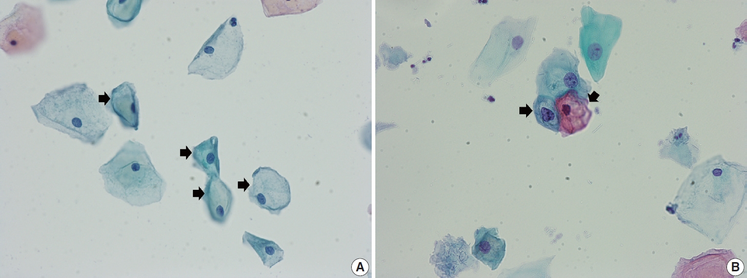
PDF - Cervical cancer screening during pregnancy presents unique challenges for cytologic interpretation. This review focuses on pregnancy-associated cytomorphological changes and their impact on diagnosis of cervical intraepithelial neoplasia (CIN) and cervical cancer. Pregnancy-induced alterations include navicular cells, hyperplastic endocervical cells, immature metaplastic cells, and occasional decidual cells or trophoblasts. These changes can mimic abnormalities such as koilocytosis, adenocarcinoma in situ, and high-grade squamous intraepithelial lesions, potentially leading to misdiagnosis. Careful attention to nuclear features and awareness of pregnancy-related changes are crucial for correct interpretation. The natural history of CIN during pregnancy shows higher regression rates, particularly for CIN 2, with minimal risk of progression. Management of abnormal cytology follows modified risk-based guidelines to avoid invasive procedures, with treatment typically deferred until postpartum. The findings reported in this review emphasize the importance of considering pregnancy status in cytological interpretation, highlight potential problems, and provide guidance on differentiating benign pregnancy-related changes from true abnormalities. Understanding these nuances is essential for accurate diagnosis and proper management of cervical abnormalities in pregnant women.
-
Citations
Citations to this article as recorded by
- HPV in Pregnancy: Implications for Screening, Vaccination, and Maternal–Fetal Health
Suman Kumar, Swati, Swati Salila, Akanksha Raj, Pratima Gupta, Neha Sharad, Nidhi Chaudhary
Journal of Pregnancy.2026;[Epub] CrossRef - The significance of biological samples from pregnant women in cervical intraepithelial neoplasia
Xue Mi, Maharjan Rashmi, Zangyu Pan, Di Wu, Jinwei Miao
Frontiers in Medicine.2025;[Epub] CrossRef - Oncologic and pregnancy outcomes of cervical high-grade intraepithelial lesions and delivery mode
Olga P. Matylevich, Ilya A. Tarasau, Sviatlana Y. Shelkovich, Aliaksandr F. Martsinkevich
Academia Oncology.2025;[Epub] CrossRef
- HPV in Pregnancy: Implications for Screening, Vaccination, and Maternal–Fetal Health
- Non-conventional dysplastic subtypes in inflammatory bowel disease: a review of their diagnostic characteristics and potential clinical implications
- Won-Tak Choi
- J Pathol Transl Med. 2021;55(2):83-93. Published online March 9, 2021
- DOI: https://doi.org/10.4132/jptm.2021.02.17

- 11,632 View
- 461 Download
- 29 Web of Science
- 32 Crossref
-
Abstract
PDF
- The early detection and grading of dysplasia is the current standard of care to minimize mortality from colorectal cancer (CRC) in patients with inflammatory bowel disease. With the development of advanced endoscopic resection techniques, colectomy is now reserved for patients with invisible/flat dysplasia (either high-grade [HGD] or multifocal low-grade dysplasia) or endoscopically unresectable lesions. Although most pathologists are familiar with the morphologic criteria of conventional (intestinal type) dysplasia, the most well-recognized form of dysplasia, an increasing number of diagnostic material has led to the recognition of several different morphologic patterns of epithelial dysplasia. The term “non-conventional” dysplasia has been coined to describe these changes, but to date, the recognition and full appreciation of these novel forms of dysplasia by practicing pathologists is uneven. The recognition of these non-conventional subtypes is becoming increasingly important, as some of them appear to have a higher risk of developing HGD or CRC than conventional dysplasia or sporadic adenomas. This review describes the morphologic characteristics of all seven non-conventional subtypes that have been reported to date as well as our current understanding of their clinicopathologic and molecular features that distinguish them from conventional dysplasia or sporadic adenomas.
-
Citations
Citations to this article as recorded by- Recent updates and debates on basal crypt dysplasia, serrated epithelial change, and p53 immunostaining in inflammatory bowel disease
Dorukhan Bahceci, Won-Tak Choi
Human Pathology.2026; 169: 105959. CrossRef - Updated pathologic classification of inflammatory bowel disease–associated colorectal dysplasia: A guide for clinicians
Noam Harpaz, Robert D Odze
Inflammatory Bowel Diseases.2026;[Epub] CrossRef - Diagnostic Significance and Reproducibility of Non-conventional Dysplasia and Serrated Epithelial Change in Inflammatory Bowel Disease
Won-Tak Choi, Gregory Y. Lauwers
Surgical Pathology Clinics.2026;[Epub] CrossRef - Aberrant p53 immunohistochemical staining is uncommon in serrated epithelial change, regardless of association with dysplasia
Dorukhan Bahceci, Gregory Y Lauwers, Won‐Tak Choi
Histopathology.2026;[Epub] CrossRef - Morphological subtypes of colorectal low-grade intraepithelial neoplasia: diagnostic reproducibility, frequency and clinical impact
Corinna Lang-Schwarz, Maike Büttner-Herold, Stephan Burian, Ramona Erber, Arndt Hartmann, Moritz Jesinghaus, Kateřina Kamarádová, Carlos A Rubio, Gerhard Seitz, William Sterlacci, Michael Vieth, Simone Bertz
Journal of Clinical Pathology.2025; 78(2): 103. CrossRef - “Artificial histology” in colonic Neoplasia: A critical approach
Gavino Faa, Matteo Fraschini, Luca Didaci, Luca Saba, Mario Scartozzi, Enrico Orvieto, Massimo Rugge
Digestive and Liver Disease.2025; 57(3): 663. CrossRef - Examination of non-conventional dysplasias adjacent to colorectal adenocarcinoma in patients with IBD
Szintia Almási, Zsófia Balajthy, Bence Baráth, Zsófia Krisztina Török, Panna Szaszák, Tamás Lantos, Bence Kővári, Anita Sejben
Pathology and Oncology Research.2025;[Epub] CrossRef - Clinical Characteristics, Management, and Outcomes of Colitis-Associated Colorectal Cancer and the Comparison With Sporadic Colorectal Cancer in Taiwan
Hsin-Yun Wu, Meng-Tzu Weng, Jen-Wei Chou, Hsu-Heng Yen, Chun-Chi Lin, Feng-Fan Chiang, Chen-Shuan Chung, Wei-Chen Lin, Chen-Wang Chang, Puo-Hsien Le, Chia-Jung Kuo, Ching-Pin Lin, Wen-Hung Hsu, Chiao-Hsiung Chuang, Tzung-Jiun Tsai, I-Che Feng, Shu-Chen We
Clinical and Translational Gastroenterology.2025; 16(2): e00798. CrossRef - Dysplasia in Pediatric Patients with Inflammatory Bowel Disease Shows Distinct Clinicopathologic Features Compared With that in Adult Patients
Dorukhan Bahceci, Shaomin Hu, Xiaoyan Liao, Lindsay Alpert, Hwajeong Lee, Huaibin Mabel Ko, Adam L. Booth, Gregory Y. Lauwers, Won-Tak Choi
Modern Pathology.2025; 38(6): 100735. CrossRef - Nonconventional dysplasia in patients with inflammatory bowel disease and colorectal adenocarcinoma: a case-cohort study
Siri A Urquhart, Namratha Pallipamu, Hima Varsha Voruganti, Bhavana Baraskar, Pratyusha Muddaloor, Arshia K Sethi, Renisha Redij, Keirthana Aedma, Keerthy Gopalakrishnan, Shivaram Poigai Arunachalam, Kelli N Burger, Douglas W Mahoney, Blake A Kassmeyer, R
Journal of Crohn's and Colitis.2025;[Epub] CrossRef - Cutting Edge: A Comprehensive Guide to Colorectal Cancer Surgery in Inflammatory Bowel Diseases
Ionut Eduard Iordache, Lucian-Flavius Herlo, Razvan Popescu, Daniel Ovidiu Costea, Luana Alexandrescu, Adrian Paul Suceveanu, Sorin Deacu, Gabriela Isabela Baltatescu, Alina Doina Nicoara, Nicoleta Leopa, Andreea Nelson Twakor, Andrei Octavian Iordache, L
Journal of Mind and Medical Sciences.2025; 12(1): 6. CrossRef - Inflammatory bowel disease‐associated serrated lesions with dysplasia are frequently associated with advanced neoplasia: supporting a unified classification approach
Dorukhan Bahceci, Anita Sejben, Lindsay Yassan, Gregory Miller, Xiaoyan Liao, Huaibin Mabel Ko, Marcela Salomao, Masato Yozu, Gregory Y. Lauwers, Won‐Tak Choi
Histopathology.2025; 87(3): 408. CrossRef - Whole-Exome Sequencing Analysis of Inflammatory Bowel Disease-Associated Serrated Dysplasia
Zsófia Balajthy, Szintia Almási, Tamás Lantos, Levente Kuthi, Georgios Deftereos, Won-Tak Choi, Anita Sejben
International Journal of Molecular Sciences.2025; 26(12): 5704. CrossRef - Interobserver variability in the histologic evaluation of serrated epithelial change in inflammatory bowel disease among gastrointestinal pathologists: a comparison of two different definitions
Dorukhan Bahceci, Rish K Pai, Ian Brown, Joseph Misdraji, M Priyanthi Kumarasinghe, Sanjay Kakar, Gregory Y Lauwers, Dongliang Wang, Won‐Tak Choi
Histopathology.2025; 87(4): 606. CrossRef - A pilot evaluation of the artificial intelligence system CAD-EYE to optically characterise lesions in inflammatory bowel disease surveillance
Sherman Picardo, Shankar Menon, Kenji So, Kannan Venugopal, Wendy Cheng, Krish Ragunath
Therapeutic Advances in Gastrointestinal Endoscopy.2025;[Epub] CrossRef - Hyperplasticus polypusszerű átalakulás gyulladásos bélbetegség diagnózisának felállításakor
Ádám Ferenczi, Anita Sejben
Orvosi Hetilap.2025; 166(31): 1230. CrossRef - Recently described types of dysplasia associated with IBD: tips and clues for the practising pathologist
Zahra Alipour, Kristen Stashek
Journal of Clinical Pathology.2024; 77(2): 77. CrossRef - Nonconventional Dysplasia is Frequently Associated With Goblet Cell Deficient and Serrated Variants of Colonic Adenocarcinoma in Inflammatory Bowel Disease
Andrew Xiao, Masato Yozu, Bence P. Kővári, Lindsay Yassan, Xiaoyan Liao, Marcela Salomao, Maria Westerhoff, Anita Sejben, Gregory Y. Lauwers, Won-Tak Choi
American Journal of Surgical Pathology.2024; 48(6): 691. CrossRef - Increased Active Inflammation in the Colon is Not a Reliable Predictor of an Elevated Risk of Dysplasia in Patients With Primary Sclerosing Cholangitis and Ulcerative Colitis
Ruth Zhang, Dongliang Wang, Gregory Y. Lauwers, Won-Tak Choi
American Journal of Surgical Pathology.2024; 48(9): 1154. CrossRef - Dysplasia Detected in Patients With Serrated Epithelial Change Is Frequently Associated With an Invisible or Flat Endoscopic Appearance, Nonconventional Dysplastic Features, and Advanced Neoplasia
Dorukhan Bahceci, Lindsay Alpert, Tanner Storozuk, Xiaoyan Liao, Masato Yozu, Maria Westerhoff, Bence P. Kővári, Gregory Y. Lauwers, Won-Tak Choi
American Journal of Surgical Pathology.2024; 48(10): 1326. CrossRef - Difficulties in diagnosis of non-conventional dysplasia in inflammatory bowel disease
Kh. M. Akhrieva, A. S. Tertychnyy, N. V. Pachuashvili, L. S. Urusova
Bulletin of the Medical Institute "REAVIZ" (REHABILITATION, DOCTOR AND HEALTH).2024; 14(3): 21. CrossRef - Hypermucinosus és kehelysejtszegény, gyulladásos bélbetegséghez társult, non-conventionalis dysplasia colorectalis adenocarcinoma mellett
Szintia Almási, Bence Baráth, Panna Szaszák, Bence Kővári, Anita Sejben
Orvosi Hetilap.2023; 164(51): 2039. CrossRef - DNA content abnormality frequently develops in the right/proximal colon in patients with primary sclerosing cholangitis and inflammatory bowel disease and is highly predictive of subsequent detection of dysplasia
Ruth Zhang, Peter S. Rabinovitch, Aras N. Mattis, Gregory Y. Lauwers, Won‐Tak Choi
Histopathology.2023; 83(1): 116. CrossRef - Non‐conventional dysplasia is frequently associated with low‐grade tubuloglandular and mucinous adenocarcinomas in inflammatory bowel disease
Fahire Goknur Akarca, Masato Yozu, Lindsay Alpert, Bence P Kővári, Lei Zhao, Marcela Salomao, Xiaoyan Liao, Maria Westerhoff, Gregory Y Lauwers, Won‐Tak Choi
Histopathology.2023; 83(2): 276. CrossRef - The yield of dysplasia and serrated lesions in a single-centre tertiary inflammatory bowel disease cohort
Fiona Yeaman, Lena Thin
Therapeutic Advances in Gastroenterology.2023;[Epub] CrossRef - MYC overexpression in inflammatory bowel disease-associated conventional dysplasia and association of subsequent low-grade dysplasia in follow-up biopsies
Yuanxin Liang, Yansheng Hao, Yiqin Xiong, Minghao Zhong, Dhanpat Jain
Pathology - Research and Practice.2023; 248: 154642. CrossRef - Characteristics, Reporting, and Potential Clinical Significance of Nonconventional Dysplasia in Inflammatory Bowel Disease
Won-Tak Choi
Surgical Pathology Clinics.2023; 16(4): 687. CrossRef - Using of endoscopic polypectomy in patients with diagnosed malignant colorectal polyp – The cross-sectional clinical study
Vladislava Stojic, Natasa Zdravkovic, Tamara Nikolic-Turnic, Nebojsa Zdravkovic, Jelena Dimitrijevic, Aleksandra Misic, Kristijan Jovanovic, Stefan Milojevic, Jelena Zivic
Open Medicine.2023;[Epub] CrossRef - And the story goes on: non-conventional dysplasia of the colorectum
Lavisha S. Punjabi, Yi Neng Lai, Anjula Thomas
Journal of Pathology and Translational Medicine.2022; 56(2): 109. CrossRef - Clinicopathologic features of undetected dysplasia found in total colectomy or proctocolectomy specimens of patients with inflammatory bowel disease
Dorukhan Bahceci, Gregory Y Lauwers, Won‐Tak Choi
Histopathology.2022; 81(2): 183. CrossRef - Increased Risk of Non-conventional and Invisible Dysplasias in Patients with Primary Sclerosing Cholangitis and Inflammatory Bowel Disease
Ruth Zhang, Gregory Y Lauwers, Won-Tak Choi
Journal of Crohn's and Colitis.2022; 16(12): 1825. CrossRef - Increased histologic inflammation is an independent risk factor for nonconventional dysplasia in ulcerative colitis
Eric D. Nguyen, Dongliang Wang, Gregory Y. Lauwers, Won‐Tak Choi
Histopathology.2022; 81(5): 644. CrossRef
- Recent updates and debates on basal crypt dysplasia, serrated epithelial change, and p53 immunostaining in inflammatory bowel disease
Case Study
- Fibrocartilaginous mesenchymoma with an unusual location in the rib
- Sun-Ju Oh
- J Pathol Transl Med. 2021;55(1):75-78. Published online December 3, 2020
- DOI: https://doi.org/10.4132/jptm.2020.10.08
- 6,685 View
- 121 Download
- 2 Web of Science
- 3 Crossref
-
Abstract
PDF
- Fibrocartilaginous mesenchymoma is a rare bone tumor, with fewer than 35 cases reported in the literature since 1984. This tumor usually occurs in the long bones of children and adolescents. In the current case, the tumor affected a rib. A 17-year-old boy presented with a mass in the right fifth rib. Radiologic findings revealed an osteolytic mass with cortical destruction and calcification; en bloc resection was performed. The tumor showed three distinct histologic features: bland spindle cell proliferation, benign cartilage nodules, and epiphyseal plate-like enchondral ossification. The pathologic diagnosis was fibrocartilaginous mesenchymoma. The patient remains free of disease 1 year after the surgery. Pathological diagnosis of fibrocartilaginous mesenchymoma can be challenging, especially when the tumor occurs in an unusual site. When any fibro-osseous lesion with a cartilaginous component is encountered, the possibility of fibrocartilaginous mesenchymoma should be considered because of its locally aggressive behavior.
-
Citations
Citations to this article as recorded by- Fibrocartilaginous mesenchymoma: a case report and a literature review
A. A. Karyagina, V. Yu. Roshchin, I. V. Sidorov, D. M. Konovalov
Pediatric Hematology/Oncology and Immunopathology.2025; 23(3): 158. CrossRef - Fibrocartilaginous mesenchymoma of the rib with atypical imaging features
Rashed Al-Khudairi, Danielle Forster, Sofina Begum, Alexandra Rice, Adrienne M Flanagan, Fernanda Amary, Paul O’Donnell
BJR|Case Reports.2025;[Epub] CrossRef - Fibrocartilaginous mesenchymoma of pelvis—a potential diagnostic pitfall
Monalisa Hui, Shantveer G. Uppin, Ramakrishna Narayanan, K. Nageshwara Rao, B. Aravind Kumar
Skeletal Radiology.2023; 52(4): 791. CrossRef
- Fibrocartilaginous mesenchymoma: a case report and a literature review
Original Articles
- Indirect pathological indicators for cardiac sarcoidosis on endomyocardial biopsy
- Myung-Jin Cha, Jeong-Wook Seo, Seil Oh, Eun-Ah Park, Sang-Han Lee, Moon Young Kim, Jae-Young Park
- J Pathol Transl Med. 2020;54(5):396-410. Published online July 29, 2020
- DOI: https://doi.org/10.4132/jptm.2020.06.10
- 9,101 View
- 121 Download
- 11 Web of Science
- 11 Crossref
-
Abstract
PDF
 Supplementary Material
 - Background
The definitive pathologic diagnosis of cardiac sarcoidosis requires observation of a granuloma in the myocardial tissue. It is common, however, to receive a “negative” report for a clinically probable case. We would like to advise pathologists and clinicians on how to interpret “negative” biopsies.
Methods
Our study samples were 27 endomyocardial biopsies from 25 patients, three cardiac transplantation and an autopsied heart with suspected cardiac sarcoidosis. Pathologic, radiologic, and clinical features were compared.
Results
The presence of micro-granulomas or increased histiocytic infiltration was always (6/6 or 100%) associated with fatty infiltration and confluent fibrosis, and they showed radiological features of sarcoidosis. Three of five cases (60%) with fatty change and confluent fibrosis were probable for cardiac sarcoidosis on radiology. When either confluent fibrosis or fatty change was present, one-third (3/9) were radiologically probable for cardiac sarcoidosis. We interpreted cases with micro-granuloma as positive for cardiac sarcoidosis (five of 25, 20%). Cases with both confluent fibrosis and fatty change were interpreted as probable for cardiac sarcoidosis (seven of 25, 28%). Another 13 cases, including eight cases with either confluent fibrosis or fatty change, were interpreted as low probability based on endomyocardial biopsy.
Conclusions
The presence of micro-granuloma could be an evidence for positive diagnosis of cardiac sarcoidosis. Presence of both confluent fibrosis and fatty change is necessary for probable cardiac sarcoidosis in the absence of granuloma. Either of confluent fibrosis or fatty change may be an indirect pathological evidence but they are interpreted as nonspecific findings. -
Citations
Citations to this article as recorded by- Unmasking Cardiac Sarcoidosis: Integrating Multimodal Imaging with Histochemical and Ultrastructural Analysis
Jakub Kancerek, Damian Świerczek, Wiktoria Baron, Marcin Rojek, Piotr Lewandowski, Romuald Wojnicz
International Journal of Molecular Sciences.2026; 27(7): 2969. CrossRef - Diagnostic Value of Comprehensive Echocardiographic Assessment Including Speckle-Tracking in Patients with Sarcoidosis Versus Healthy Controls: A Systematic Review and Meta-Analysis
Hritvik Jain, Maryam Shahzad, Muneeba Ahsan, Rahul Patel, Jagjot Singh, Ramez M. Odat, Aman Goyal, Raveena Kelkar, Nishad Barve, Hina Farrukh, Raheel Ahmed
Diagnostics.2025; 15(6): 708. CrossRef - Advances in cellular and tissue-based imaging techniques for sarcoid granulomas
Junwoo Kim, Girish Dwivedi, Berin A. Boughton, Ankur Sharma, Silvia Lee
American Journal of Physiology-Cell Physiology.2024; 326(1): C10. CrossRef - Lipomatous Metaplasia Is Associated With Ventricular Tachycardia Recurrence Following Ablation in Patients With Nonischemic Cardiomyopathy
Lingyu Xu, Mirmilad Khoshknab, Juwann Moss, Ronald D. Berger, Jonathan Chrispin, David Callans, Francis E. Marchlinski, Stefan L. Zimmerman, Yuchi Han, Natalia Trayanova, Benoit Desjardins, Saman Nazarian
JACC: Clinical Electrophysiology.2024; 10(6): 1135. CrossRef - Cardiac Sarcoidosis: A Comprehensive Clinical Review
András Vereckei, Zsuzsanna Besenyi, Viktória Nagy, Bence Radics, Hajnalka Vágó, Zsigmond Jenei, Gábor Katona, Róbert Sepp
Reviews in Cardiovascular Medicine.2024;[Epub] CrossRef - Cardiac sarcoidosis: phenotypes, diagnosis, treatment, and prognosis
Jukka Lehtonen, Valtteri Uusitalo, Pauli Pöyhönen, Mikko I Mäyränpää, Markku Kupari
European Heart Journal.2023; 44(17): 1495. CrossRef - Cardiac sarcoidosis: a comprehensive review of risk factors, pathogenesis, diagnosis, clinical manifestations, and treatment strategies
Hussain Haider Shah, Syeda Alishah Zehra, Aliza Shahrukh, Radeyah Waseem, Tooba Hussain, Muhammad Sheheryar Hussain, Fareeha Batool, Muhammad Jaffer
Frontiers in Cardiovascular Medicine.2023;[Epub] CrossRef - Histology of Cardiac Sarcoidosis with Novel Considerations Arranged upon a Pathologic Basis
Shu Kato, Yasuhiro Sakai, Asako Okabe, Yoshiaki Kawashima, Kazuhiko Kuwahara, Kazuya Shiogama, Masato Abe, Hiroyasu Ito, Shin’ichiro Morimoto
Journal of Clinical Medicine.2022; 11(1): 251. CrossRef - Cardiac sarcoidosis: A multimodal approach to reach the diagnosis
Nicolas Piriou, Patrick Bruneval
International Journal of Cardiology.2021; 323: 264. CrossRef - Value of 3D mapping‐guided endomyocardial biopsy in cardiac sarcoidosis
Danielle M. Haanschoten, Ahmet Adiyaman, Nils A. ‘t Hart, Piet L. Jager, Arif Elvan
European Journal of Clinical Investigation.2021;[Epub] CrossRef - Cardiac Sarcoidosis: A Clinical Overview
Ana Carolina Alba, Shyla Gupta, Lakshmi Kugathasan, Andrew Ha, Alejandro Ochoa, Meyer Balter, Alvaro Sosa Liprandi, Maria Inés Sosa Liprandi
Current Problems in Cardiology.2021; 46(10): 100936. CrossRef
- Unmasking Cardiac Sarcoidosis: Integrating Multimodal Imaging with Histochemical and Ultrastructural Analysis
- CpG Island Methylation According to the Histologic Patterns of Early Gastric Adenocarcinoma.
- Junjeong Choi, Mee Yon Cho, So Young Jung, Khalilullah Mia Jan, Hyun Soo Kim
- Korean J Pathol. 2011;45(5):469-476.
- DOI: https://doi.org/10.4132/KoreanJPathol.2011.45.5.469
- 5,082 View
- 22 Download
- 2 Crossref
-
Abstract
PDF
- BACKGROUND
Although the importance of aberrant DNA methylation in the development of gastric adenocarcinoma has been described, the mechanism of pathogenesis has not been revealed yet. We quantitatively analyzed methylation of four CpG islands and one repetitive DNA element, according to the histologic features of adenocarcinoma with precursor lesions.
METHODS
We divided the cases as adenocarcinoma with intestinal type precursors (type A, n=19 cases) and adenocarcinoma with diffuse type precursors (type B, n=19 cases). We micro-dissected tumor cells and matched non-neoplastic gastric mucosa from the hematoxylin and eosin-stained slides.
RESULTS
A total of 20 CpG sites of long interspersed nucleotide element-1 (LINE1), RAR-related orphan receptor alpha (RORA), Kruppel-like factor 7 (KLF7), mutL homolog 1 (MLH1), MINT25, and CD133 were analyzed. Methylation was determined by bisulfate-pyro-sequencing, and hypomethylation of LINE1 and CD133 was noted in the tumors, compared to the levels in the non-neoplastic gastric mucosa (p=0.014 and p=0.015, respectively). A statistically different methylation pattern of CpG sites at CD133 and KLF7 was noted only in type B lesions, compared to that in matched non-neoplastic gastric mucosa (p=0.027 and p=0.043, respectively).
CONCLUSIONS
Given that aberrant methylation occurs in a relatively early phase of carcinogenesis, different patterns of methylation may determine the carcinoma phenotype. However, further large-scale study is required to clarify the significance of this difference. -
Citations
Citations to this article as recorded by- Molecular function of Krüppel-like factor 7 in biology
Yi Mao, Yuechan Chen, Zhiwei Zhang
Acta Biochimica et Biophysica Sinica.2023; 55(5): 713. CrossRef - DNA methylation status of a distinctively different subset of genes is associated with each histologic Lauren classification subtype in early gastric carcinogenesis
YOSEP CHONG, KHALILULLAH MIA-JAN, HOON RYU, JAMSHID ABDUL-GHAFAR, JIJGEE MUNKHDELGER, SAYAMAA LKHAGVADORJ, SO YOUNG JUNG, MIRA LEE, SUN-YOUNG JI, EUNHEE CHOI, MEE-YON CHO
Oncology Reports.2014; 31(6): 2535. CrossRef
- Molecular function of Krüppel-like factor 7 in biology
- Expressions of MIB-1, p53 and CEA in Endocervical Glandular Lesions.
- Mi Jin Kim, Young Gi Lee, Dong Sug Kim
- Korean J Pathol. 2001;35(1):41-47.
- 2,201 View
- 64 Download
-
Abstract
PDF
- BACKGROUND
Endocervical glandular lesions include glandular atypia (GA), endocervical glandular dysplasia (EGD), adenocarcinoma in situ (AIS), and invasive adenocarcinoma (IA). The diagnosis of malignant glandular lesions is occasionally difficult to distinguish from benign mimickers, and the morphologic features of EGD remain unsettled.
METHODS
Immunohistochemical stains for MIB-1, p53 and CEA were performed on 81 cases of paraffin-embedded endocervical glandular lesions including 22 IA, 15 AIS, 15 EGD, 13 GA, 8 microglandular hyperplasia (MGH) and 8 tubal metaplasia (TM).
RESULTS
The MIB-1 labelling index of IA was 59.68%, 69.53% for AIS, 26.60% for EGD, 16.03% for benign. p53 overexpression was noted in 4 (18%) cases of IA, 3 (20%) of AIS, but none of EGD and benign lesions. It was Interesting to note that one case of MGH showed p53 staining in low intensity. Diffuse strong cytoplasmic CEA positivity was present in all of IA and AIS, whereas seven (47%) of 15 EGD and 12 (41%) of 29 benign lesions showed focal cytoplasmic CEA positivity. There were significant differences in MIB-1 and CEA immunostainings among the adenocarcinoma, EGD, and benign glandular lesions. Adenocarcinoma was closely related to p53 overexpression, although occurring in a low percentage of the cases.
CONCLUSION
MIB-1 immunostaining can be useful in differentiating among endocervical adenocarcinoma, endocervical glandular dysplasia and benign glandular lesions. p53 overexpression might be helpful in the diagnosis of adenocarcinoma.
- Congenital Cystic Disease of the Kidney overview and a classification.
- Mee Joo, Yeon Mee Kim, Chong Jai Kim, Yeon Lim Suh, Jeong Wook Seo, Je Geun Chi
- Korean J Pathol. 1997;31(3):233-243.
- 2,049 View
- 22 Download
-
Abstract
PDF
- The congenital renal cystic disease encompasses a complex group of pathologic and clinical entities. We retrospectively reviewed 42 cases of congenital renal cystic lesions classified into four Potter types in a series of 2,063 consecutive autopsies from 1981 to 1996. According to our study based on morphologic, clinical, genetic features and associated anomalies, type I and III are relatively compatible with Potter's original definition. However, it was reasonable that type II and IV are classified to the same group because of: 1) very similar histologic findings representing dysplastic kidney, 2) many associated anomalies, 3) no evidence of inheritance, and 4) presence of a combined type. Syndrome associated cysts, such as Meckel-Gruber syndrome, were also separately classified. If the dysplastic evidence was insufficient for diagnosis to the dysplastic kidney in type II and IV, then these cases would be better classified into a cystic disease associated with congenital hydronephrosis. We propose a classification of the congenital cystic disease of the kidney to be: 1) dysplastic kidney, 2) cystic disease associated with congenital hydronephrosis, 3) polycystic kidney, and 4) syndromic cystic disease.
- An Image Analytical Study on the Structural Spectrum of Intestinal Metaplasia-Dysplasia-Carcinoma of the Stomach.
- Sang Woo Juhng, Dong Ha Park, Ji Shin Lee, Kyu Hyuk Cho
- Korean J Pathol. 1993;27(1):50-57.
- 2,234 View
- 14 Download
-
Abstract
PDF
- Intestinal metaplasia and dysplasia of the stomach have been stressed as precursors of gastric carcinoma of the intestinal type, although their preneoplastic nature is still debated. In this study, the cytomorphometric and cytokinetic spectra of the suggested preneoplastic and neoplastic lesions of the stomach were investigated. From the resected stomachs of early gastric carcinoma of intestinal type, areas of normal, intestinal metaplasia, dysplasia, and carcinoma were selected. They were immunostained for proliferating cell nuclear antigen, counterstained with propidium iodide, and various nuclear parameters were measured by image analysis. Normal and intestinal metaplastic mucosae differed by the localization of proliferation zone, but not by nuclear profile area, circular shape factor, and proliferation index. In dysplasia, proliferation zone covered large parts of the dysplastic area. Nuclear profile area and proliferation index were larger whereas circular shape factor was smaller than in normal or intestinal metaplasia. Carcinomatous lesion had diffuse proliferation activity, the largest nuclear profile area and proliferating index, and circular shape factor in-between those of normal or intestinal metaplasia and dysplasia. The above results showed a structural spectrum among normal of intestinal metaplasia, dysplasia, and carcinoma of intestinal type in cytomorphometric and cytokinetic terms. The structural spectrum raises the possibility that dysplasia of the stomach is a preneoplastic lesion.
- Grading System for Gastric Epithelial Proliferative Diseases Standardized Guidelines proposed by Korean Study Group for Pathology of Digestive Diseases.
- Hoguen Kim, So Young Jin, Ja June Jang, Woo Ho Kim, Sang Yong Song, Kyu Rae Kim, Eun Sil Yu, Hyung Sik Shin, Han Kyeom Kim, Jin Hee Sohn, Eun Kyung Hong, Youn Wha Kim, Jin Sook Jeong, Chang Jin Kim, Shin Eun Choi, In Suh Park, Chan Il Park, Yong Il Kim
- Korean J Pathol. 1997;31(5):389-400.
- 2,319 View
- 42 Download
-
Abstract
PDF
- The assessment of epithelial changes in gastric mucosal biopsies has been one of the major problems caused by inconsistencies in and disagreements about nomenclature and interpretation. To resolve these issues, members of the Study Group for Pathology of Digestive Diseases reviewed microslides of 50 gastric lesions showing varying degrees of mucosal abnormality and reached the following consensus; 1) the proliferating gastric epithelium can be divided into hyperplastic and neoplastic; 2) the term "dysplasia" is reserved for the microscopic epithelial changes that are unequivocally neoplastic; 3) Biopsy specimens can be categorized as regenerative(negative for dysplasia), indefinite(questionable dysplasia), positive(positive for dysplasia) and overt carcinoma; 4) The positive category can be divided into two subgroups, high grade dysplasia and low grade dysplasia. Criteria for each grade are presented and discussed. We offer these guidelines for establishing the correct diagnosis of the gastric mucosal biopsy specimens and for prospective studies.
Case Report
- Achondrogenesis Type 2: An autopsy case.
- Joon Mee Kim, Young Chae Chu, Soo Kee Min, Hee Jeung Cha, Je Geun Chi
- Korean J Pathol. 1997;31(5):482-488.
- 2,531 View
- 33 Download
-
Abstract
PDF
- Achondrogenesis type 2 is a lethal form of congenital skeletal dysplasia characterized by severe short-limbed dwarfism, decreased vertebral ossification and normal ossification of the skull. We report an autopsy case of achondrogenesis type 2 in a female fetus terminated at 29 weeks of gestation. External morphology revealed a relatively large head, short upper and lower extremities, short neck, and distended abdomen. The x-ray finding showed normal calvarial ossification, hypoplastic ilium and unossified ischium, and metaphyseal flares of the femur and tibia. Histologically, chondrocytes were large and irregular with increased vascularity.
Original Articles
- Hyperplasia, Metaplasia, and Dysplasia of the Gallbladder Correlation to Gallbladder Adenocarcinoma.
- Hee Jin Chang, Jung Il Suh
- Korean J Pathol. 1997;31(6):527-537.
- 3,159 View
- 70 Download
-
Abstract
PDF
- The correlation of metaplasia to dysplasia and carcinoma in the gallbladder has attracted the attention of many investigators. We mapped and examined a total of 263 cholecystectomized gallbladders to analyze the mucosal changes in the carcinogenesis of the gallbladder. Stones were present in 59.7%, hyperplasia in 28.5%, metaplasia in 55.5% (gastric 37.6%, intestinal 17.9%), dysplasia in 17.1% (low grade 9.1%, high grade 8%) and carcinoma in 7.6%. Metaplasia was more frequently identified in the stone-positive group (62.4%) than in the stone-negative group (45.3%) (P<0.05). Especially, the incidence of intestinal metaplasia was significantly higher in the stone-positive group. Dysplasia and carcinoma were more frequent in the metaplasia-positive group (dysplasia 26.7%, carcinoma 11%) than in the metaplasia-negative group (dysplasia 5.1%, carcinoma 3.4%) (P<0.05). Their incidences were significantly higher in the intestinal metaplasia than in the gastric metaplasia. Forty four percent of the dysplasia-positive cases were associated with carcinoma in the adjacent mucosa but carcinoma was absent in the dysplasia-negative cases. Hyperplasia did not reveal any significant correlation with metaplasia, dysplasia and carcinoma. These results suggest that gallstone is causally related to the metaplasia in the gallbladder and the metaplasia-dysplasia- carcinoma sequence exists in the gallbladder.
- Pathologic Analysis of 71 Cases of Cerebral Cortical Dysplasia.
- Sang Pyo Kim, Seung Che Cho
- Korean J Pathol. 1997;31(9):815-822.
- 2,177 View
- 18 Download
-
Abstract
PDF
- Cortical dysplasia (CD) is considered to be a malformative lesion of the neocortex which exhibits a spectrum of pathologic changes reflecting a disturbance in the process of its development. CD is recently recognized as a major cause of intractable epilepsy with non-neoplastic lesions. Mischel et al. proposed that CD can be graded mild, moderate and severe with regard to nine specific microscopic abnormalities: mild CD consists of 1) cortical laminar disorganization, 2) single heterotopic white matter neurons, 3) neurons in the cortical molecular layer, 4) persistent remnants of the subpial granular cell layer, and 5) marginal glioneuronal heterotopia; moderate CD displays 6) polymicrogyria and 7) white matter neuronal heterotopia; severe CD phows 8) neuronal cytomegaly with associated cytoskeletal abnormalities and 9) balloon cell change. We reassessed 71 cases of cortical dysplasia to elucidate the proportion and histologic features of each group, using Mischel's grading system. CD was most frequently found in the temporal lobe with 50 cases (70%). Mild CD was predominently seen and was noted in 61 cases (86%) Cortical laminar disorganization and single heterotopic white matter neurons were identified in all mild CD cases. Neurons in the cortical molecular layer, persistent subpial granular cell layer, and marginal glioneuronal heterotopia were also noted in case numbers 40, 3, and 1 of mild CD, respectively. Moderate CD was composed of 2 cases with polymicrogyria, and the remaining 8 cases had severe CD. All moderate and severe CD were associated with the various histological features of mild CD. Thirty eight cases (51%) of CD showed dual pathology, composed of both CD and hippocampal sclerosis, and 5 cases of dysembryoplastic neuroepithelial tumor also had CD. Neurofilament immunostain revealed disarray of abnormally beaded axons in CD. We believe that the grading system of CD is very important to the evaluation and classification of CD.
Case Reports
- Arrhythmogenic Right Ventricular Dysplasia/Cardiomyopathy: Report of an autopsy case.
- Tae Yub Kim, Young Min Kim, Jae Gul Chung, Gyung Yub Gong, Su Kil Park, In Chul Lee, Joo Ryung Huh
- Korean J Pathol. 1997;31(11):1233-1236.
- 1,939 View
- 10 Download
-
Abstract
- A 35-year-old man was admitted with a 20 day history of generalized edema and muscular weakness of the lower extremities. He was alert with a pale puffy face and an ejection murmur was heard at the cardiac apex. The electrocardiogram disclosed low voltage, first degree atrioventricular block, and a right bundle branch block. During the hospitalization an intractable diastolic hypotension developed, which measured 0 mmHg at the lowest point. At that time the echocardiogram revealed a dilated, akinetic right ventricle. Eventually a multiorgan failure developed and an autopsy following his death presented a fibrofatty replacement of the right ventricular myocardium. This might be a case of an arrhythmogenic right ventricular dysplasia/cardiomyopathy, which is usually characterized clinically by a ventricular tachycardia and may cause a sudden death in young adults.
- Liposclerosing Myxofibrous Tumor in Tibia: A Case Report and Review of the Literature.
- Jung Woo Choi, Young Seok Lee, Ju Han Lee, Han Kyeom Kim, Bom Woo Yeom, Jong Sang Choi, Hong Chul Lim, Chul Hwan Kim
- Korean J Pathol. 2005;39(3):207-210.
- 2,744 View
- 48 Download
-
Abstract
PDF
- Liposclerosing myxofibrous tumor (LSMFT) is a benign fibro-osseous lesion that is characterized by a complex mixture of histologic elements, including its fibrous dysplasia-like features and its lipoma, myxofibroma, xanthoma and pseudo-Paget's bone patterns. However, this lesion is considered by some researchers as a variant of fibrous dysplasia or as the non-specific end result of degenerative change, while it is considered by others as a definite clinicopathologic entity. Here, we report on a case of LSMFT occurring in tibia, which is a very uncommon location for this tumor, and we review the related literatures. The case presented here shares features with those described for LSMFT, except for the location of this tumor. We believe that more studies on a larger scale that compare LSMFT with other benign bone lesions, including fibrous dysplasia, are required to clarify the origin and behavior of this lesion.
- Angiodysplasia Arising in the Bowels: Two cases report.
- Soo Kee Min, Hee Jeung Cha, Joon Mee Kim, Young Chae Chu
- Korean J Pathol. 1997;31(12):1308-1313.
- 2,218 View
- 19 Download
-
Abstract
PDF
- Gastrointestinal angiodysplasia is a distinct disease entity which causes frequent gastrointestinal bleeding. It predominantly arises at the stomach and duodenum in the upper gastrointestinal tract and cecum and ascending colon in the lower gastrointestinal tract. The general histological finding of the angiodysplasia is a submucosal vascular ectasia and tortuosity. We have experienced two cases of the intestinal angiodysplasia. The first case occurred on a jejunum in a 22-year-old woman who had anemia. The second case occurred on a sigmoid colon in a 59-year-old man who had constipation. In addition to the general histologic finding of the angiodysplasia, the microscopic findings of the first case revealed some capillary hemangioma-like areas; and in the second case, there was a marked ischemic change and the thickening of the wall.
Original Article
- Cytoskeletal Changes in Cortical Dysplasia.
- Min Young Lee, Jae Hun Chung, Young Jong Woo, Hyoung Ihl Kim, Min Cheol Lee
- Korean J Pathol. 2000;34(4):300-309.
- 2,055 View
- 11 Download
-
Abstract
PDF
- Cortical dysplasia is a cause of intractable epilepsy and a candidate for surgical resection to control epileptic attacks. The neuronal cytomegaly and balloon cell change are the diagnostic hallmarks of cortical dysplasia. Little research has been performed about the normal-sized dysplastic neuron which has complex arborizing dendrites and lacks in its polarity. The aim of this study was to define the histopathologic characteristics of the neurons in cortical dysplasia. Twelve cases of cortical dysplasia who underwent partial lobectomy for intractable seizures were selected and immunohistochemical staining for NF-M/H, MAP2, tau, and ubiquitin was performed. The perikarya and dendrite of dysplastic neurons were more intensely labeled with antibodies for the high and medium molecular weight neurofilament proteins (NF-M/H) than normal neurons. Immunoreactivity with the MAP2 antibody expressed mainly within the somatodendritic regions was present in the dysplastic or normal neurons without any significant difference in intensity. The complex arborizing dendrites of dysplastic neurons were easily identified due to pronounced immunoreactivity within the somatodendritic regions. Immunoreactivity with the primary antibody against tau and ubiquitin was present in the normal-looking neurons as well as the dysplastic neurons. This study suggests that the dysplastic neurons in cortical dysplasia are accompanied by changes of cytoskeletal neurofilaments, and the immunohistochemical stains for NF-M/H, MAP2, tau, and ubiquigin are useful to detect them.
Case Report
- Multicystic Renal Dysplasia with Ipsilateral Ectopic Ureteral Orifice and Seminal Vesicle Cyst: A case report.
- Hyun Jin Son, Joo Heon Kim, Myoung Jae Kang
- Korean J Pathol. 2000;34(4):310-313.
- 2,348 View
- 12 Download
-
Abstract
PDF
- Renal dysplasia results from aberrant metanephric histogenesis caused fundamentally by a defect in inducer tissue or responding tissue. Dysplastic kidneys vary tremendously in gross and microscopic appearance but are characterized by abnormal organization and a mixed population of primitive structures, such as fetal or immature cartilage, dysplastic ducts, immature tubules, and undifferentiated mesenchyme. We report a case of unilateral multicystic renal dysplasia associated with an ipsilateral ectopic ureteral orifice entering a seminal vesicle cyst in a 33-year-old man. He was admitted due to primary infertility which had developed three years ago. The his semen analysis revealed oligospermia. No evidence of a family history of renal dysplasia was reported. Microscopic examination showed that the entire kidney was composed of cysts lined by flattened cells, dysplastic ducts and immature tubules surrounded by collars of spindle cells, primitive mesenchyme, and a few aberrantly formed glomeruli.
Original Articles
- Radiation-Induced Changes in Cervico-vagical Smears of Uterine Cervical Cancer Patients.
- Sung Hye Park, In Sun Kim, Seung Yong Paik, Hyo Sook Park, No Won Park
- Korean J Pathol. 1988;22(3):268-276.
- 2,685 View
- 54 Download
-
Abstract
PDF
- Nowday, ionizing radiation is one of the methods eradicating the uterine cervical malignancy. However radiation alone or in combination with surgery have an effect on normal tissue as well as the malignant cells, and their changes have been well described in other countries. Unfortunately, the history of radiation modality for cancer treatment is relatively short and the reports about radiation induced changes are limited in our country. We evaluated the radiation-induced changes in cervico-vaginal smears of 107 uterine cervical cancer patients obtained from March, 1985 to October, 1987. Most patients had been received 5,400 Rads of external radiation and intracavitary radiation. Patient's age ranged from 30 to 67 years old. Of 107 cases, 24 cases were normal, 72 cases showed benign radiation changes, 7 cases revealed radiation dysplasia, and residual and recurrent carcinomas found in one and 3 cases, respectively. Cytoplasmic and nuclear enlargement were the most common and noted in 57 and 38 cases, respectively. Vacuolization and polychromasia of the cytoplasm were identified in 43 and 30 cases, respectively. The most common histiocytic change was multinucleation, which was found in about one third. The radiation changes of the cytoplasm and nuclear enlargement persisted for a long time after completion of radiation, however, nuclear degeneration and multinucleation gradually disappeared after 6 months. The inflammation in background prolonged for a long time but degeneration disappeared after 6 months. The biologic significance of post-radiation dysplasia could not evaluated because of short follow up period.
- Intrauterine Infection as a Cause of the Neonatal Pulmonary Injury and Bronchopulmonary Dysplasia.
- Jin Haeng Chung, Jeong Wook Seo
- Korean J Pathol. 2000;34(6):431-436.
- 2,005 View
- 12 Download
-
Abstract
PDF
- The pathogenetic role of intrauterine infection to the neonatal pulmonary injury and bronchopulmonary dysplasia was assessed by studying the interleukin-6 (IL-6) level in the umbilical cord blood and the early morphologic changes of the neonatal lung. Patients were grouped into bronchopulmonary dysplasia (4 cases), chorioamnionitis without chronic lung injury (4 cases), and 6 cases without morphologic evidence of chronic lung injury or placental inflammation. IL-6 level of umbilical cord blood was higher in babies with bronchopulmonary dysplasia (17.7 pg/ml) compared to those with chorioamnionitis (4.7 pg/ml) or those with morphologically normal lung and placenta (6.2 pg/ml). Morphologic parameters of neonatal pulmonary injury were hyaline membrane, terminal bronchiole inflammation, terminal bronchiole regeneration, alveolar collapse and fibroblastic proliferation. Bronchiolar regeneration was the most peculiar feature seen in the lung with bronchopulmonary dysplasia. Alveolar collapse and interstitial fibroblastic reaction were commonly seen in bronchopulmonary dysplasia. The postnatal age at death was higher in those with bronchopulmonary dysplasia, although the occurrence of the morphologic changes was related with the chronicity of those lesions. These findings suggest that intrauterine infection is an aggravating factor for the neonatal pulmonary injury and bronchopulmonary dysplasia, although the early stage of the lung injury is not a definitive indicator for the progressive pulmonary damage leading to the bronchopulmonary dysplasia.
- Multicystic Renal Dysplasia associated with Ectopic Ureteral Orifice.
- Shin Mong Kang, Yong Koo Park, Ju Hie Lee, Soo Eung Chai, Moon Ho Yang
- Korean J Pathol. 1988;22(3):331-335.
- 2,305 View
- 12 Download
-
Abstract
PDF
- A case of unilateral multicystic renal dysplasia was reported in view of rarity, and a review of literature 23-year-old female was admitted to Kyung Hee University Hospital with a complaint of urinary incontinence. An excretory urogram revealed non-visualization of the right kidney. The right ureter had opening at the 2 cm above the vaginal introitus. The resected right kidney revelaed multilobulated outer surface with multiloculated cystic struture on cut surface. The associated anomaly was uterine didelphy. The condition was treated by nephroureterectomy.
- Beckwith-Wiedemann Syndrome with Unusual Sialoadenomegaly.
- Hye Seung Han, Seung Sook Lee, Suk Keun Lee, Je G Chi
- Korean J Pathol. 1996;30(10):939-942.
- 2,266 View
- 19 Download
-
Abstract
PDF
- Beckwith-Wiedemann syndrome is a rare clinical entity characterized by exomphalos, macroglossia, macrosomia, and renal hyperplasia/dysplasia. Although its entity is established, its etiology and obligatory features have not been settled. We report an autopsy case with the unusual involvement of the salivary gland. This infant was born to a 37-year-old mother as a normal full-term spontaneous delivery. At 11 days of age she developed with purulent eye discharge and weak sucking, and died suddenly. At autopsy the baby weighed 2,630 gm and the head circumference was 35 cm. She showed thick and prominent skin folds, bilateral aural fissures, macroglossia, hepatomegaly, cardiomegaly, dysmorphic kidneys, and nesidioblastosis. Both kidneys showed dysplastic tubules and hyperplastic cortical tissue enclosing the medulla. In this case there were characteristic findings in major and minor salivary glands with both acinar and ductal hyperplasia, and hypertrophy of mammary glands. Besides, she had generalized depletion of subcutaneous fat, immature buccal fat, patent ductus arteriosus, hyperlobation of the right lung, two accessory spleens, and hyperplasia of basophils and chromophobes in the pituitary gland. The lungs showed diffuse interstitial pneumonia and multiple fibrin thrombi. There were no adrenal cytomegaly, umbilical hernia and exophthalmos.
Case Report
- Multiple Fibromuscular Dysplasia of Arteries with Aneurysm Formation: A case report.
- Soo Min Kang, Sang Yong Song, Yong Il Kim, Sung Kwon Kim, Jung Sang Lee, Sang Joon Kim
- Korean J Pathol. 1992;26(3):288-292.
- 2,026 View
- 13 Download
-
Abstract
PDF
- Multiple involvement of arterial fibromuscular dysplasia is unusal and such a case is quite limited in the literature. We present a case of multiple arterial fibromuscular dysplasia with aneurysm formation in a 38-year-old non-hypertensive man. Angiographic study showed multiple aneurysmal dilatations of the right renal, superior mesenteric, celiac axis, and left colic arteries. The resected right kidney demonstrated an aneurysm of the main renal artery and extensive infarction in the midportion of the renal parenchyma. The segmentally resected superior mesenteric artery disclosed a similar nature of aneurysm occluded by recent thrombi. Microscopically, all the resected arterial walls showed both intimal and medial fibroplasia together with periadventitial fibrosis resulting in marked narrowing of the lumen and subsequent thrombus formation.
Original Articles
- Studies of CD44s and CD44v6 Expressions and Correlation with Clinicopathologic Parameters in Gastric Carcinoma.
- Zhu Hu Li, Zhen Hua Lim, Ho Jong Jeon
- Korean J Pathol. 2003;37(5):320-324.
- 2,352 View
- 14 Download
-
Abstract
PDF
- BACKGROUND
The aim of this study is to elucidate the relationship between the CD44s and CD44v6 expression level and the biological characteristics of a gastric carcinoma.
METHODS
CD44s and CD44v6 expression was investigated in 56 gastric carcinomas, 18 dysplasias, and 22 normal mucosae by immunohischemical staining.
RESULTS
The CD44s and CD44v6 expression rates in gastric carcinomas, dysplasia, and normal mucosae were 80.3% and 83.9%, 72.2% and 77.8%, and 13.6% and 4.5%, respectively. Statistical analysis showed significant difference after comparing a gastric carcinomas and dysplasia to the normal mucosae (p<0.001). The CD44s and CD44v6 expression rates in the cases with invasion to the muscle proper and serosa were 60.7% and 57.1%, and 82.4% and 88.2%, respectively. Both showed a significant statistical difference compared to the expression rates in the cases with invasion to the mucosae and submucosae. The CD44s and CD44v6 expression rates in gastric carcinomas with a lymph node metastasis showed a statistically significant difference compared to those without a lymph node metastasis (p<0.001 and p<0.01, respectively). CD44s and CD44v6 were also expressed in the normal basal cells around gastric carcinomas.
CONCLUSIONS
The CD44s and CD44v6 expression showed a significant relationship with gastric carcinogenesis, toward an aggressive biologic behavior.
- Liver Cell Dysplasia: Analysis of 141 cases with reference to histopathologic Characterization and proliferative activity.
- Sang Yong Song, Yong Il Kim
- Korean J Pathol. 1992;26(4):338-347.
- 1,946 View
- 12 Download
-
Abstract
PDF
- Liver cell dysplasia of Anthony(LCD) is a common association in hepatocellular carcinoma(HCC)-bearing liver and has been regarded as a premalignant condition with strong linkage to hepatitis B virus infection and cirrhosis. A total of 189 surgically resected livers [HCC(168 cases), cholangiocarcinoma(3 cases), metastatic carcinoma(3 cases), and non-neoplastic lesions(15 cases)] were reviewed to elucidate the nature of LCD by means of light microscopic examination, in situ hybridization method for HBV DNA and expression of proliferatin cell nuclear antigen(PCNA) using immunohistochemical technique. LCD was present in 141 cases(74.6%), and its prevalence rate was independently significant in HCCs with or without cirrhosis than other groups. There was no difference in mean age, although LCD-positive group was younger than its negative counterpart. Association rate of LCD in HCC-cirrhosis group was statistically significant than the non-cirrhotic group, and higher histological grading of LCD was correlated well with wider distribution pattern and clustering. Seropositivity of HBsAg was not correlated with presence of LCD or with histological grading. In situ hybridization techique using HBV DNA probe demonstrated fine granular stainable particles even in LCD cells. Immunohistochemical study for PCNA revealed that the proliferative activity of LCD was lower than that of the cirrhotic cell. With the above results it is concluded that LCD reflects neither a regenerating condition nor a premalignant lesion but suggest a reactive change.
- Diagnostic Significance of the CEA, AgNORs and PCNA in the Gastric Dysplasia and Adenocarcinoma.
- Weon Cheol Han, Hyung Bae Moon
- Korean J Pathol. 1995;29(1):61-67.
- 2,108 View
- 15 Download
-
Abstract
PDF
- This study aimed to differentiate gastric mucosal lesions such as the inflammatory gastric mucosa, gastric dysplasia and adenocarcinoma, using the CEA(carcinoembryonic antigen), AgNORS(Nucleolar organizer regions) and PCNA(proliferating cell nuclear antigen) stains. The tissue samples were taken from 30 cases of inflammatory gastric mucosa (19 gastritis and 11 regenerative hyperplasia), 28 cases of gastric dysplasia (9 mild dysplasia, 10 moderate dysplasia and 9 severe dysplasia) and 21 cases of gastric adenocarcinoma. The CEA was expressed in 16 of 21 adenocarcinomas(76%), but in neither inflammatory nor dysplastic gastric mucosae. The mean number of AgNORs per nucleus was 1.54 in inflammatory gastric mucosa, 1.80 in gastric dysplasia, and 1.88 in adenocarcinoma. The number of AgNORs was increased in dysplasia and adenocarcinoma compared to the inflammatory gastric mucosa without statistical significance. The percentage of the PCN A positive cells was 35.2% in inflammatory gastric mucosa, 44.1 % in gastric dysplasia, and 69.0% in gastric adenocarcinoma. The positivity of the PCNA was significantly increased in adenocarcinoma compared to the inflammatory gastric mucosa and dysplasia. In conclusion, the frequency of the CEA positive staining was increased in the gastric adenocarcinoma, and so CEA stain will be able to provide an additive method for the differential diagnosis between severe dysplasia and adenocarcinoma of the stomach.
- A Study on the Cell Kinetics of the Dysplastic Epithelium in the Stomach.
- Jong Hee Nahm, Kyu Hyuk Cho
- Korean J Pathol. 1989;23(1):29-35.
- 1,980 View
- 11 Download
-
Abstract
PDF
- This study was designed to evaluate the biological behavior of the dysplastic lesion of the stomach by applying immunohistochemical method for bromodeoxyuridine (BrdUrd). The results obtained were as follows. 1) In most hyperplastic and dysplastic lesions, the proliferative cell zones, loci of BrdUrd-labelled cells, were found in the upper later of the mucosa, whereas they were confined to the neck zone in the normal gastric mucosa. 2) The labelling indices (LIs), percentages of BrdUrd-labelled cells, were 11.0% to 13.6% in the normal gastric mucosa, and were 14.3% to 17.9%, 16.4% to 19.2% and 17.4% to 20.7% in the simple hyperplasia, in the atypical hyperplasia and in the dysplasia, respectively. These findings suggested that proliferative potential in hyperplasia and dysplasia were greater than that in normal gastric mucosa, the higher the grade of dysplasia being, the greater the proliferative potentials.
- PAX2 Expression in Renal Dysplasia.
- Yoon Hee Lee, Woo Hee Jung, Soon Won Hong, Hyeon Joo Jeong
- Korean J Pathol. 2007;41(2):69-78.
- 2,358 View
- 24 Download
-
Abstract
PDF
- BACKGROUND
Renal dysplasia is the abnormal development of the kidney. The condition is usually detected in childhood along with other urinary tract anomalies, but can remain unnoticed until adulthood. It was recently reported that a PAX2 gene mutation plays a major role in the development of renal dysplasia. The aim of this study was to examine the expression of PAX2 in dysplastic kidneys of children and adults.
METHODS
A total of 30 cases diagnosed with renal dysplasia after a nephrectomy were examined. PAX2 expression was evaluated using immunohistochemistry. Apoptosis was detected using an Apop Tag detection kit.
RESULTS
In the dysplastic kidneys, there was strong PAX2 expression in the epithelia of the primitive ducts in both children and adults, but the degree was significantly lower in adults (p=0.007). However, the mesenchyme surrounding the primitive ducts of children showed stronger staining for the smooth muscle actin antibody and trichrome than the adults. The apoptosis index was significantly higher in the primitive duct epithelia than in the surrounding normal collecting duct epithelia (p=0.000).
CONCLUSIONS
PAX2 is overexpressed in the primitive ducts of renal dysplasia, which is sustained until adulthood and is associated with increased apoptosis. However, a decrease in PAX2 expression in the dysplastic epithelia and mesenchymal cuff of adults suggest a gradual regression of the dysplastic elements with time.
- Renal Dysplasia: A Clinicopathologic Review of Six Cases.
- Gil Hyun Kang, Jong Ok Kim, Bum Kyung Kim, Kwang Sun Suh
- Korean J Pathol. 1997;31(1):34-39.
- 2,146 View
- 16 Download
-
Abstract
PDF
- Renal dysplasia results from aberrant histogenesis in metanephric differentiation. It is characterized morphologically by abnormal organization and a persistence of primitive structures, such as cartilage, undifferentiated mesenchyme, and immature tubules. Six cases of renal dysplasia from five children and one adult are reviewed. Five patients were female and one patient was male. The chief complaint was urinary incontinence in four patients, dysuria in one patient, and the sixth patient suffered from vesicoureteral reflux. No evidence of family history of renal dysplasia in any patient was seen. According to Risdon's classification, three cases were hypoplastic dysplasia, one case was dysplasia in a duplex system, one case was dysplasia in a triplex system, and one case was dysplasia with vesicoureteral reflux. The ipsilateral ectopic ureteral orifice was identified in four patients, two of which drained into a Gartner's duct cyst, and the orifice was suggested in one patient. On histologic examination, all cases showed primitive ducts surrounded by concentrically arranged primitive mesenchyme. Nests of metaplastic cartilage were observed within the stroma in three of the six cases.
- Osteochondrodysplasia Pathologic study of 29 autopsy cases.
- Yi Kyeong Chun, Yee Jeong Kim, Sung Ran Hong, Min Suk Kim, Je G Chi
- Korean J Pathol. 1999;33(1):32-41.
- 2,235 View
- 27 Download
-
Abstract
PDF
- Osteochondrodysplasia is a heterogeneous group of disorders appearing short limbed dwarfism. Because many of these entities are lethal and hereditary, an accurate diagnosis is mandatory. The purpose of this study is to define the clinicopathologic features and radiologic findings of osteochondrodysplasia. We reviewed 29 autopsy cases of congenital short limbed dwarfism, consisting of thanatophoric dysplasia (TD) (12 cases), osteogenesis imperfecta (OI) (12 cases), asphyxiating thoracic dysplasia (ATD) (3 cases), short-rib-polydactyly syndrome (SRPS) (1 case) and hypochondrogenesis (1 case). The gestational age ranged from 16 to 41 weeks. Of 6 fetuses that were born alive, 3 were ATD, 2 were TD and 1 was hypochondrogenesis. TD was frequently complicated by hydramnios. Of 8 cases studied chromosomally, only 1 showed chromosomal abnormality -46XY, inv 9. Intrauterine growth retardation was frequently associated with OI. Pulmonary hypoplasia was present in 23 cases (79%), including all cases of ATD, SRPS and hypochondrogenesis, 11 in TD and 7 in OI. Other associated anomalies were present in 17 cases (59%).
- The Observation of Histologic Changes of Major Intrahepatic Bile Duct Epithelium in the Resected Liver Tissue with Hepatolithiasis.
- Woon Sup Han, Sae Kyung Choi, Sun Hee Sung
- Korean J Pathol. 2001;35(1):20-25.
- 2,392 View
- 35 Download
-
Abstract
PDF
- BACKGROUND
The relationship between hepatoliths and cholangiocarcinoma is etiologically unclear. However, histogenetic sequencing with hyperplasia, dysplasia and carcinoma can occur in the bile ducts of hepatolithiasis.
METHODS
We studied 55 cases of hepatolithiasis and examined the specimens of resected liver tissue with a microscope. The growth patterns of bile duct epithelium were divided into four types: flat, tufting, micropapillary and papillary. The dysplasia was also divided into low-grade dysplasia (LGD) and high-grade dysplasia (HGD).
RESULTS
Of 55 cases of hepatolithiasis, 30 cases (54.6%) were of the flat pattern, 13 cases (23.6%) the micropapillary pattern, and 11 cases (20%) the tufting pattern. Epithelial hyperplasia was noted in only 36 cases (65.5%) in the large bile ducts, but dysplastic changes were found in 19 cases. Of 19 cases of dysplasia, LGD was present in 14 cases (25.5% of total 55 cases) an HGD in 5 cases (9% of total 55 cases). The epithelial hyperplasia showed histologic growth of the flat pattern in 29 cases out of 36 cases. But LGD (14 cases) had 6 cases of the tufting pattern and 7 cases of the micropapillary pattern. HGD (5 cases) revealed 4 cases of the micropapillary pattern with one case of the tufting pattern.
CONCLUSION
This study suggests that sequences of hyperplasia, low-grade dysplasia and high-grade dysplasia can play a role in the carcinogenesis of bile duct epithelium in hepatolithiasis with the histologic pattern changing from flat to micropapillary growth.
 First
First Prev
Prev


