 E-submission
Search
- Page Path
- HOME > Search
- Colorectal adenocarcinoma with enteroblastic differentiation: diagnostic challenges of a rare case encountered in clinical practice
- Evi Abada, Ifeoma C. Anaya, Othuke Abada, Anthony Lebbos, Rafic Beydoun
- J Pathol Transl Med. 2022;56(2):97-102. Published online January 21, 2022
- DOI: https://doi.org/10.4132/jptm.2021.10.28
- 9,242 View
- 218 Download
- 11 Web of Science
- 12 Crossref
-
 Abstract
 PDF
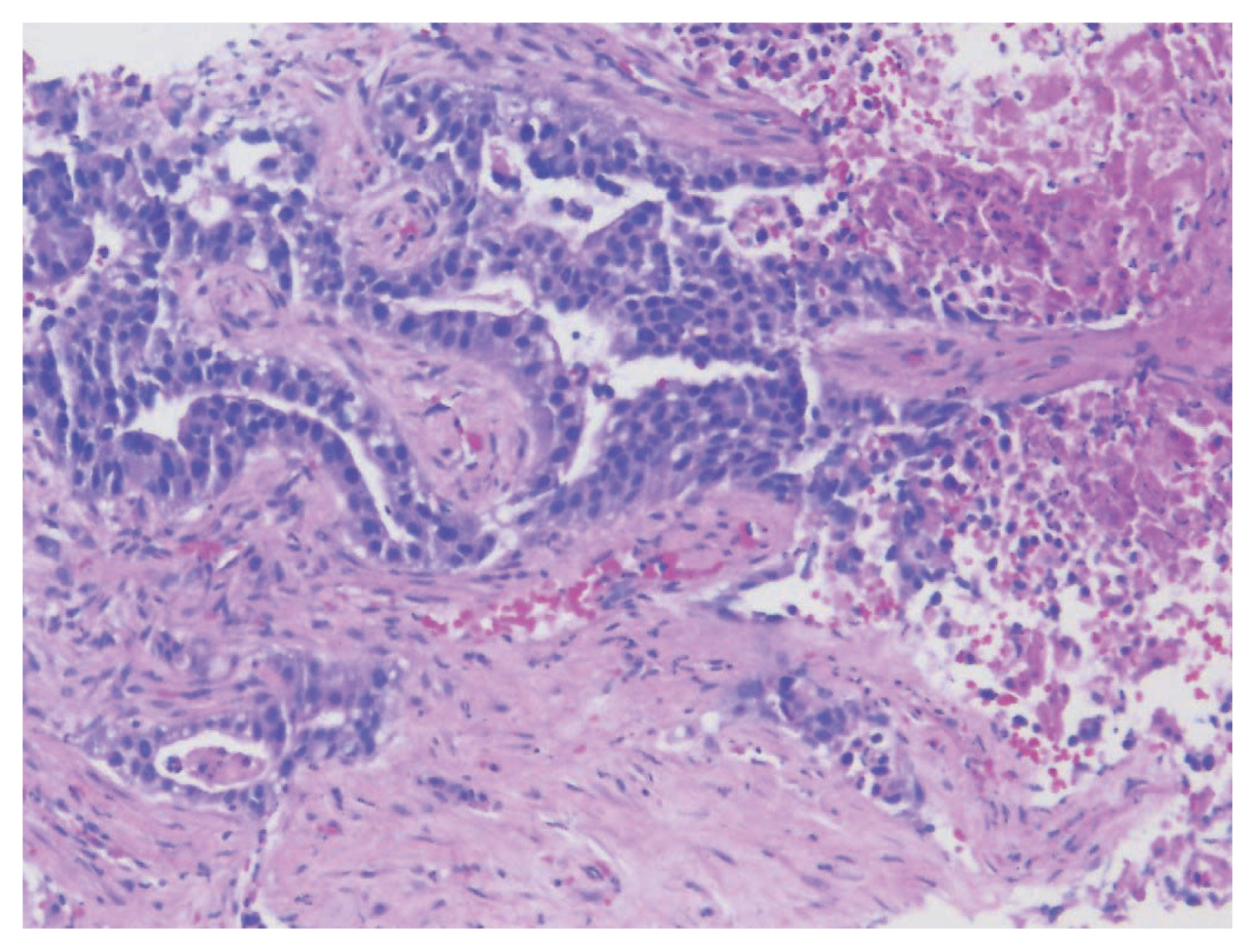
PDF - Colorectal adenocarcinoma with enteroblastic differentiation (CAED) is a rare subtype of colonic adenocarcinoma characterized by increased α-fetoprotein (AFP) production and the expression of at least one enteroblastic marker including AFP, glypican 3 (GPC3), or Spalt like transcription factor 4 (SALL4). We report a case of a 26-year-old female who presented with low back pain and constipation which persisted despite supportive measures. Imaging revealed multiple liver lesions and enlarged retroperitoneal nodes. Tumor markers including AFP were markedly elevated. On biopsy, samples from the liver revealed infiltrating glands lined by columnar-type epithelium with mostly eosinophilic granular to focally clear cytoplasm. By immunohistochemistry, the tumor showed immunoreactivity with AFP, hepatocyte antigen, GPC3, SALL4, CDX2, SATB2, and cytokeratin 20. A colonoscopy performed subsequently revealed a mass in the sigmoid colon and biopsy of this mass revealed a similar histology as that seen in the liver. A diagnosis of CAED was made, following the results of gene expression profiling by the tumor with next-generation sequencing which identified pathogenic variants in MUTYH, TP53, and KDM6A genes and therefore supported its colonic origin. Cases such as this underscores the use of ancillary diagnostic techniques in arriving at the correct diagnosis in lesions with overlapping clinicopathologic characteristics.
-
Citations
Citations to this article as recorded by
- Colorectal Adenocarcinoma with Enteroblastic Differentiation: A Report of Two Cases
Yusuke Nakamura, Hiroyuki Fukuda, Yasuo Ishida
Nihon Daicho Komonbyo Gakkai Zasshi.2026; 79(2): 82. CrossRef - Raised alpha fetoprotein in cirrhotic liver with colorectal liver metastasis mimicking hepatocellular carcinoma: case report and review of literature
Aboje Adugba, Dominic Obotu Ayegba, Mansour Al Moundhri, Kareem Al Rezk, Rawan AlMallah, Ramesh Babu Telugu, Abdallah Al Farai
Diagnosis.2026;[Epub] CrossRef - Enteroblastic and Hepatoid Colorectal Carcinomas are Aggressive Cancers With a Distinctive Immunophenotype, While Clear Cell Carcinoma Appears to Represent Clear Cell Change in Conventional Colorectal Cancer
Fahad Khan, Alexandros D. Polydorides, John D. Paulsen, Wei Chen, Jennifer Vazzano, Aaron Huber, David Papke, Nima Sharifai, Kristen Stashek, Ignacio Ruz-Caracuel, Daniela Allende, Kelsey McHugh, Jinru Shia, Teri A. Longacre, Deepti Dhall, Elias Makhoul,
American Journal of Surgical Pathology.2026;[Epub] CrossRef - Exploring the Multifunctional Role of Alpha-Fetoprotein in Cancer Progression: Implications for Targeted Therapy in Hepatocellular Carcinoma and Beyond
Hyunjung Kim, Minji Jang, Eunmi Kim
International Journal of Molecular Sciences.2025; 26(10): 4863. CrossRef - Rectal adenocarcinoma with a yolk sac tumor component: A rare case report and review of the literature
Sato Nishida, Tomohiro Takeda, Tatsuya Shonaka, Shoichiro Mizukami, Masahide Otani, Mizuho Ohara, Chikayoshi Tani, Kimiharu Hasegawa, Yuki Kamikokura, Mishie Tanino, Hideki Yokoo
International Cancer Conference Journal.2025; 15(1): 138. CrossRef - SALL4 in gastrointestinal tract cancers: upstream and downstream regulatory mechanisms
Tairan Wang, Yan Jin, Mengyao Wang, Boya Chen, Jinyu Sun, Jiaying Zhang, Hui Yang, Xinyao Deng, Xingyue Cao, Lidong Wang, Yuanyuan Tang
Molecular Medicine.2024;[Epub] CrossRef - Gastric adenocarcinoma with enteroblastic differentiation in a
67-year-old man in Korea: a case report
Hae Rin Lee, Gwang Ha Kim, Dong Chan Joo, Moon Won Lee, Bong Eun Lee, Kyung Bin Kim
The Ewha Medical Journal.2024;[Epub] CrossRef - Colorectal adenocarcinoma with clear cell changes: immunohistological and molecular findings in three cases
Andreas Gocht, Carsten Heidel, Jutta Kirfel, Rita Vesce, Pamela Lazar-Karsten, Helen Pasternack, Madelaine Melzer, Phillip Hildebrand, Nicole Warkentin, Hendrik Schimmelpenning, Verena-Wilbeth Sailer
Virchows Archiv.2024; 485(3): 569. CrossRef - Ureteral Metastasis of Colonic Adenocarcinoma with Enteroblastic Differentiation: A Rare Case to be Distinguished from Clear Cell Adenocarcinoma of the Urinary Tract
Hiroshi Minato, Akane Yoshikawa, Sho Tsuyama, Kazuyoshi Katayanagi, Kengo Hayashi, Yusuke Sakimura, Hiroyuki Bando, Tomohiro Hori, Yosuke Kito
International Journal of Surgical Pathology.2023; 31(8): 1553. CrossRef - Beyond liver cancer, more application scenarios for alpha-fetoprotein in clinical practice
Chenyu Ma, Yuexinzi Jin, Yuhan Wang, Huaguo Xu, Jiexin Zhang
Frontiers in Oncology.2023;[Epub] CrossRef - AIEgens assisted label free DNA supersandwich immunoassay for ultrasensitive α-fetoprotein detection
Xiaowen Ou, Jingman Dai, Yiting Huang, Xiaoqin Xiong, Zhi Zheng, Xiaoding Lou, Fan Xia
Giant.2022; 11: 100110. CrossRef - Rectal carcinoma with dual differentiation toward enteroblastic and neuroendocrine features arising in a patient with ulcerative colitis: a case report
Takako Kihara, Ryuichi Kuwahara, Kurando Kusunoki, Tomohiro Minagawa, Yuki Horio, Motoi Uchino, Hiroki Ikeuchi, Seiichi Hirota
World Journal of Surgical Oncology.2022;[Epub] CrossRef
- Colorectal Adenocarcinoma with Enteroblastic Differentiation: A Report of Two Cases
- Fallopian Metaplastic Papillary Tumour: An Atypical Transdifferentiation of the Tubal Epithelium?
- Miguel Fdo. Salazar, Isaías Estrada Moscoso, Lorena Troncoso Vázquez, Nubia Leticia López García, Paola Andrea Escalante Abril
- J Pathol Transl Med. 2015;49(2):148-155. Published online March 12, 2015
- DOI: https://doi.org/10.4132/jptm.2014.10.15
- 10,974 View
- 60 Download
- 3 Web of Science
- 3 Crossref
-
Abstract
PDF
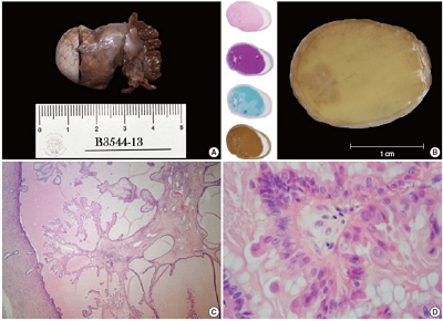
- A metaplastic papillary tumor of the Fallopian tube is an extremely uncommon condition, with odd and confusing features that make it difficult to categorize as benign or borderline. Here, we summarize all the published cases to date and document the case of a 41-year-old woman diagnosed with this alteration after her last childbirth and ensuing tubal ligation. One of the tubes was bulky and filled with a caramel-like substance encircling a blurry spot. Light microscopy detailed a slender stalk covered by eosinophilic, columnar plump cells, showing atypical nuclei and focal budding. Mitotic figures were absent. The immunohistochemistry panel was positive for pan-cytokeratin, epithelial membrane antigen, cyclin D1, and hormone receptors. Additionally, a proliferation index of less than 5% was rated using Ki-67. The true nature of this tumor (reactive vs neoplastic) is uncertain. Nonetheless, its association with pregnancy suggests an adaptive change, likely similar to the atypical transdifferentiation proposed for Arias-Stella reaction.
-
Citations
Citations to this article as recorded by- Ungewöhnliche Proliferation des Eileiters
Angelina Vlaški, Vanessa Neukunft, Andrea Maria Gassel, Frederick Klauschen, Doris Mayr
Die Pathologie.2025; 46(1): 56. CrossRef - Fallopian tube papilloma
Shashank Mishra, Prerna Guleria
Indian Journal of Pathology and Microbiology.2021; 64(3): 608. CrossRef - Metaplastic papillary tumour of the fallopian tube, a rare entity, analysed by next‐generation sequencing
Sandra Sunitsch, Julia Reisinger, Luca Abete, Karl Kashofer, Peter Regitnig
Histopathology.2020; 76(6): 923. CrossRef
- Ungewöhnliche Proliferation des Eileiters
- Oncocytic Lipoadenoma: A Rare Case of Parotid Gland Tumor and Review of the Literature
- Chen-lin Chi, Tseng-tong Kuo, Li-yu Lee
- J Pathol Transl Med. 2015;49(2):144-147. Published online March 12, 2015
- DOI: https://doi.org/10.4132/jptm.2014.02.10
- 11,782 View
- 69 Download
- 8 Web of Science
- 9 Crossref
-
Abstract
PDF
- Oncocytic lipoadenoma is a rare tumor, with only 18 cases having been reported since the first in 1998. We encountered a case of oncocytic lipoadenoma presenting as a slowly growing parotid mass in a 71-year-old man. This tumor is characteristically comprised of a mixture of oncocytes and adipocytes. The present case is one of five reported cases of oncocytic lipoadenoma showing sebaceous differentiation. The results of immunohistochemical study with DOG1 antibody supported the origination of this tumor in the striated duct.
-
Citations
Citations to this article as recorded by- Multimodal Imaging of Oncocytic Lipoadenoma Arising from the Parotid Deep Lobe with Medial Extension into the Parapharyngeal Space: A Case Report with Histopathologic Findings and Literature Review
Jong-Uk Lee, Hye Jin Baek, Kwang Ho Choi, Eun Cho, Hyo Jung An
Diagnostics.2026; 16(9): 1366. CrossRef - Oncocytic lipoadenoma of the parotid gland: a case report and a review of the literature
Jood K Alotaibi, Turki Mohammed Almuhaimid, Ghada Abdallah Moumneh
Journal of Surgical Case Reports.2024;[Epub] CrossRef - Oncocytic sialolipoma of parotid gland: Case report and literature review
VenuPatel Sureja, KoyyeRavindranath Tagore
Indian Journal of Pathology and Microbiology.2023; 66(3): 591. CrossRef - Complex Component of Oncocytic and Non-Oncocytic Lipoadenomas in the Parotid Gland: A Case Report
Fuyuki Sato, Takashi Nakajima, Takashi Sugino
Diagnostics.2021; 11(8): 1478. CrossRef - Oncocyitic lipoadenoma of the parotid gland
Renato PIANTANIDA, Alberto CARANTI, Adele CHIESA, Jessica BARIZZI, Ulrike PERRIARD, Filippo BARUCCA, Antonio PELLANDA
Otorinolaringologia.2021;[Epub] CrossRef - A case of oncocytic lipoadenoma of the submandibular gland and its diagnostic cytology challenges
Khaled A. Murshed, Ammar Khalafalla, Belal Alani, Hanan Farghaly, Moustafa Alkhalil
Diagnostic Cytopathology.2020; 48(4): 364. CrossRef - An extremely rare case of giant oncocytic adenolipoma of the parotid gland
Dipesh Shakya, Ajit Nepal
Clinical Case Reports.2020; 8(12): 2390. CrossRef - A rare cause of primary hyperparathyroidism: Parathyroid lipoadenoma
Sabri Özden, Servet Güreşci, Barış Saylam, Gül Dağlar
Auris Nasus Larynx.2018; 45(6): 1245. CrossRef - Oncocytic osteolipoadenoma of the submandibular gland
Domenico Corradi, Rodolfo Monaco, Giulia D'Angelo, Paola Bini, Teore Ferri, Enrico M Silini
Histopathology.2016; 69(1): 148. CrossRef
- Multimodal Imaging of Oncocytic Lipoadenoma Arising from the Parotid Deep Lobe with Medial Extension into the Parapharyngeal Space: A Case Report with Histopathologic Findings and Literature Review
- Development of Six Tumors in a Sebaceus Nevus of Jadassohn: Report of a Case
- Serap Gozel, Melahat Donmez, Noyan Can Akdur, Hulya Yikilkan
- Korean J Pathol. 2013;47(6):569-574. Published online December 24, 2013
- DOI: https://doi.org/10.4132/KoreanJPathol.2013.47.6.569
- 11,439 View
- 96 Download
- 22 Crossref
-
Abstract
PDF
Nevus sebaceus of Jadassohn is a congenital cutaneous hamartoma comprised of multiple skin structures. It has the potential to develop into variety of neoplasms of various epidermal adnexal origins. While multiple tumors may occasionally arise, it is unusual for more than four tumors to arise simultaneously within a single sebaceus nevus. Here in, we report a case of a 70-year-old woman with six neoplastic proliferations including a syringocystadenoma papilliferum, pigmented trichoblastoma, tubular apocrine adenoma, sebaceoma, tumors of follicular infundibulum and superficial epithelioma with sebaceus differentiation arising in a long standing nevus sebaceus on the scalp. Our case is extraordinary because a single nevus sebaceus contained six neoplastic proliferations with differentiation toward the folliculosebaceous-apocrine unit.
-
Citations
Citations to this article as recorded by- Melanotrichoblastoma Arising on Nevus Sebaceous: A Rare Occurence
Apaopa J. Thekho, Deepika Uikey, Shanta Passi, V. Ramesh
Indian Journal of Dermatology.2025; 70(2): 105. CrossRef - Co-occurrence of Tubular Apocrine Adenoma and Syringocystadenoma Papilliferum over the Hypogastrium: A Rare Case Report
R Raghunatha Reddy, Mukunda Ranga Swaroop, Yogesh Devaraj, Greeshma Jagadish, Namratha Govindaraju
Clinical Dermatology Review.2025; 9(1): 69. CrossRef - Tumor of follicular infundibulum – reappraisal in a series of 28 patients with critical review of the literature
Michael Wilk, Bettina G. Zelger, Bernhard Zelger
JDDG: Journal der Deutschen Dermatologischen Gesellschaft.2024; 22(2): 223. CrossRef - Tumor des follikulären Infundibulums – Neubewertung in einer Serie von 28 Patienten mit kritischer Analyse der Literatur
Michael Wilk, Bettina G. Zelger, Bernhard Zelger
JDDG: Journal der Deutschen Dermatologischen Gesellschaft.2024; 22(2): 223. CrossRef - Adnexal neoplasms of the eye
Roman Drozdowski, Jane M. Grant-Kels, Madina Falcone, Campbell L. Stewart
Clinics in Dermatology.2024; 42(4): 321. CrossRef - Melanotrichoblastoma: sixth case report in the literature
Juliana Polizel Ocanha-Xavier, José Cândido Caldeira Xavier-Júnior
Anais Brasileiros de Dermatologia.2023; 98(6): 871. CrossRef - Multiple secondary neoplasms in nevus sebaceus excision
Travis S. Dowdle, David A. Mehegran, Dylan Maldonado, Cort D. McCaughey
Baylor University Medical Center Proceedings.2022; 35(2): 241. CrossRef - Congenital tumors arising from nevus sebaceous in 2 neonates
Lynette Wei Yi Wee, Bori Born, Sharon Mun Yee Wong, Hui-Ling Chia, Sithach Mey, Suresh Chandran, Mark Jean Aan Koh
JAAD Case Reports.2022; 21: 70. CrossRef - Development of seven secondary neoplasms in a nevus sebaceous: a case report and literature review
Yi-Wen Kuo, Jung-Chia Lin, Wei-Hsuan Tsai
Archives of Craniofacial Surgery.2022; 23(2): 83. CrossRef - Multiple rare neoplasms arising from the nevus sebaceous of the scalp: A case report
Deepthi Shetty, Anilkumar Desai, Niranjan Kumar, Dinesh U.S., Aditya Agnihotri, Saurav Bhaduri
Gulhane Medical Journal.2022; 64(2): 197. CrossRef - Syringocystadenoma Papilliferum and Basal Cell Carcinoma Arising in Nevus Sebaceous
Jingjing Jiang, Yujuan Chen, Qi He, Jiao Yang, Zhengzhong Zhang, Hao Yang, Huan Zhang, Chuan Yang
Clinical, Cosmetic and Investigational Dermatology.2022; Volume 15: 2021. CrossRef - Eyelid trichoblastoma – A case series
Gunja Chowdhury, Meghana Tanwar, Usha Kim, Shanthi R. Krishnan
Journal of Clinical Ophthalmology and Research.2021; 9(3): 123. CrossRef - Trilogy Revisited
Anand Bardia, Debajyoti Chatterjee, Keshavamurthy Vinay
Indian Dermatology Online Journal.2021; 12(4): 577. CrossRef - Trichilemmoma coexisting with sebaceous nevus
AngooriG Rao, VangaliS Reddy, M Tejal, M Divya
Indian Dermatology Online Journal.2020; 11(2): 253. CrossRef - Syndromic sebaceous nevus: current findings
Oumama El Ezzi, Anthony S. de Buys Roessingh, Michèle Bigorre, Guillaume Captier
International Journal of Dermatology.2018; 57(5): 599. CrossRef - Syringocystadenoma papilliferum and trichoblastoma arising in the nevus sebaceous
Feifei Wang, Yatong Wu, Zhancai Zheng, Yanping Bai
Indian Journal of Pathology and Microbiology.2018; 61(1): 106. CrossRef - Dermoscopic Analysis of Nevus Sebaceus of Jadassohn: A Study of 13 Cases
Awatef Kelati, Hanane Baybay, Salim Gallouj, Fatima Zahra Mernissi
Skin Appendage Disorders.2017; 3(2): 83. CrossRef - Secondary neoplasms arising from nevus sebaceus: A retrospective study of 450 cases in Taiwan
Ming‐Chun Hsu, Jau‐Yu Liau, Jin‐Liern Hong, Yin Cheng, Yi‐Hua Liao, Jau‐Shiuh Chen, Yi‐Shuan Sheen, Jin‐Bon Hong
The Journal of Dermatology.2016; 43(2): 175. CrossRef - A Histological Snapshot of Hypothetical Multistep Progression From Nevus Sebaceus to Invasive Syringocystadenocarcinoma Papilliferum
Vishwas Parekh, Cesar E. Guerrero, Charles F. Knapp, Craig A. Elmets, Kristopher M. McKay
The American Journal of Dermatopathology.2016; 38(1): 56. CrossRef - Trichoblastoma, syringocystadenoma papilliferum, desmoplastic trichilemmoma and tumor of the follicular infundibulum with signet‐ring cells, all arising in nevus sebaceus
Emilie Dore, Megan H. Noe, Brian L. Swick
Journal of Cutaneous Pathology.2015; 42(9): 645. CrossRef - Ceruminous adenoma (ceruminoma) arising in a nevus sebaceus of Jadassohn within the external auditory canal of a 3 year-old boy – A case report
Elżbieta Niemczyk, Kazimierz Niemczyk, Jadwiga Małdyk, Lidia Zawadzka-Głos
International Journal of Pediatric Otorhinolaryngology.2015; 79(11): 1932. CrossRef - Fehlbildungen und Nävi des behaarten Kopfes
V. Behle, H. Hamm
Der Hautarzt.2014; 65(12): 1022. CrossRef
- Melanotrichoblastoma Arising on Nevus Sebaceous: A Rare Occurence
- Myxoid Liposarcoma with Cartilaginous Differentiation: A Case Study with Cytogenetical Analysis
- Hyunchul Kim, Won Hwangbo, Sangjeong Ahn, Suhjin Kim, Insun Kim, Chul Hwan Kim
- Korean J Pathol. 2013;47(3):284-288. Published online June 25, 2013
- DOI: https://doi.org/10.4132/KoreanJPathol.2013.47.3.284
- 10,083 View
- 43 Download
- 3 Crossref
-
Abstract
PDF
Myxoid liposarcoma is a subtype of liposarcoma. This specific subtype can be identified based on its characteristic histological and cytogenetical features. The tumor has a fusion transcript of the
CHOP andTLS genes, which is caused by t(12;16)(q13;p11). Most of the fusion transcripts that have been identified fall into three categories, specifically type I (exons 7-2), type II (exons 5-2), and type III (exons 8-2). A total of seven myxoid liposarcomas associated with the rare phenomenon of cartilaginous differentiation have been documented in the literature. Currently, only one of these cases has been cytogenetically analyzed, and the analysis indicated that it was a type IITLS-CHOP fusion transcript in both the typical myxoid liposarcoma and cartilaginous areas. This study presents a second report of myxoid liposarcoma with cartilaginous differentiation, and includes a cytogenetical analysis of both the myxoid and cartilaginous areas.-
Citations
Citations to this article as recorded by- Myxoid liposarcoma with nuclear pleomorphism: a clinicopathological and molecular study
Naoki Kojima, Takashi Kubo, Taisuke Mori, Kaishi Satomi, Yuko Matsushita, Shintaro Iwata, Yasushi Yatabe, Koichi Ichimura, Akira Kawai, Hitoshi Ichikawa, Akihiko Yoshida
Virchows Archiv.2024; 484(1): 71. CrossRef - The Conundrum of Dedifferentiation in a Liposarcoma at a Peculiar Location: A Case Report and Literature Review
Ana-Maria Ciongariu, Adrian-Vasile Dumitru, Cătălin Cîrstoiu, Bogdan Crețu, Maria Sajin, Dana-Antonia Țăpoi, Aminia-Diana Ciobănoiu, Adrian Bejenariu, Andrei Marin, Mariana Costache
Medicina.2023; 59(5): 967. CrossRef - Myxoid liposarcoma with cartilaginous differentiation showing DDIT3 rearrangement
Kayo Suzuki, Taketoshi Yasuda, Kenta Watanabe, Takeshi Hori, Masahiko Kanamori, Tomoatsu Kimura
Oncology Letters.2017;[Epub] CrossRef
- Myxoid liposarcoma with nuclear pleomorphism: a clinicopathological and molecular study
- Gastric Adenocarcinoma of Fundic Gland Type: Report of Three Cases
- Eun Su Park, Young Eun Kim, Cheol Keun Park, Takashi Yao, Ryoji Kushima, Kyoung-Mee Kim
- Korean J Pathol. 2012;46(3):287-291. Published online June 22, 2012
- DOI: https://doi.org/10.4132/KoreanJPathol.2012.46.3.287
- 13,261 View
- 117 Download
- 25 Crossref
-
Abstract
PDF
Recently, fundic gland type gastric adenocarcinoma (GA-FG) has been reported as a new entity. This report describes GA-FG among Koreans for the first time. From March 2008 to July 2010 we identified only three cases of GA-FG out of over 6,000 GAs resected by endoscopy or surgery. Cell differentiation by mucin proteins, pepsinogen-I, and H+/K+-ATPase was evaluated. All three cases were male patients and diagnosed as early stage GA. Histologically, GA-FGs were well-differentiated adenocarcinoma with pale gray-blue, basophilic columnar or cuboidal cells and mildly enlarged nuclei, resembling chief cells. All three cases were positive for pepsinogen-I and were classified as gastric mucin phenotype. Among three histologic subtypes of GA-FG, since tumors were mainly composed of chief cells, our three cases were classified as chief cell predominant type. In conclusion, GA-FG is very rare among Koreans and pepsinogen-I and MUC6 expression are typical immunohistochemical findings in GA-FG suggesting differentiation toward fundic glands.
-
Citations
Citations to this article as recorded by- Endoscopic Submucosal Dissection of Early Gastric Adenocarcinoma of Fundic Gland Type: A Case Report
Ming Zhong, Wei Wei, Huang Zhong, Hang Gong, Tingyu Wang
Revista Española de Enfermedades Digestivas.2025;[Epub] CrossRef - Oxyntic Gland Neoplasms - From Adenoma to Advanced Gastric Cancer: A Review of 29 Cases
Gi Hwan Kim, Jun Su Lee, Jeong Hoon Lee, Young Soo Park
Journal of Gastric Cancer.2024; 24(4): 378. CrossRef - Transcriptome analysis reveals the essential role of NK2 homeobox 1/thyroid transcription factor 1 (NKX2-1/TTF-1) in gastric adenocarcinoma of fundic-gland type
Kazushi Fukagawa, Yu Takahashi, Nobutake Yamamichi, Natsuko Kageyama-Yahara, Yoshiki Sakaguchi, Miho Obata, Rina Cho, Nobuyuki Sakuma, Sayaka Nagao, Yuko Miura, Naoki Tamura, Daisuke Ohki, Hiroya Mizutani, Seiichi Yakabi, Chihiro Minatsuki, Keiko Niimi, Y
Gastric Cancer.2023; 26(1): 44. CrossRef - Clinicopathological Features and the Prevalence of Oxyntic Gland Neoplasm: A Single-center Retrospective Study
Hikari Asahara, Toshitatsu Takao, Yumiko Asahara, Masakyo Asahara, Douglas Motomura, Hiroya Sakaguchi, Tetsuya Yoshizaki, Nobuaki Ikezawa, Madoka Takao, Yoshinori Morita, Takashi Toyonaga, Masato Komatsu, Ryoji Kushima, Yuzo Kodama
Internal Medicine.2023; 62(19): 2763. CrossRef - Clinicopathological features of gastric adenocarcinoma of fundic gland type
Bao-Zhen Guo, Zhen-Zhen Liu, Gao-Fei Shen, Fei Zhu, Hui-Fen Lian, Xin Li, Jun-Yi Zheng, Jin-Peng Li, Shui-Miao Deng, Rui Huang
World Chinese Journal of Digestology.2023; 31(6): 244. CrossRef - Endoscopic Resection for Gastric Adenocarcinoma of the Fundic Gland Type: A Case Series
Hwa Jin Lee, Gwang Ha Kim, Dong Chan Joo, Moon Won Lee, Bong Eun Lee, Kyungbin Kim
The Korean Journal of Gastroenterology.2023; 81(6): 259. CrossRef - Gastric adenocarcinoma of the fundic gland type: A review of the literature
Zhiyong Zhai, Wei Hu, Zhaoyu Huang, Zemin Chen, Sicun Lu, Wei Gong
JGH Open.2023; 7(12): 812. CrossRef - Clinicopathological features of early stage gastric adenocarcinoma of fundic gland type
Huan Zhang, Shuyan Wang, Yongping Zhang, Fusang Ye, Chunnian Wang
Medicine.2022; 101(2): e28469. CrossRef - Gastric Adenocarcinoma of Fundic Gland Type Treated by Endoscopic Submucosal Dissection
Yong Bo Park, Gwang Ha Kim, Kyungbin Kim, Tae Kyoung Ha, Guk Bin Park, Young Min Kwak
The Korean Journal of Helicobacter and Upper Gastrointestinal Research.2021; 21(1): 82. CrossRef - Gastric epithelial neoplasm of fundic-gland mucosa lineage: proposal for a new classification in association with gastric adenocarcinoma of fundic-gland type
Hiroya Ueyama, Takashi Yao, Yoichi Akazawa, Takuo Hayashi, Koichi Kurahara, Yumi Oshiro, Masayoshi Yamada, Ichiro Oda, Shin Fujioka, Chiaki Kusumoto, Masayoshi Fukuda, Kunihisa Uchita, Tomohiro Kadota, Yasuhiro Oono, Kazuhisa Okamoto, Kazunari Murakami, Y
Journal of Gastroenterology.2021; 56(9): 814. CrossRef - Endoscopic resection is a suitable initial treatment strategy for oxyntic gland adenoma or gastric adenocarcinoma of the fundic gland type
Masaya Iwamuro, Chiaki Kusumoto, Masahiro Nakagawa, Sayo Kobayashi, Masao Yoshioka, Tomoki Inaba, Tatsuya Toyokawa, Shinichiro Hori, Shouichi Tanaka, Kazuhiro Matsueda, Takehiro Tanaka, Hiroyuki Okada
Scientific Reports.2021;[Epub] CrossRef - A series of five patients with oxyntic gland adenoma: Deciphering the clinical and histological features of these rare gastric polyps
Jerry C. Nagaputra, Tracy Jie Zhen Loh, Sangeeta Mantoo, Rafay Azhar, Vikneswaran Namasivayam, Wei Qiang Leow
Human Pathology Reports.2021; 26: 300566. CrossRef - Gastric adenocarcinoma of the fundic gland: A review of clinicopathological characteristics, treatment and prognosis
Xiang-yu Meng, Guang Yang, Cheng-ji Dong, Ru-yi Zheng
Rare Tumors.2021;[Epub] CrossRef - Gastric adenocarcinoma of the fundic gland type: clinicopathological features of eight patients treated with endoscopic submucosal dissection
Chengfang Li, Xinglong Wu, Shuang Yang, Xiaorong Yang, Jin Yao, Hong Zheng
Diagnostic Pathology.2020;[Epub] CrossRef - Multiple gastric adenocarcinoma of fundic gland type: A case report
Ou Chen, Ze-Yong Shao, Xiong Qiu, Guang-Ping Zhang
World Journal of Clinical Cases.2019; 7(18): 2871. CrossRef - Gastric Adenocarcinoma of the Fundic Gland Type
Mark A Benedict, Gregory Y Lauwers, Dhanpat Jain
American Journal of Clinical Pathology.2018; 149(6): 461. CrossRef - Oxyntic Gland Adenoma Treated by Endoscopic Mucosal Resection
In Ji Song, Jin Woo Joo, Jun Chul Park, Sung Kwan Shin, Yong Chan Lee, Sang Kil Lee
The Korean Journal of Helicobacter and Upper Gastrointestinal Research.2017; 17(2): 94. CrossRef - Chief cell‐predominant gastric polyps: a series of 12 cases with literature review
Karen Chan, Ian S Brown, Trevor Kyle, Gregory Y Lauwers, Marian Priyanthi Kumarasinghe
Histopathology.2016; 68(6): 825. CrossRef - Twelve-year natural history of a gastric adenocarcinoma of fundic gland type
Yoshinori Sato, Takashi Fujino, Akira Kasagawa, Ryo Morita, Shun-ichiro Ozawa, Yasumasa Matsuo, Tadateru Maehata, Hiroshi Yasuda, Masayuki Takagi, Fumio Itoh
Clinical Journal of Gastroenterology.2016; 9(6): 345. CrossRef - Clinicopathological features of gastric adenocarcinoma of the fundic gland (chief cell predominant type) by retrospective and prospective analyses of endoscopic findings
Takashi Chiba, Katsuaki Kato, Takayuki Masuda, Shuichi Ohara, Noriyuki Iwama, Takenobu Shimada, Daisuke Shibuya
Digestive Endoscopy.2016; 28(7): 722. CrossRef - Gastric Adenocarcinoma of the Fundic Gland Type Treated by Endoscopic Mucosal Resection: A Case Report and Review of the Literature
Eleanor Lewin, Philip Daroca, Sanjay Sikka, Tong Wu, Yukihiro Nakanishi
Case Reports in Pathology.2016; 2016: 1. CrossRef - Gastric adenocarcinoma of the fundic gland (chief cell-predominant type): A review of endoscopic and clinicopathological features
Masaki Miyazawa, Mitsuru Matsuda, Masaaki Yano, Yasumasa Hara, Fumitaka Arihara, Yosuke Horita, Koichiro Matsuda, Akito Sakai, Yatsugi Noda
World Journal of Gastroenterology.2016; 22(48): 10523. CrossRef - Oxyntic gland adenoma endoscopically mimicking a gastric neuroendocrine tumor: A case report
Tae-In Lee
World Journal of Gastroenterology.2015; 21(16): 5099. CrossRef - Oxyntic gland polyp/adenoma
Rajkumar Vajpeyi, Jyoti Dekate
Diagnostic Histopathology.2014; 20(11): 446. CrossRef - Gastric adenocarcinoma of fundic gland type with unusual behavior
Tetsuya Ueo, Hirotoshi Yonemasu, Tetsuya Ishida
Digestive Endoscopy.2014; 26(2): 293. CrossRef
- Endoscopic Submucosal Dissection of Early Gastric Adenocarcinoma of Fundic Gland Type: A Case Report
- Dedifferentiated Extraskeletal Myxoid Chondrosarcoma of the Masticator Space: A Case Report.
- Geunyoung Jung, Kyung Ja Cho, Seung Ho Choi, Mi Jung Kim
- Korean J Pathol. 2011;45:S101-S105.
- DOI: https://doi.org/10.4132/KoreanJPathol.2011.45.S1.S101
- 4,984 View
- 36 Download
- 3 Crossref
-
Abstract
PDF
- We describe a 69-year-old woman who presented with a dedifferentiated extraskeletal myxoid chondrosarcoma arising in the left masticator space. Computed tomography and magnetic resonance imaging revealed a 5 cm sized mass in the left masticator space. Histologically, the tumor consisted of two distinct areas. The less cellular area was a low-grade extraskeletal myxoid chondrosarcoma, composed of strands or cords of uniform spindle cells and abundant myxoid stroma. The more cellular, dedifferentiated area corresponded to a high grade myxofibrosarcoma, consisting of anaplastic tumor cells in myxoid stroma and geographic necrosis. The tumor cells of the former area were positive for S-100 protein, microtubule-associated protein-2 (MAP-2) and class III beta-tubulin, but negative for cytokeratin, smooth muscle actin, and desmin. The tumor cells in the latter, pleomorphic area showed MAP-2 and beta-tubulin immunoreactivity with a high Ki-67 labeling index. Based on its histologic and immunohistochemical features, the tumor was considered a dedifferentiated extraskeletal myxoid chondrosarcoma.
-
Citations
Citations to this article as recorded by- Extraskeletal myxoid chondrosarcoma of the parotid gland
NyimiBushabu Fidele, Wu Tianfu, Bing Liu, Yanfang Sun, Zhao Yifang
Annals of Maxillofacial Surgery.2019; 9(2): 439. CrossRef - Myxoid chondrosarcoma of maxilla: A rare case report
Hiralal Ash, Ajoy Kumar Shahi, Kabita Chatterjee, Dipankar Samaddar
Journal of Oral and Maxillofacial Surgery, Medicine, and Pathology.2016; 28(3): 273. CrossRef - Maxillo-facial Extraskeletal Myxoid Chondrosarcoma: A Case Report and Discussion
Ratnadeep Ganguly, Abhishek Mukherjee
The Korean Journal of Pathology.2011; 45(6): 639. CrossRef
- Extraskeletal myxoid chondrosarcoma of the parotid gland
- Diagnostic Utility of the JAZF1/JJAZ1 Gene Fusion in Endometrial Stromal Sarcomas and Their Histologic Variants.
- Sang Ryung Lee, Joon Seon Song, Ga Hye Kim, Jene Choi, Hyung Kyoung Kim, Yonghee Lee, Kyu Rae Kim
- Korean J Pathol. 2011;45(5):498-505.
- DOI: https://doi.org/10.4132/KoreanJPathol.2011.45.5.498
- 3,974 View
- 32 Download
-
Abstract
PDF
- BACKGROUND
The diagnosis of endometrial stromal sarcoma (ESS) is often difficult in cases showing diverse histological differentiation or in undifferentiated endometrial sarcoma (UES). Recently, JAZF1/JJAZ1 gene fusion has been described as a defining feature of low-grade ESS (LGESS). However, its prevalence is variably reported, and the diagnostic utility has rarely been examined for cases showing various histological differentiation.
METHODS
To test the diagnostic utility of JAZF1/JJAZ1 gene fusion in difficult cases, we compared the prevalence of the JAZF1/JJAZ1 fusion gene in LGESS with and without histological differentiation.
RESULTS
The JAZF1/JJAZ1 fusion transcript was detected in 18 of 21 LGESS (85.7%), including 14 classical LGESS (93%), four LGESS with diverse histological differentiation (67%), and two with UES (28.6%). Positive cases included two LGESS with sex cord-like differentiation, one with osseous differentiation, and two UES. LGESS showing smooth muscle differentiation revealed the fusion transcript only in the classic area. Direct sequencing analysis of two LGESS revealed a previously reported breakpoint at t(7;17)(p15;q21).
CONCLUSIONS
The JAZF1/JJAZ1 fusion gene was identified in a significant proportion of LGESS showing secondary histological differentiation except in cases with smooth muscle differentiation. Thus, this fusion gene may be useful to confirm the diagnosis in difficult cases of LGESS.
- Growth Differentiation Factor 5 (GDF5) Core Promoter Polymorphism Is Not Associated with Susceptibility to Osteoarthritis of the Knee in the Korean Population.
- Zhang Cao, Hwa Sung Lee, Jae Hwi Song, Jeong Whan Yoon, Yong Kyu Park, Suk Woo Nam, Jung Young Lee, Won Sang Park
- Korean J Pathol. 2010;44(4):404-409.
- DOI: https://doi.org/10.4132/KoreanJPathol.2010.44.4.404
- 5,632 View
- 36 Download
- 10 Crossref
-
Abstract
PDF
- BACKGROUND
Osteoarthritis (OA) is a common disease characterized by degenerating joint cartilage in the knee, hip, and hand. A functional single nucleotide polymorphism (SNP) +104T/C; rs143383 in the 5' untranslated region of the growth differentiation factor 5 (GDF5) gene was recently associated with susceptibility to OA in the Japanese and Chinese populations.
METHODS
To investigate whether this association is present in the Korean population, the frequency of the polymorphism was investigated in 276 patients with knee OA and 298 healthy subjects as controls. Polymorphism analysis was performed by amplifying the core promoter region of the GDF5 gene and digesting it with the BsiEI restriction enzyme.
RESULTS
The frequency of the TT, CT, and CC genotypes was 54.3% (150/276), 41.7% (115/276), and 4.0% (11/276), respectively, in patients with OA, and 53.4% (159/298), 37.9% (113/298), and 8.7% (26/298), respectively, in healthy controls. No significant differences in genotypic or allelic frequencies of the +104T/C SNP of the GDF5 gene were observed between patients with OA and controls. Also, no significant differences in allelic and genotypic frequencies were found when the individuals were stratified by age and gender.
CONCLUSIONS
The results suggest that the +104T/C; rs143383 GDF5 core promoter polymorphism is not a risk factor for OA in the Korean population. -
Citations
Citations to this article as recorded by- Correlation of growth differentiation factor-5 + 104T>C polymorphism with the risk of knee, hand, and hip osteoarthritis: a case-control study and meta-analysis based on 47 case-control studies
Kamran Alijanpour, Seyed Alireza Dastgheib, Leila Azizi, Amirmasoud Shiri, Mohammad Bahrami, Maryam Aghasipour, Somaye Miri, Kazem Aghili, Zinatalsadat Dehghani-Manshadi, Hossein Neamatzadeh, Sahel Khajehnoori
Nucleosides, Nucleotides & Nucleic Acids.2024; 43(10): 1215. CrossRef - The association of growth differentiation factor 5 rs143383 gene polymorphism with osteoarthritis: a systematic review and meta-analysis
Yue-peng Wang, Wen-jia Di, Su Yang, Shi-lei Qin, Yun-feng Xu, Peng-fei Han, Ke-dong Hou
Journal of Orthopaedic Surgery and Research.2023;[Epub] CrossRef - Correlation between growth differentiation factor 5 (rs143383) gene polymorphism and knee osteoarthritis: an updated systematic review and meta-analysis
Bin Jia, Yaping Jiang, Yingxing Xu, Yingzhen Wang, Tao Li
Journal of Orthopaedic Surgery and Research.2021;[Epub] CrossRef - Association between GDF5 rs143383 genetic polymorphism and musculoskeletal degenerative diseases susceptibility: a meta-analysis
Xin Huang, Weiyue Zhang, Zengwu Shao
BMC Medical Genetics.2018;[Epub] CrossRef - Association of BMP-14 rs143383 ploymorphism with its susceptibility to osteoarthritis
Yi Yin, Yan Wang
Medicine.2017; 96(42): e7447. CrossRef - Association between GDF5 +104T/C polymorphism and knee osteoarthritis in Caucasian and Asian populations: a meta-analysis based on case-control studies
Dong Jiang, Zengtao Hao, Dongsheng Fan, Wen Guo, Pengcheng Xu, Chao Yin, Shuzheng Wen, Jihong Wang
Journal of Orthopaedic Surgery and Research.2016;[Epub] CrossRef - A comprehensive meta-analysis of association between genetic variants of GDF5 and osteoarthritis of the knee, hip and hand
Rui Zhang, Jianfeng Yao, Peng Xu, Baohu Ji, James V. Luck, Brian Chin, Shemin Lu, John R. Kelsoe, Jie Ma
Inflammation Research.2015; 64(6): 405. CrossRef - Association between GDF5 rs143383 polymorphism and knee osteoarthritis: an updated meta-analysis based on 23,995 subjects
Feng Pan, Jing Tian, Tania Winzenberg, Changhai Ding, Graeme Jones
BMC Musculoskeletal Disorders.2014;[Epub] CrossRef - Association between the +104T/C polymorphism in the 5′UTR of GDF5 and susceptibility to knee osteoarthritis: A meta-analysis
SHAO-WEN HAO, QUN-HUA JIN
Molecular Medicine Reports.2013; 7(2): 485. CrossRef - A genetic association study between growth differentiation factor 5 (GDF 5) polymorphism and knee osteoarthritis in Thai population
Tulyapruek Tawonsawatruk, Theeraroj Changthong, Sarinee Pingsuthiwong, Objoon Trachoo, Thanyachai Sura, Wiwat Wajanavisit
Journal of Orthopaedic Surgery and Research.2011; 6(1): 47. CrossRef
- Correlation of growth differentiation factor-5 + 104T>C polymorphism with the risk of knee, hand, and hip osteoarthritis: a case-control study and meta-analysis based on 47 case-control studies
- Pulmonary Blastoma with Rhabdomyoblastic Differentiation: A case report with immunohistochemical and electron microscopic examination.
- Joon Mee Kim, Young Chae Chu
- Korean J Pathol. 1992;26(6):620-626.
- 2,306 View
- 14 Download
-
Abstract
PDF
- Pulmonary blastoma is a rare lung tumor composed of epithelial and mesenchymal element : the latter element may show various pattern of differentiation toward mature tissue, such as cartilage, smooth muscle, and bone. Rhabdomyoblastic differentiation in pulmonary blastoma is quire rare. In th literature, only seven cases have been reported. We report a case of pulmonary blastoma with rhabdomyoblastic differentiation which occured in a 3 year old girl. Microscopically, cytoplasmic cross-striation was present. Immunohistochemically, strong positivity for vimentin and desmin was observed. Electron microscopy demonstrated A and I bands which documented rhabdomyoblastic differentiation.
- Secretory Meningioma: A case report.
- Na Hye Myung, Je G Chi
- Korean J Pathol. 1993;27(1):64-68.
- 2,474 View
- 31 Download
-
Abstract
PDF
- Secretory meningioma is now a distinctive subtype of mostly meningotheliomatous type of meningioma, which was first defined by Alguacil-Garcia et al. It shows characteristic light-microscopic, ultrastructural, and immunohistochemical features of epithelial and secretory differentiation of meningothelial cells with accumulation of secretory material in the from of hyaline inclusions. A 38-year-old female presented with headache for about 5 months. Magnetic resonance imaging revealed a round multilobated mass, measuring 4x4x3 cm, in the right inferior frontal lobe near the skull base, with surrounding brain edema. Histologically, the tumor basically showed a pattern of meningotheliomatous meningioma but tended to deposit eosinophilic homogeneous material both in the intracellular and extracellular spaces. The shape was globular intracellularly and of variable shape and often conglomerated extracellularly. Histochemical stains revealed the material not of psammomatous but of pseudopsammomatous proteinaceous nature. On electron microscopy, there was no intracellular lumen with secretion but granular electron-dense material of variable size accumulated in the degenerated endoplasmic reticulums, suggestive of proteinaceous secretion.
- Expression of Matrix Metalloproteinase-2 and -9 in Oral Squamous Cell Carcinomas in Relation to the Histologic Invasiveness and Cellular Differentiation.
- Seong Doo Hong, San Pyo Hong, Yong Sik Kim, Jae Il Lee, Chang Yun Lim
- Korean J Pathol. 1999;33(4):243-250.
- 2,238 View
- 17 Download
-
Abstract
PDF
- A poor prognosis of oral squamous cell carcinoma (SCC) is partly due to the invasiveness and metastasis of the tumor. A key element in tumor invasion and metastasis in the degradation of extracellular matrix is matrix metalloproteinases (MMPs). This study was performed to determine the expression of MMP-2 and MMP-9 of oral SCCs with regard to the histologic invasiveness and differentiation in 5 normal oral mucosa and 36 oral SCCs. The histologic invasiveness of oral SCCs were classified into 4 grades. The differentiation of oral SCCs was divided into 3 grades. The streptavidin-biotin immunohistochemical staining, using MMP-2 and MMP-9 monoclonal antibodies, was performed to determine the expression of MMP-2 and MMP-9. The expression of MMP-2 was positive in 6 of 17 oral SCCs with weak invasiveness and was positive in 7 of 19 oral SCCs with strong invasiveness. The MMP-2 expression did not increase significantly with respect to the invasiveness of oral SCCs (P>0.05). The expression of MMP-9 was strongly positive in 6 out of 17 SCCs with weak invasiveness and was strongly positive in 14 of 19 SCCs with strong invasiveness. The MMP-9 expression increased significantly with respect to the invasiveness of oral SCCs; the stronger the expression, the stronger the invasiveness (P<0.05). The expression of MMP-9 was in 57.9% of well differentiated SCCs, 57.1% of moderately differentiated ones, and 33.3% of poorly differentiated SCCs. The expression of MMP-2 and MMP-9 did not increase significantly with respect to the histologic differentiation. We conclude that with respect to the invasiveness, the MMP-9 expression increases significantly in oral SCCs but the MMP-2 expression does not; and that with respect to the histologic differentiation, their expressions do not increase significantly. These results suggeste that MMP-9 can be used as a tool to evaluate the invasiveness of oral SCCs.
- Neuroendocrine Differentiation in Adrenal Cortical Tumor of Chidhood: A case report.
- Sang Yong Song, Seung Sook Lee, Na Hye Myung, Je G Chi
- Korean J Pathol. 1993;27(2):175-180.
- 1,995 View
- 14 Download
-
Abstract
PDF
- Although neuroendocrine differentiation is a characteristic feature of tumors of the adrenal medulla, cortical tumors may also rarely be differentiated into medullary element. Recently we experienced such a case of adrenal cortical tumor having features of both cortical and medullary tumor. The patient was an 11-year-old girl who was incidentally found to have a left adrenal mass. Laboratory results showed elevated serum cortisol, aldosterone, renin, and epinephrine with high excretion of urinary metanephrine. Urine vanillyl mandelic acid and 17-ketosteroid remained within normal limits. Histologic featuresof a 6 cm round yellowish tumor were ambiguous to decide the orgin of this neoplasm. Cortical element predominated in the tumor with minor areas of pheochromocytomatous feature. Immunohistochemically, the tumor cells were positive for vimentin, neuron specific enolase, and epithelial membrane antigen. Ultrastructural examination revealed scattered membrane bound dense core granules in the tumor cells of medullary differentiation, measuring 150~500 nm in average diameter. Cortical tumor element showed corresponding ultrastructural features. These results indicate that this is a case of adrenal cortical tumor with features of neuroendocrine differentiation.
- Primary Undifferentiated Carcinoma of the Endometrium with Small Cell and Trophoblastic Differentiation.
- Chul Hwan Kim, Seoung Hye Park, In Sun Kim, Seung Yong Paik
- Korean J Pathol. 1990;24(1):58-64.
- 2,227 View
- 19 Download
-
Abstract
PDF
- This report describes a very rare case of primary undifferentiated carcinoma of the endometrium with small cell and trophoblastic differentiation. The patient was 54-year-old woman with complaints of vaginal bleeding and palpable lower abdominal mass. The light microscopic findings revealed predominantly small cells with round nuclei, spindle cells, and large cells with hyperchromatic bizarre nuclei. Foci of syncytiotrophoblastic giant cells are scattered, especially in the hemorrhagic areas. Immunohistochemical stainging for neuron specific enolase and beta-hCG showed positive reactions to small cells and syncytiotrophoblastic giant cells, respectively. Argentaffin and argyrophil stains, however, showed negative reactions to small cells. The histogenesis of small cell undifferentiated carcinoma of the endometrium remains unclear; however, it may arise from epithelial precursors instead of neuroendocrine cells, and syncytiotrophoblastic cells may be differentiated or dedifferentiated from the undifferentiated carcinoma cells.
- Dedifferentiated Chordoma: Report of a case.
- Sang Yong Song, Mi Kyung Kim, Yong Il Kim
- Korean J Pathol. 1993;27(3):256-262.
- 2,277 View
- 27 Download
-
Abstract
PDF
- Dedifferentiated chordoma is a rare pathologic entity presenting an additional sarcomatous component in otherwise classical chordoma. It has been also emphasized that this neoplasm is classified as a distinct entity because of its different clinical settings and aggressive behavior. Dedifferentiation is a peculiar phenomenon but its histogenesis has remained controversial. A 50-yera-old man developed a huge tumor mass in the retrorectal, presacral area, featured with two histological components. The one was a typical chordoma accounted for approximately 60% of the mass and the other was made up of highly cellular, plemorphic, undifferentiated tumor cells, reminiscent partly to the cells of plemorphic malignant fibrous histiocytoma. Ultrastructural features and immunoreactivity against cytokeratin, S-100 protein and alpha-1-antichymotrypsin in both portions support that histologically different components of this neoplasm derive from the same origin. To our knowledge, this is the first case of dedifferentiated chordoma in Korea.
- Dedifferentiated Chondrosarcoma with Giant Cell-rich Sarcomatous Component Resembling Giant Cell Tumor: A Case Report.
- Pil Gyu Hwang, Jae Kyung Won, Min A Kim, Han Soo Kim, Sang Hoon Lee, Chong Jai Kim
- Korean J Pathol. 2004;38(5):345-349.
- 3,077 View
- 72 Download
-
Abstract
PDF
- Dedifferentiated chondrosarcoma is an uncommon bone tumor, defined as a tumor in which two components -a low-grade chondrosarcoma and a high-grade non-cartilaginous sarcoma-coexist with abrupt interface. We report a rare case of giant-cell rich dedifferentiated chondrosarcoma occurred in the right distal femur shaft of a 60 year-old female. The plain X-ray film showed an irregular radiolucent mass. The T2-weighted MRI revealed a heterogeneous high signal intensity. It was an irregular mass composed of bluish-white, translucent chondroid elements and yellowish solid components with extraosseous invasion. Microscopically, a low-grade chondrosarcoma and a giant-cell rich spindle cell sarcoma with areas resembling giant cell tumor were recognized with abrupt transition. Immunohistochemical staining revealed a S100 protein positivity in chondroid cells and a few spindle cells. CD68 was strongly positive in giant cells. Vimentin was positive in both components and smooth muscle actin was positive in some spindle cells. There was no cytokeratin, desmin and myogenin immunopositivity. It is important to be aware of this rare variant of dedifferentiated chondrosarcoma to avoid the misdiagnosis of more common bone tumors including giant cell tumors.
- Intra-abdominal Desmoplastic Small Round Cell Tumor Diagnosed by Lymph Node Biopsy: A case report.
- Myung Jin Ju, Kwang Min Lee, Hye Kyung Lee, Dong Kyu Chung
- Korean J Pathol. 1995;29(5):698-701.
- 2,109 View
- 10 Download
-
Abstract
- Intra-abdominal desmoplastic small round cell tumor has been described in the literature since 1989. It is characterized by the occurrence in ages less than 40 with male predominance, an intra-abdominal location, and small round to oval shaped tumor cells with divergent differentiation in the background of the desmoplastic stroma. We recently experienced this tumor in an inguinal lymph node of a 36-year-old man. It is suspected that it metastasized from a lower intra-abdominal tumor. Immunohistochemical stains for keratin, epithelial membrane antigen, vimentin, S-100 protein and neuron specific enolase were positive. This is the first documented case in Korea. Herein, we report on this tumor with a review of literature.
- Correlation between Histologic Differentiation and Prognosis of Prostate Adenocarcinoma.
- Se Jin Jang, Jung Dal Lee
- Korean J Pathol. 1990;24(3):243-253.
- 2,279 View
- 14 Download
-
Abstract
PDF
- The authors reviewed clinical data and 50 pathologic specimens from 41 patients of prostate adenocarcinoma filed in the Department of Pathology, Hanyang University school of Medicine, in order to evaluate correlation between clinical stages and histopathologic grades of prostate adenocarcinoma. Each of five currently used grading systems were compared with clinical stages of prostate adenocarcinomas. The followings results were obtained: All of the grading systems were relatively well correlated with clinical progression of the tumon. Histologic grading systems including Gleason's grading system, Gleasons scoring system and M.D. Anderson system showed better correlation than cytologic grading system of Mostofi. Gaeta gradings system regarding both histologic and cytologic aspects of the carcinoma showed good correlation to clinical stage with correlation coefficient of 0.654. Combined scoring system of cytologic and histologic grades (Mostofi-M.D. Anderson combined scoring system) showed better correlation to the clinical stage than single individual grading s system. The author conclued that Gleasons histologic grading system with cytologic characteristics of tumor cells would represent best parameter of clinical progression of the prostate adenocarcinoma.
- Intraabdominal Desmoplastic Small Cell Tumors with Divergent Differentiation: Report of two cases with immunohistochemical and ultrastructural studies.
- Young Ha Oh, Nam Hoon Kim, Joo Seob Keum, Moon Hyang Park
- Korean J Pathol. 1996;30(1):40-49.
- 2,130 View
- 20 Download
-
Abstract
PDF
- We studied two intraabdominal desmoplastic small cell tumors. The patients were two men, 37 and 23 years old, with jaundice and palpable abdominal masses. On exploratory laparotomy, each patient revealed a huge mass in the greater omentum with disseminated peritoneal seeding, measuring 32 cm and 11 cm in its greatest dimension, respectively. The tumor involved the diaphragm, rectal shelf, and cul de sac in case 1, and it involved the porta hepatis, retroperitoneum, and serosal surface of the ascending and transverse colon in case 2. Omentectomy of the huge mass and satellite masses was performed in each patient. Both tumors showed nearly the same histopathologic features. The histologic pattern was suggestive of a metastatic small cell carcinoma, but there was no specific, single primary site. The tumors consisted of variably sized, discrete islands of epithelial-like small cells in dense desmoplastic stroma. The tumor cells revealed divergent epithelial, mesenchymal, and neural differentiation by histologic, immunohistochemical, and electron microscopic observations. Only one cycle of chemotherapy including cisplatin and VP-16 was given in case 1 because of a subsequent hepatic problem, who, thereafter, showed massive intraabdominal recurrent tumors 6 months after diagnosis. In case 2, the poor condition of the patient had made chemotherapy and radiotherapy impossible. Case 2 died of disseminated intravascular coagulation following progressive cachexia 7 months after diagnosis.
- Choriocarcinoma of the Colon.
- Youn Mee Kim, Mee Youn Cho, Soon Won Hong, Soon Hee Jung
- Korean J Pathol. 1997;31(8):794-797.
- 2,318 View
- 41 Download
-
Abstract
PDF
- Choriocarcinoma of the gastrointestinal tract is rare. Among them, that of the stomach is the most common. Six cases of choriocarcinoma of the colon were found in the review of the literature. All of these previously reported cases had multiple metastatic foci in the liver, lung, lymph nodes and the prognosis seemed to be very poor. Therefore we think that choriocarcinoma of the colon should be distinguished from conventional adenocarcinoma. A 66-year old female patient, described in this case, was operated on under the impression she was suffering from acute appendicitis. The resected ascending colon revealed extensive hemorrhagic necrosis and perforation with fibrous adhesion in the cecum. On the cut section, the mural tumorous thickening was not definite. Histologically, the tumor showed a focus of typical adenocarcinoma arising from glandular epithelial cells, which were transformed into highly anaplastic tumor cells. There were frequent vascular invasions of tumor cells, similar to syncytiotrophoblasts. In the immunohistochemical stains, both glandular and highly anaplastic tumor cells reacted with cytokeratin. The glandular cells were also reactive for carcinoembryonic antigen (CEA) and anaplastic tumor cells for human chorionic gonadotrophin (hCG). This is the first report of choriocarcinoma of the colon in Korea. We describe this case with a review of the literature.
- A Case of Combined Hepatocellular and Cholangiocarcinoma with Neuroendocrine Differentiation and Sarcomatoid Transformation: A Case Report.
- Mi Jung Kim, Hyun Lyoung Koo, Seung Kyu Lee, Jae Y Ro, Eunsil Yu
- Korean J Pathol. 2005;39(2):125-129.
- 2,096 View
- 15 Download
-
Abstract
PDF
- We report here on a case of combined hepatocellular and cholangiocarcinoma (CHC) with neuroendocrine differentiation and sarcomatoid transformation. A 59-year-old male who had had HBV-associated chronic liver disease presented with hepatic masses. The explanted liver showed three small masses, two in the right lobe and one in the left lobe. The largest one in the right lobe was a 2.0 cm sized binodular mass,consisting of a yellowish tan nodule and an abutting reddish brown nodule. Microscopically, the reddish brown nodule was a cholangiocarcinoma (CC) showing neuroendocrine differentiation and sarcomatoid tranformation. The yellowish tan nodule and the remaining two masses were hepatocellular carcinoma (HCC)s. On immunohistochemistry, both the adenocarcinoma and spindle sarcomatoid cells were positive for pancytokeratin, but only the adenocarcinoma cells were positive for chromogranin and carcinoembryonic antigen (CEA). Mitotic and Ki67 labeling indices as well as p53 immunopositivity were significantly increased only in the CC component. We report here on the first case of CHC in which the CC displayed neuroendocrine differentiation and sarcomatoid transformation with high mitotic and Ki67-labeling indices, as well as having p53 overexpression.
- Immunohistochemical and Ultrastructural Study of Fibroblast Differentiation.
- Chae Hong Suh
- Korean J Pathol. 1996;30(2):106-114.
- 2,122 View
- 18 Download
-
Abstract
PDF
- The histogenesis of the myofibroblast continues to be a controversial issue. The most popular view is that the myofibroblast is derived directly from the fibroblast. The important role of myofibroblasts in the synthesis of collagen and in wound contraction was demonstrated initially in granulation tissue in experimental animals. Four settings are recognized in which myofibroblasts are the principal proliferative cells: reparative responses, pseudoneoplastic disorders, stromal response to neoplasia, and true neoplasms, both benign and malignant. To identify of fibroblastic cells with smooth muscle differentiation features in the nonneoplastic and neoplastic lesions, we examined a variety of histological, immunohistochemical and ultrastructural features of 7 cases of granulation tissue, 7 of hypertrophic scar, 10 of chronic persistent hepatitis, 10 of chronic active hepatitis, 7 of liver cirrhosis, 7 of fibromatosis, 42 of cervical intraepithelial neoplasia, 14 of microinvasive carcinoma, 14 of invasive carcinoma, 7 of fibroma, 20 of fibrosarcoma and 72 of malignant fibrous histiocytoma. Antibodies against alpha-smooth muscle actin and desmin were used in a biotin-streptavidin procedures. The results of immunohistochemical and electron microscopical examinations yielded virtually identical findings. The identification of fibroblastic cells with smooth muscle cell differentiation features in the desmoplastic reactions of carcinomas, fibroma, fibrosarcoma and malignant fibrous histiocytoma offers also novel diagnostic and prognostic perspectives, that might help in evaluating preneoplastic lesions and malignant lesions. So degree of proliferative myofibroblasts was helpful diagnostic aid in differentiation of chronic persistent hepatitis, chronic active hepatitis and liver cirrhosis.
- Uterine Tumor Resembling Ovarian Sex-Cord Tumor: A Case Report of the Cytologic Finding.
- Insun Kim, Eun Mee Han, Woon Yong Jung, Ju Han Lee, Bum Woo Yeom
- J Pathol Transl Med. 2003;14(2):71-75.
- 2,079 View
- 14 Download
-
Abstract
PDF
- Uterine stromal tumors with features of ovarian sex-cord differentiation are relatively rare. The neoplasms composed of sex cord-like components in more than 50% of the tumor are classified as group II. We report the cytologic findings of a case of uterine tumor resembling ovarian sex-cord tumor. The cervical smears of a 62-year-old woman with submucosal tumor showed loose aggregates of spindle cells as well as glandular or tubular structures of round cells with a distinct cell membrane and a prominent small nucleolus. Because uterine stromal tumor can have sex cord differentiation, its possibility should be considered in the interpretation of cervical smears.
- Immunohistochemical Expression of Neuron Specific Enolase-Positive Cells in Gastric Adenocarcinomas.
- Ghee Young Choe, Yong Il Kim
- Korean J Pathol. 1991;25(4):291-304.
- 2,050 View
- 13 Download
-
Abstract
PDF
- In order to correlate the frequency of neuroendocrine cells with pathologic parameters in gastric adenocarcinomas, immunoperoxidase staining for neuron specific enolase was performed on 250 consecutive cases of surgically resected gastric adenocarcinomas(201 advanced gastric carcinomas[AGCs], 49 early gastric carcinomas[EGCs] and 2 cases of gastric carcinoid tumors. Of the 252 cases of gastric carcinomas, pure exocrine carcinomas were 174 cases(69%), pure neuroendocrine(NE) carcinomas 2 cases(0.8%), mixed exocrine and NE carcinomas 32 cases(12.7%), and exocrine carcinomas with occasional NE cells 44 cases(17.5%). The frequency of gastric carcinomas with NSE-positive cells increased with age proportionally. NSE positivity was higher in polypoid or fungating tumors(AGC Borrmann type I, II, EGC I and IIa) than ulcerative or scirrhous tumors. There was no significant difference in frequency of NSE-positive cells by histologic type and differentiation of gastric adenocarcinomas. The above findings reflect that most gastric carcinomas are heterogeneous in their constituents and suggest that both exocrine and neuroendocrine carcinomas are the expression of the extreme ends of the exocrine-endocrine differentiation spectrum based on the assumption that they develop from the pluripotent stem cells differentiating into both exocrine endocrine carcinomas.
- Expression of Proliferating Cell Nuclear Antigen and p53 Protein in Ovarian Epithelial Tumors.
- Jong Jae Jung, Jong Hee Nahm, Chang Soo Park
- Korean J Pathol. 1998;32(3):193-200.
- 2,161 View
- 11 Download
-
Abstract
PDF
- p53 gene mutation is commonly accepted to be associated with loss of negative cell cycle control and progression of tumors. The proliferative activity of tumor cells is considered to be a valuable indicator of tumor aggressiveness. This study is intended to compare p53 protein expression with cell proliferation rates in the ovarian epithelial tumors according to the various clinicopathological parameters. Immunohistochemistry using monoclonal p53 antibody (DO-1) and PCNA antibody (PC10) was applied to 56 cases of ovarian epithelial tumors including 17 cases of borderline tumor. The results were as follows. Both immunohistochemical staining of PCNA and p53 protein showed positive reactions confined to the nuclei of tumor cells. There were significant differences of p53 protein expression rates between borderline malignancies (11.8%) and cystadenocarcinomas (56.4%) of ovary. The expression rate of p53 protein was not significantly different according to the differentiation and the stage, but the cases of strong positive reaction to p53 protein were more frequently noted in the poorly differentiated and advanced staged tumors. The PCNA indices of p53 strong positive cases were higher than those of p53 weak positive cases. In summary, p53 protein and PCNA expression may be used as an adjuvant in differentiating borderline lesions from carcinomas of ovary and predicting their biological behaviors.
- Expressions of Id-1 and Id-2 in Hyperplastic Thyroid Tissue and Thyroid Carcinoma.
- Young A Kim, Young Joo Park, Do Joon Park, Seong Hoe Park, Ji Eun Kim
- Korean J Pathol. 2006;40(1):60-65.
- 8,007 View
- 18 Download
-
Abstract
PDF
- BACKGROUND
Id proteins are a family of helix-loop-helix proteins and are regarded to be negative regulators of cell differentiation. In general, Id-1 and Id-2 expressions are upregulated during tumor development and progression in a variety of neoplasms, and these expressions may be associated with aggressive tumor behavior. However, little is known about the roles of Id-1 and Id-2 in thyroid neoplasms.
METHODS
The expressions of Id-1 and Id-2 were assessed immunohistochemically in 310 normal, hyperplastic, and neoplastic thyroid tissues using tissue microarrays.
RESULTS
Normal thyroid tissues rarely expressed Id-1 or Id-2. Moreover, whilst Id-1 expression was more elevated in malignant thyroid tissue than in hyperplastic thyroid tissue, Id-2 expression was more variable. No significant differences were observed between histologic subtypes of thyroid carcinomas with respect to Id-1 or Id-2 expression. Follicular adenomas showed higher expressions of Id-1 and Id-2 than thyroid carcinomas. No significant association was found between clinicopathological parameters and Id-1 expression, though Id-2 expression was significantly reduced in metastatic, stage IV tumors.
CONCLUSION
The expressions of Id-1 and Id-2 were elevated in hyperplastic and neoplastic thyroid tissues. However, neither appears suitable as a marker of malignancy or an aggressive phenotype, although Id-2 expression in advanced thyroid carcinomas may reflect a favorable prognosis.
- Malignant Brenner Tumor: Report of a case.
- Kyeong Mee Park, So Young Park, Yeon Lim Suh
- Korean J Pathol. 1994;28(4):405-408.
- 2,131 View
- 21 Download
-
Abstract
PDF
- Brenner tumors constitute about l.5~2.5% of all primary ovarian neoplasms and are almost always benign. It appears to derive from the surface epithelium of the ovary which undergoes metap1asia to form the urothelial-like components. we experienced a case of malignant Brenner tumor with adenocarcinoma and squamous cell carcinoma patterns in a 57-year-old woman. It was partly cystic tumor and contained a 4cm-sized gray yellow, lobulated or papillary solid mass, projecting from the cystic wall. Ultrastructurally, the solid mass was composed of malignant urothelial-like cells with focal glandular differentiation.
- Gagtric Adenocarcinoma with Choriocarcinomatous and Hepatoid Differentiation: Report of a case.
- Kyeong Cheon Jung, Woo Ho Kim, Yong Il Kim, Kook Jin Choe
- Korean J Pathol. 1994;28(4):409-413.
- 2,119 View
- 14 Download
-
Abstract
PDF
- Association of the hepatoid and choriocarcinomatous components in adenocarcinoma of the stomach is extremely unusual and raises a possibility of new approach understand the histogenesis of gastric hepatoid adenocarcinoma. This paper describes a Borrmann type III adenocarcinoma of the stomach with both choriocarcinomatous and hepatoid components in composite tumor pattern in a 50-year-old man. Tubular arrangement of differentiated embryonalcarcinoma was encountered in choricarcinomatous and hepatoid areas, which showed strong immunoreactivity to beta-HCG and AFP, respectively. The findings suggest that gastric adenocarcinoma may have a potential of differentiation toward embryonal carcinoma. from which both choriocarcinoma and hepatoid variant of gastric adenocarcinoma may develop by retrodifferentiation.
- Early Gastric Carcinoma with Hepatoid Differentiation: Report of a case with histotopographic analysis.
- Gyeong Hoon Kang, Chong Jai Kim, Yong Il Kim
- Korean J Pathol. 1991;25(6):594-600.
- 2,186 View
- 14 Download
-
Abstract
PDF
- A 56-year-old man received subtotal gastrectomy for an early gastric carcinoma type IIa+IIc with submucosal invasion. The tumor was made up of mixed papillo-tubular adenocarcinoma and solid carcinomatous portion, the latter comprising approximately four-fifths of the total tumor mass. The solid portion was confined within the submucosa and revealed a mixture of trabecular, compact and pelioid patterns of large polyhedra cells, resembling hepatocellular carcinoma of the liver(Edmondson-Steiner grade 2). Sinusoid-like vascular stroma of classical trabecular hepatocellular carcinoma intervened the tumor cell nests but was not associated with endothelial-cell lining. Immunohistochemical stainings with alpha-fetoprotein and alpha1-antitrypsin gave a strong reactivity in those areas of hepatoid differentiation and in the adjacent minute portion of adenocarcinoma. The findings suggest that a portion of gastric carcinoma may transdifferentiate into cells with hepatoid features along the line of endodermal lineage.
- Pineoblastoma with Neuronal Differentiation: A case report.
- Sook Guem Jeong, Hee Kyung Chang, Man Ha Huh
- Korean J Pathol. 1994;28(4):433-435.
- 2,293 View
- 22 Download
-
Abstract
PDF
- A case of pineoblastoma in a 28-year-old male is reported. A computerized tomography showed hydrocephalus and a mass in the pineal region. Histologically, the tumor is composed of regular, patternless aggregates of small round undifferentiated cells, resembling medulloblastoma-retinoblastoma group. Immunohistochemical reactivity of the neoplastic cells for neuron specific enolase and synaptophysin demonstrates neuronal differentiation. The patient underwent partial resection of the mass followed by radiotherapy. The patient had no cerebrospinal dissemination at 8 month follow-up. The pineoblastoma is a highly malignant neoplasm, one of the class of primitive neuroectodermnal tumors. The tumor is a very rare pineal parenchymal meoplasms, representing an incidence of less than 0.1% of intracranial tumors. This is the first case of pineoblastoma reported in Korea. In this report the divergent differentiation of the tumor is discussed, along with review of literatures.
- Histochemical and Immunohistochemical Properties of Endometrial and Endocervical Adenocarcinoma.
- Kyu Rae Kim, In Joon Choi
- Korean J Pathol. 1988;22(3):259-267.
- 2,191 View
- 36 Download
-
Abstract
PDF
- The histologic differentiation of endometrial and endocervical adenocarcinomas is a common diagnostic problum of clinical importance, because the staging, treatment and prognosis of these lesions are quite different. First, we examined the distribution of acid mucin in endometrial and endocervical adenocarcinoma (23 cases and 25 cases repectively), but distinguishing differences between endometrial and endocervical adenocarcinoma, especially of endometrioid type, were not observed. Secondly, the distribution of low-molecular weight cytokeratin, vimentin and carcino-embryonic antigen (CEA) by immunohistochemistry were examined in formalin-fixed tissues. CEA was present in 88% of endocervical adenocarcinomas and 34.8% of endometrial adenocarcinoma. vimentin was found in 91.3% of endometrial adenocarcinomas, in contrast with only in 16% of endocervical adenocarcinomas. This study showed that the presence of vimentin in neoplastic glands, in which CEA is negative, may be helpful in the differential diagnosis of endometrial from endocervical adenocarcinomas.
- Invasive Extramammary Paget Disease: A Report of 2 Cases with Immunohistochemical and Ultrastructural Findings.
- Kyu Rae Kim, Chong Woo You, Jeong Ho Han, Young Hyeh Ko
- Korean J Pathol. 1996;30(9):858-864.
- 2,396 View
- 42 Download
-
Abstract
PDF
- We present 2 cases of invasive extramammary Paget disease occuring in the vulva area of a 60 year old female, and in the scrotal and penile area of a 63 year old male patient. The histologically typical Paget cells were not only seen in the surface epithelium but were also involved in the outer root sheath of the hair follicles. Stromal infiltration of tumor cells into the upper dermis were present in both cases, however, no underlying primary sweat gland carcinoma was present. Metastatic foci of inguinal lymph nodes showed apocrine-type epithelium with abundant eosinophilic granular cytoplasm, which were positive for anti-CEA and GCDFP-15, as well as eccrine-type epithelium containing mucinous secretory materials in the lumen and the cytoplasm. Ultrastructural findings showed interdigitating plasma membranes with prominent desmosomes between the Paget cells, intracytoplasmic tonofibrils, intracellular tubules, lipid vacuoles, and enlarged mitochondria. Histological, immunohistochemical, and ultrastructural findings suggested that Paget cells showed both eccrine and apocrine differentiation.
- Mullerian Adenosarcoma of the Ovary with Sex Cord-Stromal Differentiation: A case report.
- Sun Hee Sung, Soon Won Hong, Kyu Rae Kim, Woo Ick Yang
- Korean J Pathol. 1992;26(2):164-170.
- 2,350 View
- 30 Download
-
Abstract
PDF
- Mullerian adenosarcoma is a tumor composed of a mixture of glandular and stromal elements in which the glandular component appear to be neoplastic but, histologically, benign with the stromal component showing varying degrees of malingancy. We report a case of ovarian m llerian adenosarcoma with sex cord stroma differentiation in the stromal components. A 57 year-old female who presented with palpable mass in the right lower abdomen had undergone through salingo-oophorectomy. Grossly, the ovary was multicystic, and partly showed a solid appearance with multiple polypoid projections into the dilated cystic spaces. On microscopic examination, the tumor consisted of benign to borderline epithelial glands that were lined by variety of mullerian epithelia and sarcomatous component with sex cord-stromal elements, which include sertoliform tubules, Leydig cell like clusters, and granulosa cells.
- Uterine Tumor Resembling Ovarian Sex-Cord Tumor: A case report.
- Il Seon Lee, Soon Bong Chung, Bang Hur, Man Ha Huh
- Korean J Pathol. 1992;26(2):180-185.
- 2,283 View
- 19 Download
-
Abstract
PDF
- The authors report a case of uterine tumor resembling ovarian sex-cord tumor in a 31-year-old woman with emphasis on immunohistochemistry. Histologically this case showed identical features to a well-recognized endometial stromal tumor except for focal epithelial-like differentiation that resembled sex-cord tumors of the ovary. The sex-cord like differentiation of tumor cells were manifested by trabeculae, plexiform cords, and gland-like pattern. We diagnosed this case, according to the features described by Clement and Scully(1976), as uterine tumor resembling ovarian sex-cord tumor, group I. Although the histogenesis of this tumor is unclarified, most authors believe that this tumor may be originated from multipotent mesenchymal cells of the uterus. On immunohistochemical stains, Desmin was uniformly reactive in epithelial-like cells and in focal areas of endometrial stromal sarcoma-like component. Vimentin was partly reactive in all tumor components, however EMA was non-reactive.
- Hepatoid Adenocarcinoma of the Stomach: A Pathologic Analysis of 14 cases.
- Gyeong Hoon Kang, Yong Il Kim
- Korean J Pathol. 1994;28(6):620-628.
- 2,215 View
- 21 Download
-
Abstract
PDF
- Hepatoid adenocarcinoma of the stomach has been designated to a primary gastric adenocarcinoma with minimum criteria of elevated serum alpha-fetoprotein and its histological resemblance to neoplastic liver cells. Of the 1,500 consecutive cases of surgically resected gastric carcinomas during a period of 4 years, we retrieved 14 cases of adenocarcinoma which met the histologic features of hepatoid growth and compared them histologically with 400 consecutive cases of non-hepatoid gastric adenocarcinomas. The patient's age ragned from 32 to 80 years(non-hepatoid group: 25 to 81 years) and their male to female ratio was 3.7 : 1(non-hepatoid group: 1.8 : 1). Grossly, five case were Borrmann type II and another five cases type III. All three cases of early gastric carcinomas were the submucosal type IIc. The remaining one was an advanced gastric carcinoma mimicking early gastric carcinoma. Microscopically, the hepatoid portions varied in growing patterns and arranged in either compact, trabecular or pseudoglandular pattern and gave an immunoreactivity to alpha-fetoprotein and alpha-1-antichymotrypsin. Regardless of the tumor stage, the hepatoid areas were located in the deeper portion of the tumor mass and grew in an expanding/nodular pattern. The associated adenocarcinomatous areas were mostly papillotubular, moderately to well differentiated, and frequently revealed clear PAS-negative cytoplasm reminiscent of the differentiated embryonal carcinoma. Tumor emboli and nodal metastasis were the frequent associations. We assume that the hepatoid adenocarcinoma may develop from gastric'adenocarcinoma through embryonal carcinomatous growth.
- Fetal Rhabdomyomatous Nephroblastoma: A case report.
- Nam Hoon Kim, Chan Pil Park, Eun Kyung Hong, Poong Man Jung, Moon Hyang Park
- Korean J Pathol. 1995;29(1):96-102.
- 2,372 View
- 51 Download
-
Abstract
PDF
- A fetal rhabdomyomatous nephroblastoma is considered to be a predominantly monophasic mesenchymal variant of Wilms' tumor, which acts less aggressively than a conventional Wilms' tumor despite its much larger size. Bilaterality of this tumor in a nine month-old girl, however, may negatively affect the overall prognosis. A radical nephrectomy for bulky masses in the left kidney and a partial nephrectomy for right kidney with five small tumor masses was performed at the same time. Two small tumor masses in the upper part of right kidney were left behind because of preserving minimal renal functional capacity. Pathological study revealed a mixed type of nephroblastoma which was composed predominantly of mesenchymal components with fetal rhabdomyomatous differentiation. After post-operative chemotherapy with vincristine, actinomycin D and adriamycin, and radiotherapy(2,130 rad), residual tumor masses became a single tumor 5 cm in diameter and well demarcated, which was resected at 15 months after first operation when the size and renal function of remained right kidney was appropriate to resect out the residual tumor. The tumor resected out at second operation was entirely composed of scattered differentiated fetal skeletal muscle cells in the fibrovascular tissue. Only a few entrapped epithelial components were seen but no blastemal cornponents were present. Follow up abdominal CT and ultrasonographic examinations revealed no evidence of tumor recurrence. The girl has developed normally without disease.
- Pineal Anlage Tumor: A case report.
- Jong Sun Choi, Hyung Jin Shin, Yeon Lim Suh
- Korean J Pathol. 2000;34(12):1029-1033.
- 2,506 View
- 62 Download
-
Abstract
PDF
- The term "pineal anlage tumor" has been recently proposed and few cases have been reported. We report the first Korean case of pineal anlage tumor in a 6-year-old girl who complained of headache and vomiting for 2 months. Brain MRI revealed a well defined, lobulated, calcifying mass in the pineal region. Tumor was totally removed. Pathological examination revealed a primitive pineal parenchymal tumor with melanotic epithelial component that was similar to histologic findings of melanotic neuroectodermal tumor of infancy, so-called retinal anlage tumor and of the developing pineal gland. The tumor was composed mostly of small, undifferentiated cells, Flexner-Wintersteiner rosettes, and ganglionic differentiation. The tumor also contained the cartilage and skeletal muscle cells.
 First
First Prev
Prev


