 E-submission
Search
- Page Path
- HOME > Search
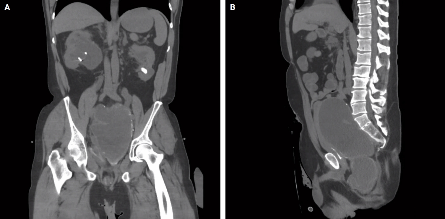
- Mucocele of the rectal stump: mucinous cystic neoplasm with low-grade dysplasia simulating low-grade appendiceal mucinous neoplasm
- Hasan Basri Aydin, Maria Faraz, A. David Chismark, Haiyan Qiu, Hwajeong Lee
- J Pathol Transl Med. 2025;59(2):139-146. Published online February 26, 2025
- DOI: https://doi.org/10.4132/jptm.2024.12.27
- 3,656 View
- 174 Download
-
 Abstract
 PDF
PDF - Mucoceles, commonly observed in the appendix, are mucin-filled, dilated structures arising from a range of etiologies. Cases associated with dysplastic or neoplastic epithelium can rupture and disseminate within the abdominopelvic cavity. Similar lesions in other parts of the colon are exceedingly rare, with only 16 colonic mucoceles having been reported. The first case of a colonic mucinous neoplasm with dysplasia resembling a low-grade appendiceal mucinous neoplasm involving rectal stump was described in 2016. Here, we present the second such case arising in the rectal stump, identified in a 44-year-old male with extensive surgical history. Microscopic examination revealed low-grade dysplastic epithelium lining the cyst and mucin dissecting into the stroma, without evidence of rupture or extramural mucin. The patient was followed for 16 months without recurrence or peritoneal disease. The exact etiology and outcome of these rare lesions remain unknown, requiring close follow-up.
- Coexisting Mucinous Cystic Neoplasm of the Pancreas and Type 1 Autoimmune Pancreatitis
- Mee-Jeong Kim, Tae Jun Song, Hyoung Jung Kim, Song-Cheol Kim, Myung-Hwan Kim, Seung-Mo Hong
- J Pathol Transl Med. 2019;53(2):125-128. Published online November 14, 2018
- DOI: https://doi.org/10.4132/jptm.2018.10.25
- 10,889 View
- 122 Download
- 3 Web of Science
- 6 Crossref
-
Abstract
PDF
- Type 1 autoimmune pancreatitis (AIP1) is an IgG4-related systemic disease that mimics tumors. We report a rare case of AIP1 accompanied by mucinous cystic neoplasm (MCN). A pancreatic lesion was incidentally detected in a woman in her 60s. After 6 years of follow-up, the lesion abruptly increased in size. Computed tomography showed a 3.5 cm unilocular cyst in the tail of the pancreas and distal pancreatectomy was performed. On microscopic examination, the cyst was lined by mucinous and non-mucinous epithelial cells with mild cytologic atypia. The surrounding stroma comprised ovarian-type spindle cells with progesterone receptor positivity. The pericystic pancreas exhibited multifocal lymphoid follicles, lymphoplasmacytic infiltrations, obliterative phlebitis, and storiform fibrosis. IgG4-positive plasma cell infiltration (215 cells high-power field) and the IgG4/IgG ratio (57%) were increased. Cases of MCN coexisting with AIP1 are extremely rare; only two such cases have been reported in the English-language literature. This third case featured low-grade MCN with AIP1.
-
Citations
Citations to this article as recorded by
- Utilizing Immunoglobulin G4 Immunohistochemistry for Risk Stratification in Patients with Papillary Thyroid Carcinoma Associated with Hashimoto Thyroiditis
Faridul Haq, Gyeongsin Park, Sora Jeon, Mitsuyoshi Hirokawa, Chan Kwon Jung
Endocrinology and Metabolism.2024; 39(3): 468. CrossRef - Histological features of autoimmune pancreatitis and IgG4-related sclerosing cholangitis with a correlation with imaging findings
Kenji NOTOHARA
Choonpa Igaku.2023; 50(1): 55. CrossRef - Imaging Features and Risk Factors of Pancreatic Cystic Lesions Complicating
Autoimmune Pancreatitis: A Retrospective Study
Bin-Bin Zhang, Xin-Meng Hou, Yu-Qi Chen, Jian-Wei Huo, Er-Hu Jin
Current Medical Imaging Reviews.2023;[Epub] CrossRef - Histological features of autoimmune pancreatitis and IgG4-related sclerosing cholangitis with a correlation with imaging findings
Kenji Notohara
Journal of Medical Ultrasonics.2021; 48(4): 581. CrossRef - 自己免疫性膵炎診療ガイドライン2020
Suizo.2020; 35(6): 465. CrossRef - Mucinous cystic neoplasm of the pancreas with type-1 autoimmune pancreatitis-like lesion
Kevin Gowing, David F. Schaeffer, Hui-Min Yang
Human Pathology: Case Reports.2019; 18: 200339. CrossRef
- Utilizing Immunoglobulin G4 Immunohistochemistry for Risk Stratification in Patients with Papillary Thyroid Carcinoma Associated with Hashimoto Thyroiditis
- Metaplastic Carcinoma with Chondroid Differentiation Arising in Microglandular Adenosis
- Ga-Eon Kim, Nah Ihm Kim, Ji Shin Lee, Min Ho Park
- J Pathol Transl Med. 2017;51(4):418-421. Published online April 4, 2017
- DOI: https://doi.org/10.4132/jptm.2016.10.06
- 9,514 View
- 114 Download
- 5 Web of Science
- 6 Crossref
-
Abstract
PDF
- Microglandular adenosis (MGA) of the breast is a rare, benign proliferative lesion but with a significant rate of associated carcinoma. Herein, we report an unusual case of metaplastic carcinoma with chondroid differentiation associated with typical MGA. Histologically, MGA showed a direct transition to metaplastic carcinoma without an intervening atypical MGA or ductal carcinoma in situ component. The immunohistochemical profile of the metaplastic carcinoma was mostly similar to that of MGA. In both areas, all the epithelial cells were positive for S-100 protein, but negative for estrogen receptor, progesterone receptor, HER2/neu, and epidermal growth factor receptor. An increase in the Ki-67 and p53 labelling index was observed from MGA to invasive carcinoma. To the best of our knowledge, this is the first case of metaplastic carcinoma with chondroid differentiation arising in MGA in Korea. This case supports the hypothesis that a subset of MGA may be a non-obligate morphologic precursor of breast carcinoma, especially the triple-negative subtype.
-
Citations
Citations to this article as recorded by- Two similar but distinct types of breast acinar cell carcinoma: evidence from histological, immunohistochemical and molecular features
Mingfang Sun, Lin Fu, Hongjiu Ren, Jian Wang, Xuyong Lin, Qingfu Zhang
Histopathology.2025; 87(6): 904. CrossRef - Elucidating the nature of acinic cell carcinoma of the breast with high-grade morphology: evidence from case report
Yunjie Ge, Xianping Wei, Jing-Nan Liu, Ping-Li Sun, Hongwen Gao
Diagnostic Pathology.2024;[Epub] CrossRef - New insights into acinic cell carcinoma of the breast: clinicopathology, origin of histology, molecular features, prognosis, and treatment
Yunjie Ge, Xianping Wei, Jing-Nan Liu, Ping-Li Sun, Hongwen Gao
Frontiers in Oncology.2024;[Epub] CrossRef - Metaplastic Matrix-Producing Carcinoma and Apocrine Lobular Carcinoma In Situ Associated with Microglandular Adenosis: A Unique Case Report
Nektarios Koufopoulos, Dionysios Dimas, Foteini Antoniadou, Kyparissia Sitara, Dimitrios Balalis, Ioannis Boutas, Alina Roxana Gouloumis, Adamantia Kontogeorgi, Lubna Khaldi
Diagnostics.2022; 12(6): 1458. CrossRef - Salivary gland-type mammary carcinoma arising in microglandular adenosis: A case report and clinicopathological review of the literature
Victoria Rico, Yukiko Shibahara, Marjorie Monteiro, Elzbieta Slodkowska, Samantha Tam, Pearl Zaki, Carlo De Angelis, Edward Chow, Katarzyna Joanna Jerzak
Cancer Treatment and Research Communications.2020; 24: 100178. CrossRef - Microglandular adenosis is an advanced precursor breast lesion with evidence of molecular progression to matrix-producing metaplastic carcinoma
Christopher J. Schwartz, Igor Dolgalev, Esther Yoon, Iman Osman, Adriana Heguy, Eleazar C. Vega-Saenz de Miera, Diana Nimeh, George Jour, Farbod Darvishian
Human Pathology.2019; 85: 65. CrossRef
- Two similar but distinct types of breast acinar cell carcinoma: evidence from histological, immunohistochemical and molecular features
- Comprehensive Cytomorphologic Analysis of Pulmonary Adenoid Cystic Carcinoma: Comparison to Small Cell Carcinoma and Non-pulmonary Adenoid Cystic Carcinoma
- Seokhwi Kim, Jinah Chu, Hojoong Kim, Joungho Han
- J Pathol Transl Med. 2015;49(6):511-519. Published online October 19, 2015
- DOI: https://doi.org/10.4132/jptm.2015.09.07
- 12,086 View
- 76 Download
- 6 Web of Science
- 6 Crossref
-
Abstract
PDF
- Background
Cytologic diagnosis of pulmonary adenoid cystic carcinoma (AdCC) is frequently challenging and differential diagnosis with small cell carcinoma is often difficult. Methods: Eleven cytologically diagnosed cases of pulmonary AdCC were collected and reviewed according to fifteen cytomorphologic characteristics: small cell size, cellular uniformity, coarse chromatin, hyperchromasia, distinct nucleolus, frequent nuclear molding, granular cytoplasm, organoid cluster, sheet formation, irregular border of cluster, hyaline globule, hyaline basement membrane material, individual cell necrosis or apoptotic body, and necrotic background. Twenty cases of small cell carcinoma and fifteen cases of non-pulmonary AdCC were also reviewed for the comparison. Results: Statistically significant differences were identified between pulmonary AdCC and small cell carcinoma in fourteen of the fifteen cytomorphologic criteria (differences in sheet formation were not statistically significant). Cellular uniformity, distinct nucleolus, granular cytoplasm, distinct cell border, organoid cluster, hyaline globule, and hyaline basement membrane material were characteristic features of AdCC. Frequent nuclear molding, individual cell necrosis, and necrotic background were almost exclusively identified in small cell carcinoma. Although coarse chromatin and irregular cluster border were observed in both, they favored the diagnosis of small cell carcinoma. Hyaline globules were more frequently seen in non-pulmonary AdCC cases. Conclusions: Using the fifteen cytomorphologic criteria described by this study, pulmonary AdCC could be successfully distinguished from small cell carcinoma. Such a comprehensive approach to an individual case is recommended for the cytologic diagnosis of pulmonary AdCC. -
Citations
Citations to this article as recorded by- Primary pulmonary adenoid cystic carcinoma: A study of clinicopathological features and molecular alterations in twenty-one cases
Zhiyuan Yao, Tong Qiu, Changlei Li, Weimao Kong, Guangqi Li, Peng Song, Guohua Wang, Wenjie Jiao
Lung Cancer.2025; 201: 108414. CrossRef - Recent developments in the pathology of primary pulmonary salivary gland‐type tumours
Julia R Naso, Anja C Roden
Histopathology.2024; 84(1): 102. CrossRef - Bronchial cytology of pulmonary adenoid cystic carcinoma – A multi-institute series with emphasis on immunocytochemistry
Joanna K.M. Ng, Ka Pang Chan, Gary M. Tse, Joshua J.X. Li
Annals of Diagnostic Pathology.2023; 64: 152132. CrossRef - Pulmonary adenoid cystic carcinoma: molecular characteristics and literature review
Zhixin Chen, Jiapeng Jiang, Ying Fan, Hongyang Lu
Diagnostic Pathology.2023;[Epub] CrossRef - Recent updates in salivary gland tumors of the lung
Anja C. Roden
Seminars in Diagnostic Pathology.2021; 38(5): 98. CrossRef - Cytology of Primary Salivary Gland-Type Tumors of the Lower Respiratory Tract: Report of 15 Cases and Review of the Literature
Chiara Saglietti, Marco Volante, Stefano La Rosa, Igor Letovanec, Marc Pusztaszeri, Gaia Gatti, Massimo Bongiovanni
Frontiers in Medicine.2017;[Epub] CrossRef
- Primary pulmonary adenoid cystic carcinoma: A study of clinicopathological features and molecular alterations in twenty-one cases
- Comparison of Cytologic Characteristics between Adenoid Cystic Carcinoma and Adenoid Basal Carcinoma in the Uterine Cervix
- Juhyeon Jeong, Seung Yeon Ha, Hyun Yee Cho, Dong Hae Chung, Jungsuk An
- J Pathol Transl Med. 2015;49(5):396-402. Published online August 17, 2015
- DOI: https://doi.org/10.4132/jptm.2015.07.08
- 11,748 View
- 97 Download
- 1 Web of Science
- 2 Crossref
-
Abstract
PDF
- Background
Adenoid cystic carcinoma (ACC) and adenoid basal carcinoma (ABC) are rare in the uterine cervix. ACC is more aggressive than ABC, thus accurate differential diagnosis is important. In this study, we identified cytologic features useful in distinguishing these two tumors for diagnosis. Methods: Three cases of ACC and five cases of ABC were selected for this study. Cervicovaginal smear slides were reviewed retrospectively, and the area, circumference, major axis, and minor axis of nuclei were measured using an image analyzer. Results: ACC displayed three-dimensional clusters with a small acini pattern. ABC displayed peripheral palisading without an acini pattern. The nuclei of ACC were more irregular and angulated than those of ABC, and the former showed a coarsely granular chromatin pattern. The nucleic area, circumference, major axis, and minor axis were 18.556±8.665 µm2, 23.320±11.412 µm, 5.664±1.537 µm, and 4.127±1.107 µm in ACC and 11.017±4.440 µm2, 15.920±5.664 µm, 4.612±1.025 µm, and 3.088±0.762 µm in the cases of ABC. All measured values showed statistically significant difference (p < .001). Conclusions: Although the nuclei of both of these tumor types were oval shaped, inferred from the ratio of minor axis to major axis (0.728 in ACC and 0.669 in ABC), the area of nuclei was approximately 1.7 times larger in ACC than in ABC. Distinguishing nucleic features, including area, morphology, and chromatin pattern, may be helpful in making a correct diagnosis. -
Citations
Citations to this article as recorded by- Adenoid basal carcinoma of the uterine cervix
Anas Mohamed, Tesfalem Korga, Ahlam Ali, Javier Laurini
International Journal of Gynecological Cancer.2025; : 101873. CrossRef - Adenoid Basal Carcinoma of the Uterine Cervix: A Case Report
Tatsuya Kanuma, Keiko Kigure, Tosio Nishimura, Yuji Ibuki, Shigeru Tsuchida, Harumi Kamiyama, Misa Iijima, Kazuto Nakamura
The KITAKANTO Medical Journal.2016; 66(1): 11. CrossRef
- Adenoid basal carcinoma of the uterine cervix
- Digital Papillary Carcinoma
- Sharon Lim, Inju Cho, Mi Ja Lee
- Korean J Pathol. 2014;48(6):438-441. Published online December 31, 2014
- DOI: https://doi.org/10.4132/KoreanJPathol.2014.48.6.438
- 10,697 View
- 50 Download
- 5 Crossref
-
PDF
-
Citations
Citations to this article as recorded by- Digital Papillary Adenocarcinoma: Uncommon Malignancy of Sweat Glands - Two Rare Cases
Neeti Goyal, Pawan Dhaman, Jasvinder Kaur Bhatia, Pragya Sharma, Prabha Shankar Mishra, Vikram Singh, Anvesh Rathore
Journal of Marine Medical Society.2025; 27(1): 103. CrossRef - Digital papillary adenocarcinoma: A case report of a rare malignant tumour with recommendations on management and follow-up
Varanindu Mudduwa, Mohammad Goodarzi, Richard Chalmers, Haitham Khashaba
International Journal of Surgery Case Reports.2025;[Epub] CrossRef - Digital Papillary Carcinoma: A Literature Review of Epidemiology, Management Strategies, and Patient Outcomes
William Liu, Rahul Nanda, David Zloty
Dermatologic Surgery.2025;[Epub] CrossRef - Digital papillary adenocarcinoma: A case report
Betty A. Kasimo, Vivian Akello, James J. Yahaya
Clinical Case Reports.2021;[Epub] CrossRef - A rare case of a digital papillary carcinoma of the hand with secondary conservative management
Rabeet Khan, Renu Irri, Effie Katsarma
Journal of Surgical Case Reports.2020;[Epub] CrossRef
- Digital Papillary Adenocarcinoma: Uncommon Malignancy of Sweat Glands - Two Rare Cases
- Cytokeratin-Positive Gastrointestinal Stromal Tumor of Biphasic Morphology: A Case Report
- Sung Sun Kim, Yoo Duk Choi, Jae Hyuk Lee, Chan Choi
- Korean J Pathol. 2014;48(5):375-378. Published online October 27, 2014
- DOI: https://doi.org/10.4132/KoreanJPathol.2014.48.5.375
- 9,299 View
- 39 Download
- 2 Crossref
-
PDF
-
Citations
Citations to this article as recorded by- CYTOKERATINS: NOT AN EPITHELIAL ENTITY ANYMORE?
Geetpriya Kaur, Devicharan Shetty, Seema Sikka, Aparna Pathak
INTERNATIONAL JOURNAL OF SCIENTIFIC RESEARCH.2022; : 15. CrossRef - Gastrointestinal stromal tumors of the stomach in a 10-year-old child
Saeed Nasher, Fayed Al-Yousofy, Faisal Ahmed
Journal of Pediatric Surgery Case Reports.2021; 74: 102044. CrossRef
- CYTOKERATINS: NOT AN EPITHELIAL ENTITY ANYMORE?
- Sebaceous Carcinoma Arising in Mature Cystic Teratoma of Ovary
- Hyo Jeong An, Yong Han Jung, Hye Kyoung Yoon, Soo Jin Jung
- Korean J Pathol. 2013;47(4):383-387. Published online August 26, 2013
- DOI: https://doi.org/10.4132/KoreanJPathol.2013.47.4.383
- 9,605 View
- 69 Download
- 12 Crossref
-
Abstract
PDF
Roughly 1% of mature cystic teratomas undergo malignant transformation. In particular, cutaneous-type adnexal neoplasms may occur in mature cystic teratomas. Sebaceous carcinomas, which arise from mature cystic teratomas, have rarely been observed, with only seven cases previously reported. Here, we present a case of a 69-year-old female who had pelvic pain for two weeks and who subsequently underwent bilateral salpingo-oophorectomy and hysterectomy. Her left ovary showed a unilocular cyst, measuring 22.0 cm in diameter, filled with sebaceous material and a few hairs. A luminally-protruding solid mass measuring 4.0 cm in diameter was also noted. Microscopic findings revealed lobular or diffusely arranged basophilic, atypical sebaceous cells connected to a typical mature cystic teratoma. Tumor cells demonstrated positive immunoreactivity for high molecular weight cytokeratin, cytokeratin 7, cytokeratin 19, epithelial membrane antigen, and carcinoembryonic antigen. Here, we present a case of sebaceous carcinoma arising from a mature cystic teratoma along with a review of previously published reports.
-
Citations
Citations to this article as recorded by- Teratoma cístico ovariano maduro com transformação maligna para carcinoma sebáceo: um relato de caso raro
Camilla Moreira Lopes, Maria Luiza Julinhaque Beraldo, Beatriz Moreira Salles Juliatto, Bruna Boeira Sobieray, Luir José Ruaro, Bibiana Quatrin Tiellet da Silva, Thais Dvulatk Marques Pançan, Janiceli Blanca Carlotto Hablich Silvestre
Femina.2026; 54(3): 251. CrossRef - How can we best manage ovarian sebaceous carcinomas arising from mature cystic teratomas?
Hong Min Shaye Peng, Sung Hock Chew, Yang Huang Grace Ng, Felicia Hui Xian Chin
BMJ Case Reports.2025; 18(2): e264651. CrossRef - Genetic Profiling of Sebaceous Carcinoma Arising from an Ovarian Mature Teratoma: A Case Report
Sumika Zaitsu, Yoko Aoyagi, Haruto Nishida, Kohei Nakamura, Mitsutake Yano, Eiji Kobayashi
International Journal of Molecular Sciences.2024; 25(12): 6351. CrossRef - Extraocular sebaceous carcinoma arising in a mature cystic teratoma of ovary: A case report and review of literature
Sara Pakbaz, Tanya Chawla, Marcus Q Bernardini, Liat Hogen, Marjan Rouzbahman
Human Pathology Reports.2022; 27: 300592. CrossRef - Sebaceous adenoma occurring within an intracranial dermoid cyst
Takashi Minamisaka, Johji Imura, Keitaro Shiraishi, Kohji Takagi, Takahiko Tomia, Sinichi Tanaka, Akira Noguchi, Takuya Akai, Kyo Noguchi, Satoshi Kuroda
Neuropathology.2022; 42(4): 289. CrossRef - Malignant transformation of mature cystic teratoma of the ovary
Doaa Atwi, Maria Kamal, Michael Quinton, Lewis A. Hassell
Journal of Obstetrics and Gynaecology Research.2022; 48(12): 3068. CrossRef - Sebaceous Carcinoma Arising in Ovarian Teratoma: First Report Associated With Germline Mismatch Repair Gene Mutation
Jacinta Murray, Patrick McIlwaine, Patrick J. Morrison, W. Glenn McCluggage
International Journal of Gynecological Pathology.2022; 41(6): 608. CrossRef - Impact of surgery and adjuvant treatment on the outcome of extraocular sebaceous carcinoma: a systematic review and individual patient's data analysis of 206 cases
Prashanth Giridhar, Lakhan Kashyap, Supriya Mallick, Ashish Dutt Upadhyay, Goura K. Rath
International Journal of Dermatology.2020; 59(4): 494. CrossRef - Mismatch repair deficiency is implicated in carcinoma arising from ovarian teratoma
Alvin Ho-Kwan Cheung, Chit Chow, Mei-Yung Yu, Wendy Wai-Tak Law, Peggy Pui-Ying Law, Paul Cheung-Lung Choi, Wei Kang, Ka-Fai To
Pathology.2019; 51(1): 67. CrossRef - Malignant transformation of an ovary mature cystic teratoma: case report and review of the literature
Elkin Fabián Dorado-Roncancio, Oscar Joel Carrillo-Garibaldi
Obstetrics & Gynecology International Journal.2019;[Epub] CrossRef - A case of ovarian clear cell carcinoma arising from ovarian mature cystic teratoma
Kazuya Maeda, Yoshito Terai, Shinichi Terada, Hiroshi Maruoka, Yuhei Kogata, Keisuke Ashihara, Yoshimichi Tanaka, Tomohito Tanaka, Hiroshi Sasaki, Satoshi Tsunetoh, Takashi Yamada, Masahide Ohmichi
Journal of Ovarian Research.2018;[Epub] CrossRef - Sebaceous carcinoma arising within an ovarian mature cystic teratoma: A case report with discussion of clinical management and genetic evaluation
Alyssa Wield, Melissa Hodeib, Mohammad Khan, Lindsay Gubernick, Andrew J. Li, Shivani Kandukuri
Gynecologic Oncology Reports.2018; 26: 37. CrossRef
- Teratoma cístico ovariano maduro com transformação maligna para carcinoma sebáceo: um relato de caso raro
- Ghost Cell Odontogenic Carcinoma Arising from Calcifying Cystic Odontogenic Tumor: A Case Report
- Zhi-Yu Zhu, Zhi-Gang Chu, Yu Chen, Wei-Ping Zhang, Di Lv, Ning Geng, Ming-Zhong Yang
- Korean J Pathol. 2012;46(5):478-482. Published online October 25, 2012
- DOI: https://doi.org/10.4132/KoreanJPathol.2012.46.5.478
- 11,197 View
- 85 Download
- 19 Crossref
-
Abstract
PDF
Ghost cell odontogenic carcinoma (GCOC) is an exceptionally rare and malignant odontogenic tumor with aggressive growth characteristics. We describe a case of GCOC which was considerably derived from a previously resected calcifying cystic odontogenic tumor (CCOT). Cellular atypia, mitotic activity, Ki-67 labeling index and matrix metalloprotease-9 positive expression rate were all increased in the currently resected specimen compared to the initial one. This is a rare case of malignant transformation of CCOT to GCOC with respect to its histopathological and immunohistochemical findings.
-
Citations
Citations to this article as recorded by- Ghost Cell Odontogenic Carcinoma of the Anterior Maxilla with ARID1A Mutation: A Case Report and Literature Review
Nasser Mohammed Almadan, Doaa Alghamdi, Meshal AlOrf, Hamed Alali, Mohammed Mohajrye
Head and Neck Pathology.2025;[Epub] CrossRef - Ghost Cell Odontogenic Carcinoma: Case Series and Literature Review
Dalja Parks, Nancy J. Zhou, Danny A. Vazquez, Matthew Fisher, Sakar Budhathoki, Jergin Chen, Shawn Iganej, Onita Bhattasali, Lester D. R. Thompson
Head and Neck Pathology.2025;[Epub] CrossRef - Late recurrence of calcifying odontogenic cyst: Report of a rare case and review of the literature
Paris Tamiolakis, Maria Georgaki, Panagiotis Christopoulos, Nikolaos G. Nikitakis
Oral Surgery.2024; 17(3): 258. CrossRef - Treatment challenges of persistent ghost cell odontogenic carcinoma: a case report and literature review
Ali Al-Sammak, Othman Rezki, Michael Pennington, Frances Manosca, Maria Cuevas-Nunez, Mohammed Qaisi, Even Greenbaum, James Murphy
Oral Surgery, Oral Medicine, Oral Pathology and Oral Radiology.2023; 136(4): e123. CrossRef - Ghost cell odontogenic carcinoma: A rare case report and review of literature
Yong Xia, Zongchang Song, Xinlei Zhang, Xinhong Guan, Guifang Tan, Yi Le, Shuang Liu, Hui Xue, Jing Li, Yajun Zhang, Jing Chen, Huajuan Jiang, Xia Jiang, Yanxia Cheng, Chuchu Zhou, Xu Sha, Jin-Xin Lou
Medicine.2023; 102(38): e35225. CrossRef - A novel parotid carcinoma with a prominent ghost cell population: a masquerading tumor or “salivary ghost cell carcinoma”?
Hiroshi Harada, Mitsuo P. Sato, Naoki Otsuki, Mao Kawamura, Akira Kurose, Takao Satou
Medical Molecular Morphology.2022; 55(1): 76. CrossRef - Dentinogenic ghost cell tumor with focal atypical features suggesting ghost cell odontogenic carcinoma: Report of a challenging diagnosis
Danielle Castex Conde, Gustavo de Souza Vieira, Pâmella de Pinho Montovani, João Pedro Roque Beserra, Mauro César Gaspar Ribeiro, Rafaela Elvira Rozza-de-Menezes, Karin Soares Cunha
Oral Oncology.2022; 124: 105524. CrossRef - Comparative Analysis Between Dentinogenic Ghost Cell Tumor and Ghost Cell Odontogenic Carcinoma: A Systematic Review
Gustavo de Souza Vieira, Pâmella de Pinho Montovani, Rafaela Elvira Rozza-de-Menezes, Karin Soares Gonçalves Cunha, Danielle Castex Conde
Head and Neck Pathology.2021; 15(4): 1265. CrossRef - Ghost cell odontogenic carcinoma of anterior mandible
Gopikrishnan Vijayakumar, Mala Kamboj, Anjali Narwal, Anju Devi
Journal of Oral and Maxillofacial Pathology.2021; 25(Suppl 1): S99. CrossRef - Ghost cell odontogenic carcinoma of the jaws: Report of two cases and a literature review
Meng-Qi Jia, Jun Jia, Li Wang, Hai-Xiao Zou
World Journal of Clinical Cases.2019; 7(3): 357. CrossRef - Ghost cell odontogenic carcinoma with suspected cholesterol granuloma of the maxillary sinus in a patient treated with combined modality therapy
You Qin, Yanwei Lu, Liduan Zheng, Hong Liu
Medicine.2018; 97(7): e9816. CrossRef - A lesion categorized between ghost cell odontogenic carcinoma and dentinogenic ghost cell tumor with CTNNB1 mutation
Yae Ohata, Kou Kayamori, Akane Yukimori, Kanako Sumikura, Toshimitsu Ohsako, Hiroyuki Harada, Kei Sakamoto, Tohru Ikeda
Pathology International.2018; 68(5): 307. CrossRef - A case of large ghost cell odontogenic carcinoma arising in the mandible
Daisuke ARAKI, Shunichi YOSHIDA, Keisuke KOYAMA, Shuutarou ISHII, Toshihiro HASEGAWA, Sadao OOYAMA
Japanese Journal of Oral and Maxillofacial Surgery.2018; 64(12): 708. CrossRef - Three-dimensional volumetric analysis of ghost cell odontogenic carcinoma using 3-D reconstruction software: a case report
João Pedro Perez Gomes, Andre Luiz Ferreira Costa, Carlos Takahiro Chone, Albina Messias de Almeida Milani Altemani, João Maurício Carrasco Altemani, Carmen Silvia Passos Lima
Oral Surgery, Oral Medicine, Oral Pathology and Oral Radiology.2017; 123(5): e170. CrossRef - Ghost cell odontogenic carcinoma transformed from dentinogenic ghost cell tumor of the maxilla after recurrences
Sase Miwako, Itoh Hiroto, Nakano Takahumi, Hayasaka Junichi, Noguchi Tadahide, Jinbu Yoshinori, Mori Yoshiyuki
Journal of Oral and Maxillofacial Surgery, Medicine, and Pathology.2017; 29(5): 438. CrossRef - Ki-67 and p53 expression in ghost cell odontogenic carcinoma: a case report and literature review
G. Del Corso, M. L. Tardio, D. B. Gissi, C. Marchetti, L. Montebugnoli, A. Tarsitano
Oral and Maxillofacial Surgery.2015; 19(1): 85. CrossRef - Integrative genomic analysis of ghost cell odontogenic carcinoma
Pinaki Bose, Erin D. Pleasance, Martin Jones, Yaoqing Shen, Carolyn Ch’ng, Caralyn Reisle, Jacqueline E. Schein, Andrew J. Mungall, Richard Moore, Yussanne Ma, Brandon S. Sheffield, Thomas Thomson, Steven Rasmussen, Tony Ng, Stephen Yip, Christopher W. Le
Oral Oncology.2015; 51(9): e71. CrossRef - Pediatric Metastatic Odontogenic Ghost Cell Carcinoma: A Multimodal Treatment Approach
Safia K. Ahmed, Masayo Watanabe, Daphne E. deMello, Thomas B. Daniels
Rare Tumors.2015; 7(2): 73. CrossRef - Predictive Factors of Potential Malignant Transformation in Recurrent Calcifying Cystic Odontogenic Tumor: Review of the Literature
Sepideh Mokhtari, Zhaleh Mohsenifar, Maedeh Ghorbanpour
Case Reports in Pathology.2013; 2013: 1. CrossRef
- Ghost Cell Odontogenic Carcinoma of the Anterior Maxilla with ARID1A Mutation: A Case Report and Literature Review
- Multifocal Renal Cell Carcinoma of Different Histological Subtypes in Autosomal Dominant Polycystic Kidney Disease
- Ki Yong Na, Hyun-Soo Kim, Yong-Koo Park, Sung-Goo Chang, Youn Wha Kim
- Korean J Pathol. 2012;46(4):382-386. Published online August 23, 2012
- DOI: https://doi.org/10.4132/KoreanJPathol.2012.46.4.382
- 11,269 View
- 77 Download
- 14 Crossref
-
Abstract
PDF
Renal cell carcinoma (RCC) in autosomal dominant polycystic kidney (ADPKD) is rare. To date, 54 cases of RCC in ADPKD have been reported. Among these, only 2 cases have different histologic types of RCC. Here we describe a 45-year-old man who received radical nephrectomy for multifocal RCC with synchronous papillary and clear cell histology in ADPKD and chronic renal failure under regular hemodialysis. The case reported herein is another example of the rare pathological finding of RCC arising in a patient with ADPKD.
-
Citations
Citations to this article as recorded by- Pellino1-mTOR/S6K1 signaling axis is a key pathogenesis for the development of polycystic kidney disease
Suhyeon Kim, Min-Hee Kim, Bo-Kyoung Ko, Kyung-Mo Kim, Naiyu Wang, Su-Mi Jo, Heounjeong Go, Eun-Ji Park, Chang-Woo Lee
Cell Death & Disease.2026;[Epub] CrossRef - Autosomal Dominant Polycystic Kidney Disease-Related Multifocal Renal Cell Carcinoma: A Narrative Iconographic Review
Consolato M. Sergi, Luis Guerra, Josef Hager
International Journal of Molecular Sciences.2025; 26(9): 3965. CrossRef - Autosomal Dominant Polycystic Kidney Disease Patients Requiring Nephrectomy: Characteristics and Surgical Considerations
Joel Ern Zher Chan, Kate S. Olakkengil, Shantanu Bhattacharjya, Santosh Antony Olakkengil
ANZ Journal of Surgery.2025; 95(7-8): 1605. CrossRef - Renal Cell Carcinoma in the Background of Autosomal Dominant Polycystic Kidney Disease: Report of Two Cases and Review of Literature
Poorva Vias, Shikha Goyal, Renu Madan, Nandita Kakkar, Ridhi Sood, Kannan Periasamy, Rajender Kumar
Indian Journal of Medical and Paediatric Oncology.2024; 45(02): 188. CrossRef - Detection of two synchronous histologically different renal cell carcinoma subtypes in the same kidney: a case report and review of the literature
Mohamed Sakr, Merhan Badran, Sarah Ahmed Hassan, Mohamed Elsaqa, Mohamed Anwar Elwany, Nevine M. F. El Deeb, Mohamed Sharafeldeen
Journal of Medical Case Reports.2024;[Epub] CrossRef - The Importance of Genetic Testing in the Differential Diagnosis of Atypical TSC2-PKD1 Contiguous Gene Syndrome—Case Series
Petronella Orosz, Zita Kollák, Ákos Pethő, András Fogarasi, György Reusz, Kinga Hadzsiev, Tamás Szabó
Children.2023; 10(3): 420. CrossRef - Autosomal dominant polycystic kidney disease coming up with an unusual presentation of renal cell carcinoma on its first encounter
Asma Shoukat Masumdar, Anitha Padmanabhan, Nitin Gadgil, Gargi Padalkar
Indian Journal of Pathology and Oncology.2023; 10(4): 417. CrossRef - Sarcomatoid renal cell carcinoma with autosomal dominant polycystic kidney disease: a case report and literature review
Yuji Hakozaki, Kiyotaka Uchiyama, Akane Yanai, Daisuke Yamada, Yuka Kamijo, Yoshitaka Ishibashi
CEN Case Reports.2021; 10(2): 199. CrossRef - CT and MRI findings of cystic renal cell carcinoma: comparison with cystic collecting duct carcinoma
Qingqiang Zhu, Jun Ling, Jing Ye, Wenrong Zhu, Jingtao Wu, Wenxin Chen
Cancer Imaging.2021;[Epub] CrossRef - Incidental occurrence of papillary renal cell carcinoma in the native kidney with autosomal dominant polycystic kidney disease after renal transplantation: A case report
Mahmoud Abbas, Melanie Pätzel, Angelika Thurn, Olaf Brinkmann, Olaf Bettendorf
Molecular and Clinical Oncology.2021;[Epub] CrossRef - Xp11.2 translocation renal cell carcinoma in the autosomal dominant polycystic kidney disease patient with preserved renal function
Hyuk Huh, Hyung Ah Jo, YongJin Yi, Seung Hyup Kim, Kyung Chul Moon, Curie Ahn, Hayne Cho Park
The Korean Journal of Internal Medicine.2017; 32(6): 1108. CrossRef - The Association between Autosomal Dominant Polycystic Kidney Disease and Renal Cell Carcinoma
Chase C. Hansen, Michael Derrick, Irfan Warriach, James Thomas Cammack, James Thomas Cammack, Werner de Riese
Open Journal of Urology.2015; 05(06): 84. CrossRef - The MSCT and MRI findings of collecting duct carcinoma
Q. Zhu, J. Wu, Z. Wang, W. Zhu, W. Chen, S. Wang
Clinical Radiology.2013; 68(10): 1002. CrossRef - Thyroid-like follicular carcinoma of the kidney in a patient with nephrolithiasis and polycystic kidney disease: a case report
Metka Volavšek, Margareta Strojan-Fležar, Gregor Mikuz
Diagnostic Pathology.2013;[Epub] CrossRef
- Pellino1-mTOR/S6K1 signaling axis is a key pathogenesis for the development of polycystic kidney disease
- Diagnostic Features of Fine Needle Aspiration Cytology of Pleomorphic Adenoma, Adenoid Cystic Carcinoma, and Mucoepidermoid Carcinoma of Salivary Gland.
- Eun Sook Nam, Won Bo Jo, Jung Ho Han, Insun Kim
- J Pathol Transl Med. 1990;1(1):60-67.
- 6,969 View
- 302 Download
-
Abstract
PDF
- The evaluate the diagnostic findings of salivary gland tumors, we reexamined aspiration cytology smears of 7 cases of pleomorphic adenoma, 3 cases of adenoid cystic carcinoma, and 3 cases of mucoepidermoid carcinoma, performed during April 1986 to March 1990, which were comfurmed by surgical excision and histologic diagnosis. The results obtained are summarized as follows : 1. All cases of pleomorphic adenoma showed branching cellular clusters of epithelial and myoepithelial cells. Acellular elements including myxomatous and chondroid components were observed. There were no cellular pleomorphism and nucleoli. Keratinizing squamous epithelial cells and keratin pearls were noted. 2. The smears of adenoid cystic carcinoma showed cell balls or cell cords containing a central hyaline core. Nuclear atypism and the nucleoli were frequently observed. There were no keratinizing squamous epithelial cells. 3. The smears of mucoepidermoid carcinoma showed mainly sheets or clusters of intermediate cells and some mucin-producing cells. Some nuclear pleomorphism was observed. Mucinous material and many inflammatory cells were present in the background.
- Simultaneous Pancreatic Serous Microcystic Adenoma and Intraductal Papillary Mucinous Tumor of the Pancreas: A Case Report.
- Hyoung Jong Kwak, Young Kon Kim, Baik Hwan Cho, Woo Sung Moon
- Korean J Pathol. 2011;45:S29-S31.
- DOI: https://doi.org/10.4132/KoreanJPathol.2011.45.S1.S29
- 3,746 View
- 24 Download
-
Abstract
PDF
- Serous cystadenomas of the pancreas account for approximately a third of pancreatic cystic neoplasms. Their coexistence with a second tumor is extremely rare. We now report a case of a serous microcystic adenoma combined with an intraductal papillary mucinous tumor of the pancreas in a 69-year-old man. Abdominal computed tomography scans demonstrated an incidental cystic mass in the body with cystic dilatation of the duct in the head of the pancreas. Central pancreatectomy with pancreatico-jejunostomy, and cyst excision of the pancreatic head were performed. Histologic examination demonstrated a serous microcystic cystadenoma in the body coexisting with an intraductal papillary mucinous adenoma in the head of the pancreas. This case study highlights the importance of careful intra-operative and pathologic examination for synchronous pancreatic tumors.
- Cystic Nephroma: A Case Report and Comparing Literature Review with Mixed Epithelial and Stromal Tumor of Kidney.
- Hyun Jung Kim, Choong Hee Noh, Giyoung Kwon, Eunah Shin, Jung Yeon Kim, Kyeongmee Park
- Korean J Pathol. 2011;45:S25-S28.
- DOI: https://doi.org/10.4132/KoreanJPathol.2011.45.S1.S25
- 3,777 View
- 44 Download
-
Abstract
PDF
- Cystic nephroma (CN) is a benign cystic neoplasm composed of mixed epithelial and stromal elements. Less than 200 cases have been reported. We had a patient, a 41-year-old woman, who had a huge typical CN. The patient was admitted for a right renal mass that was found incidentally. On laparaoscopic right nephrectomy, there was an encapsulated 7 cm multilocular cystic mass at the upper pole. Microscopically, the cystic wall was lined by a single layer of low cuboidal or hobnail epithelium without a solid area. The thin septa were composed of bland, ovarian type spindle cells. The main differential diagnoses were mixed epithelial and stromal tumor (MEST), low grade multilocular renal cell carcinoma, and tubulocystic carcinoma. The results of immunohistochemical staining were cytokeratin 7/19(+/+) and CD10(-) in lining epithelium, estrogen receptor/progesterone receptor(+/+) in stromal cells. After surgery, she was free of recurrence for 10 months. We report this rare case and compare it with other cystic renal tumors, especially MEST.
- Bronchial Brushing Cytologic Finding of Primary Pulmonary Adenoid Cystic Carcinoma Misinterpretated as Small Cell Carcinoma: A Case Report with Literature Review.
- Hyun Jung Kim, Sangbong Choi, Jieun Kwon, Jeong Yeon Kim, Kyeongmee Park
- Korean J Pathol. 2011;45(4):441-444.
- DOI: https://doi.org/10.4132/KoreanJPathol.2011.45.4.441
- 4,671 View
- 25 Download
- 2 Crossref
-
Abstract
PDF
- An adenoid cystic carcinoma is a very rare primary pulmonary neoplasm. Bronchial washing and brushing cytological findings of pulmonary adenoid cystic carcinoma have rarely been described. Here, we report the bronchial brushing cytological findings of an adenoid cystic carcinoma, finally diagnosed in a 71-year-old female patient. The low-power view showed moderate cellularity and cohesive clusters of small to medium-sized cells. The high-power view revealed distinct nuclear moldings, a coarse chromatin pattern, and inconspicuous nucleoli, which was favorable to a diagnosis of small cell carcinoma. However, apoptotic bodies, nuclear debris, and mitoses were not seen frequently. The bronchial biopsy showed solid, trabecular, and cribriform patterns in small cells. Periodic acid Schiff staining disclosed globular basement membrane-like materials, and the immunohistochemical staining revealed the presence of myoepithelial cell components, strongly suggestive of a salivary gland type tumor, compatible with an adenoid cystic carcinoma. In this report, we describe the exfoliative cytological features of a pulmonary adenoid cystic carcinoma with emphasis on some diagnostic pitfalls.
-
Citations
Citations to this article as recorded by- Bronchial cytology of pulmonary adenoid cystic carcinoma – A multi-institute series with emphasis on immunocytochemistry
Joanna K.M. Ng, Ka Pang Chan, Gary M. Tse, Joshua J.X. Li
Annals of Diagnostic Pathology.2023; 64: 152132. CrossRef - Comprehensive Cytomorphologic Analysis of Pulmonary Adenoid Cystic Carcinoma: Comparison to Small Cell Carcinoma and Non-pulmonary Adenoid Cystic Carcinoma
Seokhwi Kim, Jinah Chu, Hojoong Kim, Joungho Han
Journal of Pathology and Translational Medicine.2015; 49(6): 511. CrossRef
- Bronchial cytology of pulmonary adenoid cystic carcinoma – A multi-institute series with emphasis on immunocytochemistry
- Coexistence of Intrapulmonary Bronchogenic Cyst and Congenital Cystic Adenomatoid Malformation: A Case Report.
- Mee Hye Oh, Eun Ah Jung, Ji Hye Lee, Hyun Deuk Cho, Ki Hyun Seo, Seock Yeol Lee, Young Tong Kim
- Korean J Pathol. 2011;45(1):92-95.
- DOI: https://doi.org/10.4132/KoreanJPathol.2011.45.1.92
- 3,961 View
- 26 Download
- 1 Crossref
-
Abstract
PDF
- Congenital cystic lesions of the lung are uncommon and a conjunction of two or more lesions is very rare. We report here on a case of coexisting intrapulmonary bronchogenic cyst and congenital cystic adenomatoid malformation in a 13-year-old female with a cystic mass in the right upper lobe of the lung. Computed tomography showed a cystic lesion measuring 2.5 cm with an air fluid level and surrounding multicystic lesions in the right upper lobe. On gross examination, the cut surface showed a cystic mass containing inspissated mucinous material, and the cystic mass was surrounded by multiple small cysts. Microscopically, the larger cystic cavity was lined with pseudostratified ciliated columnar epithelium. The submucosal tissue contained mucinous glands and plates of cartilage. The surrounding smaller cysts or irregular spaces were lined with bronchiolar-type respiratory epithelium. We propose that this hybrid lung lesion may represent the missing link in a common embryologic pathway determined by the timing of mesenchymal and epithelial interactions.
-
Citations
Citations to this article as recorded by- Case 2: Coexisting Cystic Lesions of Lung in a Term Neonate: A Management Dilemma
Bichitrananda Raut, Aakriti Soni, Susanta Kumar Badatya, Satish Saluja, Manoj Modi, Arun Soni
NeoReviews.2018; 19(9): e542. CrossRef
- Case 2: Coexisting Cystic Lesions of Lung in a Term Neonate: A Management Dilemma
- Pathologic Characteristics of Ovarian Hemorrhagic Polycyst in Estrogen Receptor-alpha (ERalpha) Knockout Mice and Roles of ERalpha in Hemorrhagic Polycyst.
- Hyun Jin Son, Joo Heon Kim, Hye Kyung Lee, Mee Ja Park, Dong Wook Kang, Che Myong Ko
- Korean J Pathol. 2010;44(4):376-383.
- DOI: https://doi.org/10.4132/KoreanJPathol.2010.44.4.376
- 4,075 View
- 53 Download
-
Abstract
PDF
- BACKGROUND
Polycystic ovary syndrome (PCOS) is the most common endocrinopathy causing anovulation in women of childbearing age. It has been well established that estrogen receptor-alpha knockout (ERalphaKO) mice display several pathologic ovarian phenotypes of PCOS. The aims of this study were to determine ovarian pathology in new ERalphaKO mice using a CreloxP approach and intra-ovarian ERalpha function as regulating key aspects of PCOS.
METHODS
ERalphaKO mice, which were deficient in exon 3 of the ERalpha gene, were used. Immunohistochemical studies were done on ovaries of control and ERalphaKO mice using antibodies specific to ERalpha, ERbeta, inhibin-alpha, and alpha-smooth muscle actin (SMA), as well as histochemical staining using Sudan black-B.
RESULTS
All ovaries of ERalphaKO mice were larger than control mouse ovaries and displayed a disrupted theca-interstitial tissue organization, multiple atretic follicles and multiple hemorrhagic cysts. None of the ERalphaKO mouse ovaries showed a corpus luteum. In addition, heavy deposition of Sudan black-B positive foamy cells was seen. The theca externa of preantral immature follicles and hemorrhagic cysts showed strong expression of alpha-SMA.
CONCLUSIONS
ERalphaKO mice show hemorrhagic polycystic ovaries and hyperplasia of the theca externa. This study demonstrates that the ERalpha is the functional key to the pathogenesis of PCOS.
- Columnar Cell Lesions in Fibrocystic Change of the Breast: The Incidence and Relationship with Microcalcifications.
- Soo Im Choi, Hye Kyoung Yoon
- Korean J Pathol. 2009;43(4):301-305.
- DOI: https://doi.org/10.4132/KoreanJPathol.2009.43.4.301
- 5,942 View
- 82 Download
-
Abstract
PDF
- BACKGROUND
Columnar cell lesions (CCLs) are characterized by the presence of columnar epithelial cells lining the terminal duct lobular units of the breast and frequently found in biopsies for microcalcifications. Their incidence and relationship with other lesions and the locations of microcalcifications have not been established. METHODS: We reviewed 1,038 cases of fibrocystic change (FCC) for the degrees of CCLs and ductal proliferative change (PC) and the locations of microcalcifications. RESULTS: Among 1,038 FCC cases, CCLs were found in 18.9%, columnar cell change (CCC) in 12.5%, columnar cell hyperplasia (CCH) in 5.3% and flat epithelial atypia (FEA) in 1.1%. CCLs were found in 14.2%, 28.8%, and 40.0% of non-PC (NPC), proliferative disease (PD) without atypia and PD with atypia, respectively. Microcalcifications were found in 33.5%, 56.2%, 61.8%, and 81.8% of caese without CCLs, with CCC, CCH and FEA, respectively. Their locations were in NPC in 66.3% of the cases, PD in 14.8% of the cases or both areas in 18.8% of FCC. CONCLUSIONS: The incidence of CCLs increased according to the degree of PD without positive correlation between the degree of CCLs and PD. The frequency of microcalcifications increased according to the degree of CCLs but was statistically insignificant. There is a possibility that a needle biopsy targeting a microcalcification area might leave additional PD around the targeted area because microcalcifications were found more frequently in NPC than PD area.
- The Cytologic Features of Adenoid Cystic Carcinoma of the Uterine Cervix: A Case Report .
- Seung Yeon Ha, Hyuni Cho, Young Ha Oh, Geun Shin Lyu
- J Pathol Transl Med. 1998;9(2):207-212.
- 2,332 View
- 22 Download
-
Abstract
PDF
- Adenoid cystic carcinoma of the uterine cervix is a rare tumor accounting for less than 1% of all cervical adenocarcinoma. This tumor is characterized by aggressive biological behavior with frequent local recurrence or metastatic spread, postmenopausal onset, and occasional association with conventional squamous cell carcinoma. The cytologic diagnosis of adenoid cystic carcinoma in the uterine cervix is often difficult because of negative smear due to intact overlying mucosa, cytologic findings mimicking endometrial cells, and masquerade as squamous cell carcinoma. Recently we have experienced a case of adenoid cystic carcinoma arising in the uterine cervix, which was identified on the routine Papanicolaou smear and was histologically confirmed by the consequent biopsy. The smear showed abundant cellularity composed of relatively uniform cells. The tumor cells were arranged in small clusters, acini, naked cells, and loose sheets with abortive cribriform pattern. There were scattered globoid basement membrane-like materials and tumor diathesis. The nuclei were pleomorphic and showed hyperchromatic and coarsely granular choromatin with inconspicuous nucleoli. The punch biopsy of the uterine cervix showed typical histologic findings of adenoid cystic carcinoma characterized by tumor nests composed of hyperchromatic uniform basaloid cells, cribriform pattern, and cylindrical hyaline bodies.
- A Cystic Mesothelioma in the Inguinal Area.
- Im Joong Yoon, Nam Bok Cho, Tae Jin Lee, Mee Kyung Kim, Se Chul Kim, Kye Yong Song
- Korean J Pathol. 1997;31(3):284-287.
- 2,132 View
- 18 Download
-
Abstract
PDF
- The cystic mesothelioma is a very rare tumor which has a clinically and histologically benign nature. Here in reported is the case of a cystic mesothelioma presented as a palpable mass of the inguinal area in a 28-year-old male. Ultrasound showed a cystic tumor at the inguinal canal, and the other physical and laboratory examinations were within normal limits. Grossly, the tumor consisted of cysts containing clear serous fluid and focally solid areas. Microscopically, the tumor was encapsulated with fibrocollagenous wall, and the tumor cells were cuboidal or polygonal epithelial cells with single or multiple layers and had clear cytoplasm. Some areas showed thyroid follicle-like structures. The content of follicle-like structures showed eosinophilia in the H&E section, but positive in mucin stain. Neither cytologic atypia nor mitoses were present. Immunohistochemical staining revealed positive reaction for keratins of low molecular weight, while negative for the thyroglobulin and CEA. These findings suggested mesothelial in origin. We concluded that this tumor was primary rather than metastatic, because he had no evidence of a tumor in gastrointestinal, genitourinary tracts and scrotum.
- Adenoid Cystic Carcinoma of Skin: A case report.
- Eun Duk Chang, Young Hee Jee, Sun Moo Kim
- Korean J Pathol. 1989;23(3):378-381.
- 2,152 View
- 11 Download
-
Abstract
- A primary skin adenoid cystic carcinoma first described by Boggio in 1975, is one of the rarest type of eccrine sweat gland carcinoma. Histologically, a tumor with typical morphologic features closely resembles adenoid cystic carcinoma was found in other tissues but in the skin must be distinguished from aggressive basal cell carcinoma. The natural history of this tumor is not yet fully determined but suggests a long indolent and progressive course. We report a case of a 77-year-old male with a small skin nodule in the abdomen.
- Metastatic Adenoid Cystic Carcinoma of the Lung Diagnosed by Fine Needle Aspiration Biopsy.
- So Yeon Park, Kwang Gil Lee
- J Pathol Transl Med. 1990;1(2):175-178.
- 2,547 View
- 16 Download
-
Abstract
PDF
- A case of metastatic adenoid cystic carcinoma of the lung, originated from the trachea, was diagnosed by fine needle aspiration. Although the cytologic features of adenoid cystic carcinoma have been well described, it is easy to confuse adenoid cystic carcinoma with more common primary small cell neoplasms of the lung, i.e., small cell carcinoma, well differentiated adenocarcinoma, and carcinoid tumor of the lung. The features distinguishing adenoid cystic carcinoma from these neoplasms include 1) tight, globular, honeycomb pattern of cells, 2) acellular basement membrane material in the lumen, and 3) cells lacking true nuclear molding and having bland chromatin pattern. The morphologic feature of metastatic adenoid cystic carcinoma in this case was so distinctive as to permit a definite diagnosis by aspiration cytology.
- Sclerosing Seat Duct Carcinoma: Report of a case.
- K H Park, N H Cho, K G Lee
- Korean J Pathol. 1989;23(3):382-386.
- 1,844 View
- 18 Download
-
Abstract
PDF
- Sclerosing sweat duct carcinoma is unusual and locally aggressive neoplasm that is important to recognize since it may be confused with other benign adnexal neoplasms, particularly syringoma. Authors present a case of sclerosing sweat duct carcinoma in a 21 yearold man. The lesion was a round fresh-colored hard plaque, 1 cm in size, near the right inner eyebrow. The lesion was not fixed. Regional lymph nodes wer not palpated. Histologically, the tumor was composed of small basaloid cell nests and numerous horn cysts in the sclerotic stroma. Some of the small nests showed ductular differentation. The tumor infilturated the dermis, adjacent to the subcutaneous fat tissue and invaded a nerve fiber. Cytologic atypism of the tumor cells was not present. The tumor cell nest is positively stained with carcinoembryonic antigen.
- Localized Cystic Disease of the Kidney: A case report.
- Wan Seop Kim, Moon Hyang Park
- Korean J Pathol. 1999;33(3):210-213.
- 1,942 View
- 12 Download
-
Abstract
PDF
- Localized cystic disease of the kidney is a rare entity with the gross and microscopic features of autosomal dominant polycystic kidney disease localized to only a portion of a kidney, and negative family history. We report a case of localized cystic disease of the kidney in a 38-year-old woman who complained of intermittent right flank pain for 1 year. The resected kidney showed multiple cysts measuring up to 4.0 3.5 3.0 cm, which were scattered throughout the mid- and lower poles of the kidney. Microscopically, the cystic lesion was composed of numerous cysts of variable size, lined by flattened epithelium. The intervening septa of the cysts contained normal or compressed renal tubules and glomeruli. Neither dysgenetic tissue such as immature cartilage or primitive mesenchymal tissue nor malignant cells was identified. Localized cystic disease should be included in the differential diagnosis of cystic lesions in the kidney.
- Potter's Syndrome with Adult Polycystic Renal Disease: An autopsy case report.
- Hwa Sook Jeong, Beom Soo Park, Geon Kook Lee
- Korean J Pathol. 1997;31(4):361-365.
- 2,479 View
- 20 Download
-
Abstract
PDF
- Potter's syndrome including bilateral renal agenesis or polycystic renal disease, bilateral pulmonary hypoplasia and characteristic face was first described in 1946. Although a great number of cases of Potter's syndrome was reported, Potter's syndrome with adult polycystic kidney disease(Potter type III) was very rarely found. In this report, we described an autopsy case of Potter's syndrome having adult polycystic kidneys disease, bilateral pulmonary hypoplasia and characteristic face in conjunction with multiple hepatic cysts, features of congenital hepatic fibrosis and a pancreatic cyst. Microscopically, all cysts were lined by cuboidal epithelial cells, showing positive for epithelial membrane antigen and cytokeratins.
- Clear Cell Ependymoma.
- Jae Hee Suh, Seung Mo Hong, In Chul Lee
- Korean J Pathol. 1997;31(4):383-387.
- 3,330 View
- 70 Download
-
Abstract
PDF
- The clear cell variant of ependymoma is a rare, recently described, intracranial tumor which is composed of clear neoplastic ependymal cells. Clear cell ependymomas may share characteristic histologic features of oligodendrogliomas or central neurocytomas; striking nuclear uniformity, perinuclear halos, and numerous angulated capillaries. In contrast to oligodendrogliomas, however, clear cell ependymomas are noninfiltrating tumors with sharp boundaries. Perivascular pseudorosette formation is frequent. Oligodendrogliomas are usually nonreactive for GFAP compared to diffuse immunoreactivity of clear cell ependymoma. Central neurocytomas may also be differentiated by their immunoreactivity for synaptophysin. This is a case of clear cell ependymoma in a 40-year-old man. By computed tomography and magnetic resonance imaging scans, a well circumscribed cystic tumor with mural nodule was demonstrated in the right frontal lobe. It was 6cm in diameter and well enhanced. Histologically, it was sharply demarcated from the brain parenchyma. The cystic wall was lined by atypical ependymal cells, which "transformed" to clear cells in the solid area. The cells had uniform nuclei and perinuclear halos. Mitotic figures and necrotic foci were focally present. The cells were immunoreactive for glial fibrillary acidic protein (GFAP), while synaptophysin was negative. Electron microscopy revealed densely packed polyheadral cells with scant organelles and well developed intercellular junctions.
- Adenoid Cystic Carcinoma of the Breast: A case report.
- Eun Ha Jung, Hye Rim Park, Jin Hee Sohn
- Korean J Pathol. 1999;33(4):299-302.
- 2,008 View
- 28 Download
-
Abstract
PDF
- Adenoid cystic carcinoma (ACC) of the breast is an uncommon carcinoma accounting for less than 1% of the breast carcinoma. This type of carcinoma has a distinctive histology, such as cylindromatous, cribriform, glandlike space and solid pattern in the variable proportion. Prognosis is favorable with rare recurrence or metastasis and the guideline for therapy is not well established. We experienced a case of ACC of right breast in the 48 year-old woman. Cytologically, it showed numerous three-dimensional, well outlined cell clusters with central core of homogeneous, eosinophilic material. The mass was well demarcated, firm and measured 2.5 2.5 2 cm. Cut surface was yellowish white and granular with focal necrosis. Histologically, the tumor was composed of cylindromatous, adenoid and solid area with marked cellular atypia. Focal invasion was identified in the adipose tissue around the tumor. Histologic and nuclear grade was III by Kleer's grade. Nodal metastasis was not found.
- Solid Variant of Mammary Adenoid Cystic Carcinoma.
- Ji Eun Kwon, Yoon Hee Lee, Ju Yeon Pyo, Sang Kyum Kim, Byeong Woo Park, Woo Ick Yang
- Korean J Pathol. 2007;41(6):424-426.
- 2,104 View
- 28 Download
-
Abstract
PDF
- Adenoid cystic carcinoma (ACC) is a rare type of breast carcinoma and this tumor makes up less than 0.1% of all mammary carcinomas; ACC is known to show a relatively favorable prognosis. Among a variety of microscopic growth patterns of mammary ACC, a solid variant is the rarest and this can cause diagnostic difficulties. We present here a case of a solid variant of mammary ACC that occurred in the right breast of a 40-year-old woman who was initially diagnosed with invasive ductal carcinoma. We discuss the histological and clinical characteristics of this case.
- Fine Needle Aspiration Cytology of Solid Type Adenoid Cystic Carcinoma of Buccal Mucosa: A Case Report .
- Jeana Kim, Kyoung Mee Kim, Young Sill Kim, Anhi Lee, Sang In Shim, Byung Kee Kim
- J Pathol Transl Med. 2000;11(2):89-92.
- 2,409 View
- 39 Download
-
Abstract
PDF
- Adenoid cystic carcinoma constitutes 4 percent of all benign and malignant epithelial salivary gland tumors and is a highly malignant tumor of the salivary glands. The cytologic presentation in aspirates is usually characteristic with spherical clusters(balls) of small tumor cells filled with hyaline material. But in case of the poorly differentiated variety(solid type), it is difficult to differentiate from other tumors because sheets of small, fairly monotonous malignant cells, with somewhat larger and more conspicuous nuclei are only seen. The cytologic findings of fine needle aspiration of solid type adenoid cystic carcinoma of buccal mucosa in a 51-year-old man are presented. On cytologic findings, solid sheets of monotonous tumor cells with focal necrosis was noted on a hemorrhagic background and the characteristic cytologic features of adenoid cystic carcinoma was absent.
- Intrapulmonary Cystic Lymphangioma.
- Mi Yeong Jeon, Je Geun Chi
- Korean J Pathol. 1997;31(5):492-494.
- 2,180 View
- 20 Download
-
Abstract
PDF
- Solitary intrapulmonary cystic lymphangioma in newborn or infant is an extremely rare disease. We describe a case of solitary intrapulmonary cystic lymphangioma in a 4-month-old boy with dyspnea and tachypnea. It was in the left lower lobe and type 1 congenital cystic adenomatoid malformation was suspected radiologically. The resected cyst was unilocular with a thin wall, and was 9.5cm in size. Histologically, the cyst was lined by flattened endothelial cells supported by a minimal fibrous stroma.
- Adenoid Cystic Carcinoma of the Male Breast: A case report.
- Mi Kyung Lee, In Chul Hong, Woo Ick Yang, Sang Ho Cho
- Korean J Pathol. 1999;33(5):389-392.
- 2,197 View
- 21 Download
-
Abstract
PDF
- A 65 year-old male patient presented with a large palpable mass beneath the areola of the right breast for 7 years. The resected breast tissue was almost totally replaced by a round large solid mass (9 6 cm) with a pink-gray to yellow firm, partly nodular cut surface. Microscopically, the tumor revealed the diagnostic biphasic cellular pattern of adenoid cystic carcinoma, which consisted of both cribriform pattern of myoepithelial cells and tubular pattern of epithelial cells. On immunohistochemistry, the tumor revealed immunoreactivities for alpha-smooth muscle actin and S-100 protein in the myoepithelial cells and for AE1/AE3 in the epithelial cells. Mitoses were scarce. Multifocal lymphatic permeation and foci of perineural invasion were also found. Underlying resection margins and overlying skin were invaded by the tumor. We diagnosed this tumor as grade II adenoid cystic carcinoma according to the system utilized for the salivary gland tumors.
- Microcystic Adnexal Carcinoma: Report of a case.
- Eun Deok Chang, Young Hee Jee, Sun Moo Kim
- Korean J Pathol. 1993;27(3):290-292.
- 2,120 View
- 16 Download
-
Abstract
PDF
- Microcystic adenxal carcinoma is an unusual, locally aggressive neoplasm that has recently been recongized as a clincopathologic entity. Its histologic appearance includes both pilar and eccrine differentiation. Microscopically, the tumor consisted of small cysts and gland-like structures in superficial portion. In other area, basaloid cell nests and abortive hair follicles in the sclerotic stroma were seen. The cysts were filled with secretory eosinophilic material, which was positively stained with Periodic acid-Schiff and carcinoembryonic antigen. Immuno-peroxidase staining for carcinoembryonic antigen supported the dual differnetiation of this neoplasm. Despite the benign histologic appearance, there was deep and extensive infiltration of the subcutaneous tissue.
- Benign Cystic Mesothelioma.
- Sung Chul Lim, You Kyung Jeong, Mi Sook Lee, Yun Shin Kim, Hyun Jong Park, Sang Joon Choi
- Korean J Pathol. 1997;31(6):595-597.
- 2,207 View
- 16 Download
-
Abstract
PDF
- Benign cystic mesothelioma (BCM) is a rare mesothelial lesion that forms multicystic masses in the upper abdomen, pelvis and retroperitoneum. Although it is categorized as a benign lesion, it has a tendency to recur. It is uncertain whether the nature of this lesion is reactive or neoplastic, but many articles support the conclusion that it is reactive rather than neoplastic. The majority of cases were associated with a history of a previous abdominal or pelvic operation, or an evidence of endometriosis or a pelvic inflammatory disease, or a combination of these findings. In a 26-year-old woman we experienced a case of BCM which was incidentally discovered at cesarean delivery revealing multilocular thin and translucent walled cysts in the pelvic cavity. Microscopic examination revealed a thin cyst wall that was composed of fibrous connective tissue and lined by internal stratified and external nonstratified single cuboidal epithelia.
- Cystic Adventitial Disease of the Popliteal Artery: A case report.
- Soo Min Kang, Kyeong Cheon Jung, Je G Chi
- Korean J Pathol. 1993;27(4):418-420.
- 2,119 View
- 15 Download
-
Abstract
PDF
- Localized cystic degeneration of peripheral arteries represents and unusual cause of arterial insufficiency. It frequently occurs in patient without generalized arteriosclerosis. It has been reported in patients from age 11 to 62 years. Cystic adventitial disease is most common in the popliteal artery. At least 115 cases have been reported worldwide, but none in Korea. We report a case of cystic adventitial disease involving the left popliteal artery. This 64-year-old man presented with an 18-month history of cramping pain of sudden onset in the left calf and claudication. Angiographic findings showed a 6 cm length of luminal obliteration of the popliteal artery. Segmentally resected popliteal artery showed two longitudinally directed cystic masses measuring 3.5x1.5 cm and 2.5x1.5 cm in the adventitia. Microscopic examination revealed cystic space in the arterial adventitia compressing arterial lumen. There were a number of foamy histiocytes collected along the cystic lumen.
- Cystic Struma Ovarii Mimicking Adenomatous Goiter of the Thyroid.
- Kee Taek Jang, Je Geun Chi
- Korean J Pathol. 1997;31(7):692-694.
- 2,461 View
- 21 Download
-
Abstract
PDF
- Struma ovarii, the most common monodennal teratoma of the ovary, causes diverse problems in differential diagnosis. The literature on the pathology of struma ovarii has focused principally on the problem of formulating criteria of malignancy. In contrast, unusual gross and microscopic features of struma ovarii and its resultant problems in differential diagnosis have received relatively little attention. We report an ovarian teratoma which was almost entirely cystic, causing the diagnosis of struma to be overlooked. The removed ovarian tumor showed all the features of adenomatous goiter of the thyroid gland. The lining epithelium of the cysts was frequently flattened, and the follicles in the cyst wall were few and atrophic. The patient was a 58-year-old woman who was found to have an ovarian tumor by routine monographic examination
- A Case Report of Renal Cell Carcinoma in a Polycystic Kidney: A case report.
- Kyoung Chan Choi, Joon Hyuk Choi, Won Hee Choi
- Korean J Pathol. 1996;30(1):57-60.
- 2,141 View
- 19 Download
-
Abstract
PDF
- A forty-nine-year-old woman with polycystic disease had a right nephrectomy for what was preoperatively thought to be a polycystic disease, but at surgery turned out to be a tumor based on frozen section. Microscopic examination revealed papillary type, renal cell carcinoma with classical features of adult polycystic kidneys. Radiologic findings revealed multiple cysts in the liver. The clinical recognition of a carcinoma developing in polycystic kidneys is often difficult because of the presence of preexisting large renal masses and occasional hematuria. Renal cell carcinoma should be thought of when confronted with abdominal pain or back pain, severe hematuria, sudden dysuria or a new renal mass occurring in a patient with polycystic kidneys.
- Florid Cystic Endosalpingiosis of the Uterus: A Case Report.
- Sang Hwa Shim, Han Seong Kim, Mee Joo, Sun Hee Chang, Ji Eun Kwak
- Korean J Pathol. 2008;42(3):189-191.
- 2,427 View
- 31 Download
-
Abstract
PDF
- A 54-year-old woman presented with vaginal bleeding. On gynecologic and radiologic examinations, bilateral cystic ovarian tumors were suspected. A laparoscopic examination revealed multiple cysts involving both the uterine horns and the posterior surface of the uterus. These were removed by total abdominal hysterectomy and bilateral salpingo-oophorectomy. Microscopically, multiple cysts were identified in the subserosal layer of the uterus, and they were lined with benign tubal type epithelium surrounded by a smooth muscle wall. These features were consistent with "florid cystic endosalpingiosis". Considering the location of the lesion, the lesion has to be distinguished from other benign cystic lesions of the uterus and adnexa, including multicystic mesothelioma, serous cystadenoma and mucinous cystadenoma. Awareness of this lesion will facilitate a correct diagnosis by both the clinician and pathologist.
- Serous Cystadenoma of the Pancreas: A case report.
- Young Kyoung Bae, Woo Young Jang, Kyoung Chan Choi, Joon Hyuk Choi, Won Hee Choi
- Korean J Pathol. 1996;30(1):68-71.
- 2,582 View
- 30 Download
-
Abstract
PDF
- Serous cystadenoma of the pancreas, also known as microcystic adenoma or glycogen-rich cystadenoma, is an unusually benign tumor. It is usually large and composed microscopically of many small cysts lined by small, cuboidal or flattened cells containing abundant glycogen. It has been suggested that serous cystadenoma probably arise from the ductular cells or centroacinar cells. Herein, we report on a case of serous cystadenoma of the pancreas in a 55-year-old female. The tumor, measuring 13.5x11.5x10.0 cm, was located in the head of the pancreas and the cut surface revealed a sponge-like appearance due to innumerable tiny cysts containing clear serous fluid. Microscopic analysis showed cystic spaces lined by cuboidal cells with intracytoplasmic glycogen.
- Adenoid Cystic Carcinoma of the Esophagus: Report of a case with brief review of the literature.
- Eun Suk Koh, In Sook Kim, Tae Jung Kwon, Dong Wha Lee, Chan Sup Shim, Kihl Rho Lee
- Korean J Pathol. 1990;24(4):482-488.
- 2,134 View
- 18 Download
-
Abstract
PDF
- Adenoid cystic carcinoma of the esophagus is a rare tumor and has been considered to be counterpart of the salivery gland. The patient we experienced was a 60-year-old female who had a tumor in the lower third of the esophagus. The tumor was located in the submucosa and showed histologic features similar to those of the salivary gland. Electron micrsopic examination revealed ductal structures invested by basal lamina, and clusters of basaloid cells with tonofilaments and desmosomes. No myoepithelial cells were identified. Immunohistochemical studies for S-100 protein, cytokeratin and vimentin were performed. A few cells showed positive reaction to the S-100 protein. These findings suggest that the esophageal adenoid cystic carcinoma arises from the duct of submucosal gland.
- Fine Needle Aspiration Cytology of Cystic Hypersecretory Intraductal Carcinoma of the Breast: Report of Two Cases.
- Hee Jeong Cha, Dae Woon Eom, Jae Hee Suh
- J Pathol Transl Med. 2003;14(1):22-26.
- 2,055 View
- 10 Download
-
Abstract
- Cystic hypersecretory carcinoma of the breast is a rare variant of ductal carcinoma of breast, first described in 1984 by Rosen and Scott. Histologically, it is characterized by the formation of dilated ducts and cysts containing an eosinophilic secretory product resembling thyroid colloid. Cytologic findings show a few clusters of atypical ductal epithelial cells in amorphous proteinaceous material with cracking artifact. Differential diagnosis include mucinous carcinoma and benign mucocele-like tumor. We present two cases of fine needle aspiration cytology of cystic hypersecretory intraductal carcinoma of the breast with a review of the literature.
- Cystic Lymphangioma of the Breast in an Adult Woman.
- Kyueng Whan Min, Si Hyong Jang, Woong Na, Se Min Jang, Young Jin Jun, Ki Seok Jang, Seung Sam Paik
- Korean J Pathol. 2008;42(4):244-246.
- 2,554 View
- 23 Download
-
Abstract
PDF
- Cystic lymphangioma is also known as cystic hygroma, and this is a congenital malformation of the lymphatic system. Most lymphangiomas are present at birth and they are diagnosed by the age of 2. They are mostly located in the neck or axillary region. The breast as a site of origin is an extremely unusual location, and especially in adults. We report here on a case of cystic lymphangioma in a 36-year-old woman. Physical examination revealed a tender cystic mass in the upper outer quadrant of the right breast. Ultrasonography revealed an irregular hypoechoic mass lesion that was associated with irregular duct dilatation and several enlarged axillary lymph nodes. After the operation, the mass was revealed to be a cystic lymphangioma. Although it is very rare, cystic lymphangioma should be considered in the differential diagnosis of a breast mass in adults.
- Galactocele in a Male Child: A case report.
- Yoon Mi Jeen, Yoon Jeong Choi, Dong Wha Lee, Chan Il Park
- Korean J Pathol. 1996;30(2):164-165.
- 2,566 View
- 122 Download
-
Abstract
PDF
- We investigated a unilocular mammary cyst occurring in a two and a half year old male baby. The cyst was lined by simple columnar epithelium and filled with a milky secretory material. These histologic features were consistent with galactocele. The child had enlarged left breast since birth, but it seemed to be noncontributory as the child had neither endocrine abnormalities nor perinatal disorders. Galactocele is an uncommon breast lesion usually occuring in females following lactation. It is rarely a cause of breast enlargement.
- Cystic Lymphangioma of the Stomach: A case report.
- Soon Ae Ok, Sook Guem Jeong, Bang Hur, Man Ha Huh
- Korean J Pathol. 1993;27(6):670-672.
- 1,914 View
- 20 Download
-
Abstract
PDF
- Cystic lymphangiomas of the stomach are extremely rare. These usually present as asymptomatic, polypoid lesion consisting of cystically dilated lymphatic vessels, which are submucosal in location. We report a case of cystic lymphangioma of the stomach. The patient is a 55 year-old woman who has complained of dull pain on epigastrium for 10 years. The mass measures 6x5 cm in cross diameter and is mainly located in the subserosal layer along lesser curvature. Microscopically, the tumor reveals a large number of dilated lymphatic cysts containing serous fluid, lined by a layer of flattened endothelial cells. Ultrastructurally, lining endothelial cells show thin discontinuous basal lamina, in contrast to normal lymphatics.
- Multilocular Cystic Renal Cell Carcinoma.
- Myoung Jin Ju, Kee Tac Jang, Je Geun Chi
- Korean J Pathol. 1997;31(11):1240-1243.
- 2,042 View
- 10 Download
-
Abstract
- Multilocular cystic renal cell carcinoma is a distinct subtype of renal cell carcinoma with its pathological characteristics and good prognosis. Multilocular renal cysts and renal cell carcinoma with cystic change are important differential diagnoses. We report a case of multilocular cystic renal cell carcinoma in a 37-year-old woman who came to the hospital because of the right renal mass. The removed right kidney showed a 6x4 cm well defined cystic mass in the lower pole. On cut section there were multiple cavities in the mass, filled with serosanguineous fluid and focal yellowish solid area. Microscopically, these cysts were lined by a single layer of flat or cuboidal cells consisted of clear cytoplasm with small central nuclei. In some portions of the tumor, the clear neoplastic cells formed sheets within the septa or walls of the cysts.
- Cystic Hygroma of the Neck Pathologic study of 26 autopsy cases.
- Yeon Lim Suh, Je Geun Chi
- Korean J Pathol. 1997;31(12):1256-1263.
- 4,212 View
- 130 Download
-
Abstract
PDF
- Cystic hygroma is a congenital malformation of the lymphatic system appearing single or multiloculated fluid-filled cavities, most often around the neck. They often progress to hydrops and cause fetal death, and frequently associated with chromosomal abnormalities and other congenital malformations. The purpose of our study is to delineate the nature of cystic hygroma and determine the relationship between cystic hygroma and associated anomalies including fetal hydrops. We used data from 26 cases of cervical cystic hygroma in autopsy files of SNU Children's Hospital. Most of cystic hygroma were found in stillborn or premature infants. The fetal cases had been dead for a quite a long period since there was discrepancy between the true gestational age and the developmental age estimated from the body length. Of 26 fetuses only 2 were studied chromosomally and both of them showed 45X. Of 26 cystic hygromas 23 occurred in the posterior neck and 3 in the anterior neck. They ranged from 2.5 to 14 cm (mean: 7.9 cm). The cystic hygroma of the posterior neck consisted of two symmetric sacs on both sides and in the nape and extended to the occipital region. The cystic hygromas of the anterior neck were unilateral or bilateral, and multiloculated and extended into the adjacent cheek. Cystic hygromas of posterior neck were always associated with hydrops, while no recognizable hydrops was found in cystic hygromas of anterior neck. The cystic hygromas were larger in patients with severe hydrops than in patients with less severe hydrops. Associated abnormalities, found in 88%, included hydrops fetalis(88%), growth retardation(80%), cardiovascular anomalies(27%), horseshoe kidney(23%), skeletal anomalies(12%) and hypoxic changes(31%) in visceral organs. In summary, when a hygroma is detected during fetal life, careful sonographic examination for associated congenital anomalies, fetal karyotyping and consideration of artificial abortion are indicated.
- Cytologic Classification of Fibrocystic Disease of the Breast: A Proposal for Use of Cytologic Criteria Grading System.
- Hye Kyoung Yoon, Chan Hwan Kim, Jong Eun Joo, Shin Kwang Khang
- J Pathol Transl Med. 1994;5(2):106-112.
- 4,070 View
- 163 Download
-
Abstract
PDF
- Fine needle aspiration biopsy has been proved as a safe, accurate and cost-effective diagnostic modality in palpable breast lesions. Cytologically, fibrocystic disease can be classified into 3 categories as nonproliferative breast disease, proliferative breast disease without atypia, and proliferative breast disease with atypia. This terminology for the needle aspirates is compatible with that of diagnostic histopathology. Cytologic differentiation of nonproliferative disease from proliferative breast disease is important, since the risk of cancer development in cases of atypical hyperplasia is 4-5 times higher than that of general population. Twenty five needle aspirates of fibrocystic disease confirmed by subsequent histopathology were re-evaluated and classified into 3 categories depending on their architectural and nuclear features. In addition. these aspirates were scored according to the cytologic grading system, devised by Masood et al. and based on six cytologic criteria. Concordance rates between cytomorpholgic diagnosis and cytologic diagnosis using the cytologic criteria grading system and histologic diagnosis were 88% and 92%, respectively.
- Cystic Meningioma: A case report.
- Jae Hoon Park, So Yeon Yu, Youn Wha Kim, Yong Koo Park, Moon Ho Yang
- Korean J Pathol. 1991;25(2):153-157.
- 2,080 View
- 22 Download
-
Abstract
PDF
- Meningiomas are usually thought of as firm solid tumors and most standard references make no mention of cystic meningiomas. Although several cases of cystic meningioma have been reported in the literature and their neuroradiological features discussed, the rarity of this entity makes its preoperative diagnosis difficult. Recently, the authors encountered a case of cystic meningioma, which was thought as ependymal cyst or infarction, preoperatively. In this report the authors discussed its clinical, neuroradiological and pathological characteristics with brief reviews of the literature.
- Adenocarcinoma Arising in Type 1 Congenital Cystic Adenomatoid Malformation: A Case Report and Review of the Literature.
- Jinyoung Yoo, Sun Mi Lee, Ji Han Jung, Myeong Im Ahn, Deog Gon Cho, Seok Jin Kang, Kyo Young Lee
- Korean J Pathol. 2008;42(6):396-400.
- 2,146 View
- 18 Download
-
Abstract
PDF
- Malignancies in congenital cystic adenomatoid malformations (CCAMs) of the lung are rare. We report a 41-year-old male patient with a pulmonary cystic lesion suspicious for CCAM, unrecognized until the patient was 40 years of age, and which subsequently became more consolidated during the interval between initial presentation and surgery. Microscopic examination of the resected specimen revealed features of type 1 CCAM with a mucinous adenocarcinoma, metastatic to the mediastinal lymph nodes. This case illustrates the importance of prompt surgical resection for all suspected CCAMs, especially those discovered in adulthood.
- Squamous Cell Carcinoma Arising from Mature Cystic Teratoma of the Ovary: A report of three cases .
- Mee Joo, Han Nae Min, Yun Kyung Kang, Hye Kyung Lee, Young Chae Cho, Eung Soo Lee
- Korean J Pathol. 1999;33(12):1211-1215.
- 2,532 View
- 23 Download
-
Abstract
PDF
- Malignant transformation develops in a little less than 2% of mature cystic teratomas. A wide variety of malignant tumors may arise within benign mature cystic teratomas, and the most common of these is squamous cell carcinoma, which account for 75~85%. In general, the tumors are in an advanced stage and the prognosis is poor as most patients die within a year. However, when the tumor is confined to the ovary, they have a good prognosis and the 5-year survival rate is 63~83%. We experienced three cases of squamous cell carcinoma arising in mature cystic teratoma. Two of the carcinomas occurred in postmenopausal women: 58-(case 1) and 66-(case 2) year-old, and were confined to the ovaries. They were alive 37 months and 18 months after the operation, respectively. The third case was a 45-year-old premenopausal woman who had an extraovarian extension of the tumor and early recurrence within two months. Histologically, cases 1 and 3 were conventional well to moderately differentiated squamous cell carcinomas and case 2 showed a well-differentiated squamous cell carcinoma with exuberant proliferating trichilemmal tumor-like areas.
- Cytopathologic Features of Primary Bronchial Adenoid Cystic Carcinoma: A Case Report.
- Ji Shin Lee, Jong Soon Kim, Bok Sook Yang, Min Cheol Lee, Chang Soo Park, Sang Woo Juhng
- J Pathol Transl Med. 1995;6(1):67-70.
- 1,907 View
- 14 Download
-
Abstract
PDF
- Adenoid cystic carcinoma arising from the submucosal glands of the trachea and bronchial tree is rare. The histopathology and natural history of bronchial adenoid cystic carcinoma have been well documented, but detailed descriptions of its cytomor- phology are few. We report a case of primary bronchial adenoid cystic carcinoma in a 20-year-old female, diagnosed by bronchial brushing cytology. The cytologic specimens showed large clusters of small cells arranged around cystlike spaces containing globular basophilic material.
- Microcystic Meningioma: A case report.
- Gyeong Sin Lee, Il Seon Lee, Bang Hur, Man Ha Hur
- Korean J Pathol. 1994;28(2):185-190.
- 2,718 View
- 68 Download
-
Abstract
PDF
- Microcystic meningioma, a distinct morphological variant of meningiomas, is histologically characterized by a vacuolated appearance with multiple cystic spaces lined by vacuolated or stellate-shaped tumor cells. We report a case of microcystic meningioma occuring in right frontoparietal area of 42-year-old woman, with emphasis on differential diagnosis, along with a review of literatures. Immunohistochemically, most of the tumor cells demonstrated positive immunoreactivity for both epithelial membrane antigen and vimentin. Electron microscopy showed that the extracellular space was extensive, where eletron-lucent material was occasionally seen. The tumor cells had long cytoplasmic processes showing complex interdigitation and a large number of desmosomes.
 First
First Prev
Prev


