- Clinicopathological implications of immunohistochemical expression of TBX21, CXCR3, GATA3, CCR4, and TCF1 in nodal follicular helper T-cell lymphoma and peripheral T-cell lymphoma, not otherwise specified
-
Bogyeong Han, Sojung Lim, Jeemin Yim, Young Keun Song, Jiwon Koh, Sehui Kim, Cheol Lee, Young A Kim, Yoon Kyung Jeon
-
J Pathol Transl Med. 2024;58(2):59-71. Published online January 22, 2024
-
DOI: https://doi.org/10.4132/jptm.2024.01.04
-
-
 Abstract Abstract
 PDF PDF Supplementary Material Supplementary Material
- Background
The classification of nodal peripheral T-cell lymphoma (PTCL) has evolved according to histology, cell-of-origin, and genetic alterations. However, the comprehensive expression pattern of follicular helper T-cell (Tfh) markers, T-cell factor-1 (TCF1), and Th1- and Th2-like molecules in nodal PTCL is unclear.
Methods
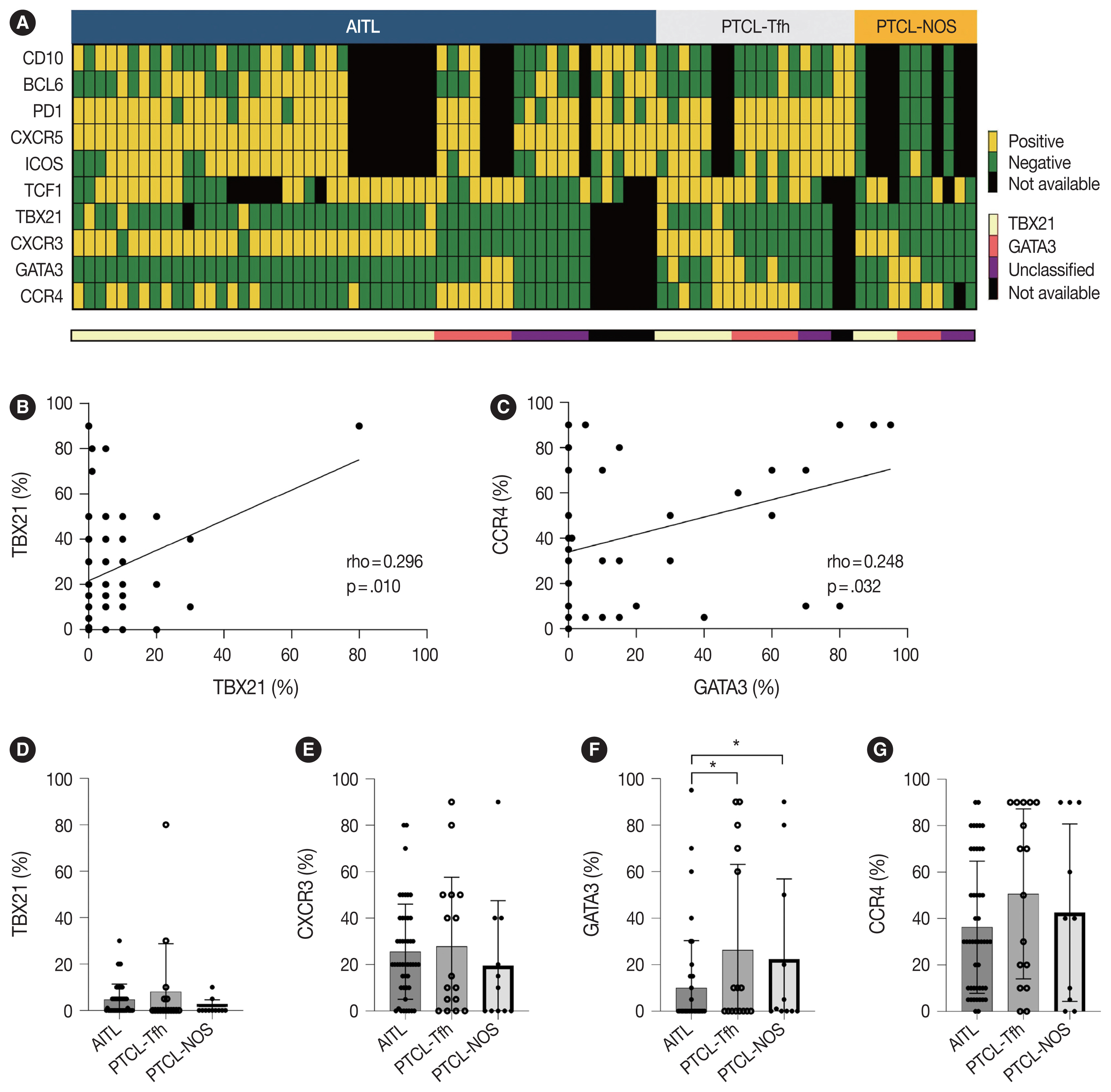
Eighty-two cases of nodal PTCL were classified into 53 angioimmunoblastic T-cell lymphomas (AITLs)/nodal T-follicular helper cell lymphoma (nTFHL)-AI, 18 PTCLs-Tfh/nTFHL–not otherwise specified (NOS), and 11 PTCLs-NOS according to the revised 4th/5th World Health Organization classifications. Immunohistochemistry for TCF1, TBX21, CXCR3, GATA3, and CCR4 was performed.
Results
TCF1 was highly expressed in up to 68% of patients with nTFHL but also in 44% of patients with PTCL-NOS (p > .05). CXCR3 expression was higher in AITLs than in non-AITLs (p = .035), whereas GATA3 expression was higher in non-AITL than in AITL (p = .007) and in PTCL-Tfh compared to AITL (p = .010). Of the cases, 70% of AITL, 44% of PTCLTfh/ nTFHL-NOS, and 36% of PTCL-NOS were subclassified as the TBX21 subtype; and 15% of AITL, 38% of PTCL-Tfh/nTFHL-NOS, and 36% of PTCL-NOS were subclassified as the GATA3 subtype. The others were an unclassified subtype. CCR4 expression was associated with poor progression-free survival (PFS) in patients with PTCL-Tfh (p < .001) and nTFHL (p = .023). The GATA3 subtype showed poor overall survival in PTCL-NOS compared to TBX21 (p = .046) and tended to be associated with poor PFS in patients with non-AITL (p = .054).
Conclusions
The TBX21 subtype was more prevalent than the GATA3 subtype in AITL. The GATA3 subtype was associated with poor prognosis in patients with non-AITL and PTCL-NOS.
- An unusual case of microsatellite instability–high/deficient mismatch repair (MSI-H/dMMR) diffuse large B-cell lymphoma revealed by targeted gene sequencing
-
Bogyeong Han, Sehui Kim, Jiwon Koh, Jeong Mo Bae, Hongseok Yun, Yoon Kyung Jeon
-
J Pathol Transl Med. 2022;56(2):92-96. Published online November 16, 2021
-
DOI: https://doi.org/10.4132/jptm.2021.10.15
-
-
7,077
View
-
256
Download
-
2
Web of Science
-
2
Crossref
-
Abstract
PDF
- Microsatellite instability-high/deficient mismatch repair (MSI-H/dMMR) status has been approved as a tissue-agnostic biomarker for immune checkpoint inhibitor therapy in patients with solid tumors. We report the case of an MSI-H/dMMR diffuse large B-cell lymphoma (DLBCL) identified by targeted gene sequencing (TGS). A 90-year-old female who presented with vaginal bleeding and a large mass in the upper vagina was diagnosed with germinal center-B-cell-like DLBCL, which recurred at the uterine cervix at 9 months after chemotherapy. Based on TGS of 121 lymphoma-related genes and the LymphGen algorithm, the tumor was classified genetically as DLBCL of EZB subtype. Mutations in multiple genes, including frequent frameshift mutations, were detected by TGS and further suggested MSI. The MSI-H/dMMR and loss of MLH1 and PMS2 expression were determined in MSI-fragment analysis, MSI real-time polymerase chain reaction, and immunohistochemical tests. This case demonstrates the potential diagnostic and therapeutic utility of lymphoma panel sequencing for DLBCL with MSI-H/dMMR.
-
Citations
Citations to this article as recorded by  - Chimeric and mutant CARD9 constructs enable analyses of conserved and diverged autoinhibition mechanisms in the CARD‐CC protein family
Jens Staal, Yasmine Driege, Femke Van Gaever, Jill Steels, Rudi Beyaert
The FEBS Journal.2024; 291(6): 1220. CrossRef - PD-L1+diffuse large B-cell lymphoma with extremely high mutational burden and microsatellite instability due to acquiredPMS2mutation
Andrew W. Allbee, James Gerson, Guang Yang, Adam Bagg
Molecular Case Studies.2023; 9(4): a006318. CrossRef
- Primary Peripheral Gamma Delta T-Cell Lymphoma of the Central Nervous System: Report of a Case Involving the Intramedullary Spinal Cord and Presenting with Myelopathy
-
Jeemin Yim, Seung Geun Song, Sehui Kim, Jae Won Choi, Kyu-Chong Lee, Jeong Mo Bae, Yoon Kyung Jeon
-
J Pathol Transl Med. 2019;53(1):57-61. Published online October 1, 2018
-
DOI: https://doi.org/10.4132/jptm.2018.08.21
-
-
6,169
View
-
156
Download
-
5
Web of Science
-
4
Crossref
-
Abstract
PDF
- Primary central nervous system lymphoma of T-cell origin (T-PCNSL) is rare, and its clinicopathological features remain unclear. Peripheral T-cell lymphoma of γδ T-cell origin is an aggressive lymphoma mainly involving extranodal sites. Here, we report a case of γδ T-PCNSL involving the intramedullary spinal cord and presenting with paraplegia. A 75-year-old Korean woman visited the hospital complaining of back pain and lower extremity weakness. Magnetic resonance imaging revealed multifocal enhancing intramedullary nodular lesions in the thoracic and lumbar spinal cord. An enhancing nodular lesion was observed in the periventricular white matter of the lateral ventricle in the brain. There were no other abnormalities in systemic organs or skin. Laminectomy and tumor removal were performed. The tumor consisted of monomorphic, medium-to-large atypical lymphocytes with pale-to-eosinophilic cytoplasm. Immunohistochemically, the tumor cells were CD3(+), TCRβF1(-), TCRγ(+), CD30(-), CD4(-), CD8(-), CD56(+), TIA1(+), granzyme B(+), and CD103(+). Epstein-Barr virus in situ was negative. This case represents a unique T-PCNSL of γδ T-cell origin involving the spinal cord.
-
Citations
Citations to this article as recorded by - B-Cell Lymphoma Intramedullary Tumor: Case Report and Systematic Review
Daniel Gregório Gonsalves, Paulo Eduardo Albuquerque Zito Raffa, Gabriela Gerenutti de Sousa, Melissa Esposito Gomes Rigueiral, Iracema Araújo Estevão, Cesar Cozar Pacheco, Roger Thomaz Rotta Medeiros, Paulo Roberto Franceschini, Paulo Henrique Pires de A
Asian Journal of Neurosurgery.2023; 18(02): 231. CrossRef - Primary intramedullary spinal cord lymphoma misdiagnosed as longitudinally extensive transverse myelitis: a case report and literature review
Huizhen Ge, Li Xu, Huajie Gao, Suqiong Ji
BMC Neurology.2023;[Epub] CrossRef - Clinicopathologic and Genetic Features of Primary T-cell Lymphomas of the Central Nervous System
Jeemin Yim, Jiwon Koh, Sehui Kim, Seung Geun Song, Jeong Mo Bae, Hongseok Yun, Ji-Youn Sung, Tae Min Kim, Sung-Hye Park, Yoon Kyung Jeon
American Journal of Surgical Pathology.2022; 46(4): 486. CrossRef - Peripheral T-Cell Lymphomas Involving the Central Nervous System: A Report From the Czech Lymphoma Study Group Registry
Heidi Mocikova, Robert Pytlík, Katerina Benesova, Andrea Janikova, Juraj Duras, Alice Sykorova, Katerina Steinerova, Vit Prochazka, Vit Campr, David Belada, Marek Trneny
Frontiers in Oncology.2022;[Epub] CrossRef
- Mucinous Cystadenoma of the Testis: A Case Report with Immunohistochemical Findings
-
Gilhyang Kim, Dohee Kwon, Hee Young Na, Sehui Kim, Kyung Chul Moon
-
J Pathol Transl Med. 2017;51(2):180-184. Published online February 13, 2017
-
DOI: https://doi.org/10.4132/jptm.2016.08.30
-
-
8,930
View
-
126
Download
-
5
Web of Science
-
8
Crossref
-
Abstract
PDF
- Mucinous cystadenoma of the testis is a very rare tumor. Herein, we report a case of mucinous cystadenoma arising in the testis of a 61-year-old man, along with a literature review. Computed tomography showed a 2.5-cm-sized poorly enhancing cystic mass. Grossly, the tumor was a unilocular cystic mass filled with mucinous material and confined to the testicular parenchyma. Histologically, the cyst had a fibrotic wall lined by mucinous columnar epithelium without atypia. Immunohistochemical staining was positive for cytokeratin 20 and CDX2, as well as focally positive for cytokeratin 7. The pathologic diagnosis was mucinous cystadenoma.
-
Citations
Citations to this article as recorded by - Review of Paratesticular Appendageal Tumors, Morphology, Immunohistochemistry, and Recent Molecular Advances
Mathew Vega, Muhammad T. Idrees
Surgical Pathology Clinics.2025; 18(1): 119. CrossRef - Cistoadenoma Mucinoso Paratesticular: Caso Interesante en el Instituto Guatemalteco de Seguridad Social
Edgar Estuardo González López, Carlos Gonzalo Estrada Pazos
Revista Guatemalteca de Urología.2023; 10(2): 16. CrossRef - Primary borderline mucinous tumor of the testis with postoperative metastasis: A rare case report
Yingyu Shi, Ling Song, Yan Luo
Radiology Case Reports.2023; 18(9): 3203. CrossRef - Case report: Misdiagnosis of primary mucinous cystadenoma of the testicle by ultrasound
Linlin Zhang, Jianyuan Xuan, Manxi Li, Mei Zhang, Yu Song, Ziang Pan, Bo Fan, Lin Lu, Hongyan Zhou, Yang Li
Frontiers in Oncology.2023;[Epub] CrossRef - Primary Borderline Mucinous Testicular Tumor: A Case Report and Literature Review
Changjuan Hao, Chunsong Kang, Xiaoyan Kang, Zhuanzhuan Yu, Tingting Li, Jiping Xue
Frontiers in Oncology.2021;[Epub] CrossRef - Ovarian-type Tumors (Mullerian Tumors) of the Testis: Clinicopathologic Findings with Recent Advances
Michelle S Lin, Alberto G Ayala, Jae Y Ro
annals of urologic oncology.2019; : 1. CrossRef - Borderline Mucinous Testicular Tumour: Diagnostic and Management difficulties
Krishan Pratap, Marlon Perera, Frances Malczewski, Rachel Esler
BMJ Case Reports.2018; 2018: bcr-2017-223787. CrossRef - Mucinous tumor arising in a giant sacrococcygeal teratoma
Fengtian Zhang, Xiaolong Yu, Jin Zeng, Min Dai
Medicine.2017; 96(47): e8759. CrossRef
- WITHDRAWN:Uterine Arteriovenous Malformation Accompanied with a Uterine Stromomyoma
-
Yul Ri Chung, Dohee Kwon, Sehui Kim, Hee Young Na, Nayoung Han, Hyo Jin Kim, Min A Kim, In Ae Park
-
Received May 13, 2016 Accepted September 23, 2016 Published online August 4, 2017
-
DOI: https://doi.org/10.4132/jptm.2016.09.24
-
-
|
 E-submission