- Adrenal hemangioblastoma
-
Joo-Yeon Koo, Kyung-Hwa Lee, Joon Hyuk Choi, Ho Seok Chung, Chan Choi
-
J Pathol Transl Med. 2022;56(3):161-166. Published online February 28, 2022
-
DOI: https://doi.org/10.4132/jptm.2021.12.28
-
-
 Abstract Abstract
 PDF PDF
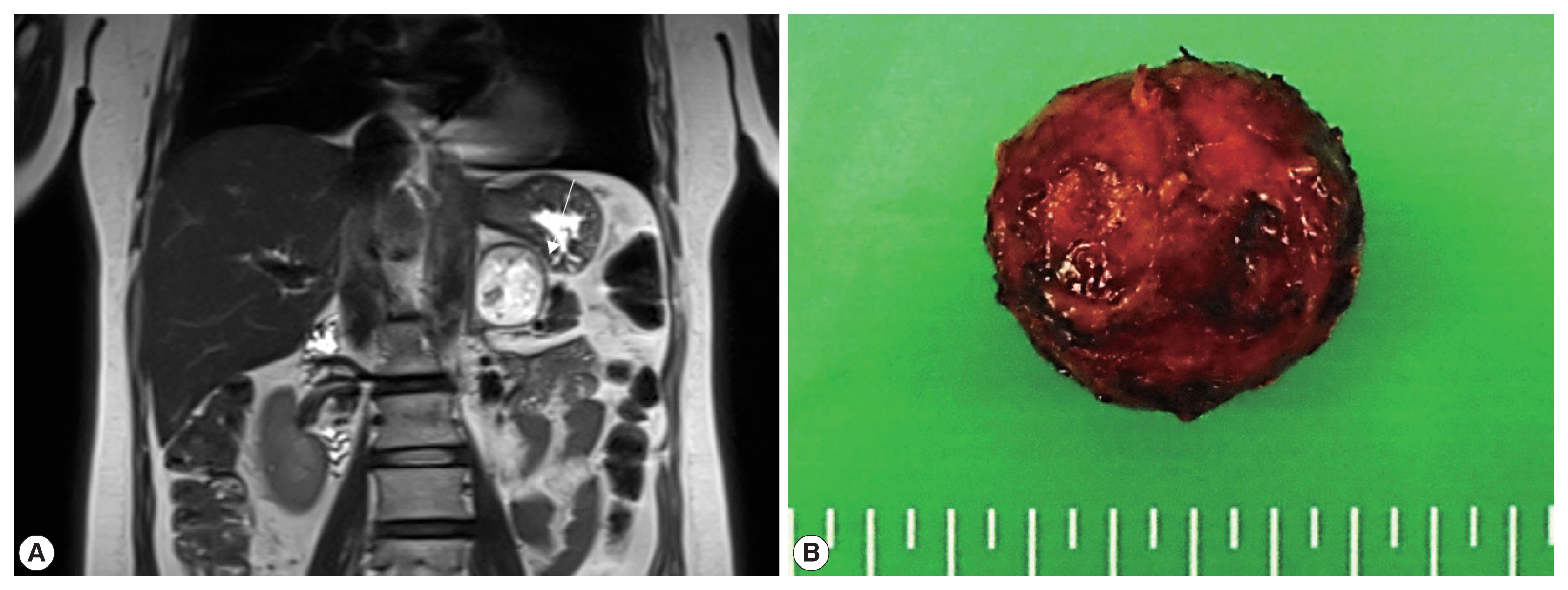
- Hemangioblastoma (HB) is a rare benign tumor that most commonly occurs in the cerebellum. HB is composed of neoplastic stromal cells and abundant small vessels. However, the exact origin of stromal cells is controversial. Extraneural HBs have been reported in a small series, and peripheral HBs arising in the adrenal gland are extremely rare. Herein, we report a case of sporadic adrenal HB in a 54-year-old woman. The tumor was a well-circumscribed, yellow mass measuring 4.2 cm in diameter. Histologically, the tumor was composed of small blood vessels and vacuolated stromal cells with clear cytoplasm. On immunohistochemical stain, the stromal cells were positive for S-100 protein, neuron-specific enolase, and synaptophysin. The tumor did not reveal mutation of VHL alleles. We herein present a case of HB of the adrenal gland and review of the literature.
- Fine Needle Aspiration Cytology of Hepatic Hydatid Cyst: A Case Study
-
Ae Ri Kim, Seok Ju Park, Mi Jin Gu, Joon Hyuk Choi, Hong Jin Kim
-
Korean J Pathol. 2013;47(4):395-398. Published online August 26, 2013
-
DOI: https://doi.org/10.4132/KoreanJPathol.2013.47.4.395
-
-
13,019
View
-
99
Download
-
9
Crossref
-
Abstract
PDF
Hydatid cysts (echinococcosis) are caused by an infestation with larval tapeworms of the genus Echinococcus. The disease is extensively distributed worldwide, and it has been rarely reported in Korea. We describe the cytologic features of a case of hepatic hydatid cyst in a 28-year-old male. Computed tomography revealed a cystic mass in the right lobe of the liver. A right hemihepatectomy was performed. The aspirated fluid from the hepatic cystic mass was clear. The smears showed protoscolices, hooklets, and a laminated membrane. -
Citations
Citations to this article as recorded by  - Delayed Diagnosis of Imported Cystic Echinococcosis and Successful Treatment With Percutaneous Drainage and Albendazole in Korea: A Case Report
Won Jun Choi, Hanna Jin, Hyeon Jae Jo, Chan Mi Lee, Chang Kyung Kang, Pyoeng Gyun Choe, Wan Beom Park, Nam Joong Kim, Min-Ho Choi
Journal of Korean Medical Science.2025;[Epub] CrossRef - Imported parasitic diseases in the Republic of Korea: status and issues
Jong-Yil Chai
Journal of the Korean Medical Association.2025; 68(1): 52. CrossRef - Chemical compounds, antioxidant and scolicidal potencies of Thymus fontanesii essential oil
Sidi Mohammed Ammar Selles, Belkacem Tahar Belhamiti, Mokhtaria Kouidri, Amar Ait Amrane, Yamina Kadari, Zohra Kaddour, Souad Kabrit
Experimental Parasitology.2024; 257: 108699. CrossRef - Complicated Liver Cystic Echinococcosis—A Comprehensive Literature Review and a Tale of Two Extreme Cases
Valentin Calu, Octavian Enciu, Elena-Adelina Toma, Radu Pârvuleţu, Dumitru Cătălin Pîrîianu, Adrian Miron
Tomography.2024; 10(6): 922. CrossRef - A rare case of hydatid cyst of the neck with concurrent pulmonary hydatid disease
Amarendra Kumar Shukla, Amrutha Peter, Veerendra Arya, Vineet Dwivedi, Manish Kumar Gupta, Nimish Rai, Pawan Tiwari, Jitendra Kishore Bhargava
Journal of Parasitic Diseases.2022; 46(4): 941. CrossRef - Hepatic Hydatid Cyst: A Case Report
Wan Chul Kim, Jae Uk Shin, Su Sin Jin
The Korean Journal of Gastroenterology.2021; 77(1): 35. CrossRef - An Imported Case of Disseminated Echinococcosis in Korea
Dong Hoon Shin, Hae Chan Jo, Jeong-Han Kim, Kang Il Jun, Wan Beom Park, Nam-Joong Kim, Min-Ho Choi, Chang Kyung Kang, Myoung-don Oh
The Korean Journal of Parasitology.2019; 57(4): 429. CrossRef - Cytology of hydatid cyst mimicking intra‐abdominal sarcoma, diagnosed by fine‐needle aspiration
Busra Ozbek, Nadir Paksoy
Diagnostic Cytopathology.2018; 46(4): 362. CrossRef - Clinical Update on Parasitic Diseases
Min Seo
Korean Journal of Medicine.2013; 85(5): 469. CrossRef
- Proposal for a Standardized Pathology Report of Gastroenteropancreatic Neuroendocrine Tumors: Prognostic Significance of Pathological Parameters
-
Mee-Yon Cho, Jin Hee Sohn, So Young Jin, Hyunki Kim, Eun Sun Jung, Mi-Jung Kim, Kyoung-Mee Kim, Woo Ho Kim, Joon Mee Kim, Yun Kyung Kang, Joon Hyuk Choi, Dae Young Kang, Youn Wha Kim, Eun Hee Choi
-
Korean J Pathol. 2013;47(3):227-237. Published online June 25, 2013
-
DOI: https://doi.org/10.4132/KoreanJPathol.2013.47.3.227
-
-
14,138
View
-
145
Download
-
12
Crossref
-
Abstract
PDF
- Background
There is confusion in the diagnosis and biological behaviors of gastroenteropancreatic neuroendocrine tumors (GEP-NETs), because of independently proposed nomenclatures and classifications. A standardized form of pathology report is required for the proper management of patients. MethodsWe discussed the proper pathological evaluation of GEP-NET at the consensus conference of the subcommittee meeting for the Gastrointestinal Pathology Study Group of the Korean Society of Pathologists. We then verified the prognostic significance of pathological parameters from our previous nationwide collection of pathological data from 28 hospitals in Korea to determine the essential data set for a pathology report. ResultsHistological classification, grading (mitosis and/or Ki-67 labeling index), T staging (extent, size), lymph node metastasis, and lymphovascular and perineural invasion were significant prognostic factors and essential for the pathology report of GEP-NET, while immunostaining such as synaptophysin and chromogranin may be optional. Furthermore, the staging system, either that of the 2010 American Joint Cancer Committee (AJCC) or the European Neuroendocrine Tumor Society (ENETS), should be specified, especially for pancreatic neuroendocrine neoplasms. ConclusionsA standardized pathology report is crucial for the proper management and prediction of prognosis of patients with GEP-NET.
-
Citations
Citations to this article as recorded by - Analysis of Prognostic Risk Factors of Endoscopic Submucosal Dissection (ESD) and Curative Resection of Gastrointestinal Neuroendocrine Neoplasms
Yuan Si, ChaoKang Huang, JingBin Yuan, XianHui Zhang, QingQiang He, ZhiJin Lin, Ling He, ZhongXin Liu, Yuvaraja Teekaraman
Contrast Media & Molecular Imaging.2022;[Epub] CrossRef - Standardization of the pathologic diagnosis of appendiceal mucinous neoplasms
Dong-Wook Kang, Baek-hui Kim, Joon Mee Kim, Jihun Kim, Hee Jin Chang, Mee Soo Chang, Jin-Hee Sohn, Mee-Yon Cho, So-Young Jin, Hee Kyung Chang, Hye Seung Han, Jung Yeon Kim, Hee Sung Kim, Do Youn Park, Ha Young Park, So Jeong Lee, Wonae Lee, Hye Seung Lee,
Journal of Pathology and Translational Medicine.2021; 55(4): 247. CrossRef - Preoperative diagnosis of well‐differentiated neuroendocrine tumor in common hepatic duct by brush cytology: A case report
Jiwoon Choi, Kyong Joo Lee, Sung Hoon Kim, Mee‐Yon Cho
Diagnostic Cytopathology.2019; 47(7): 720. CrossRef - Primary renal well-differentiated neuroendocrine tumors: report of six cases with an emphasis on the Ki-67 index and mitosis
Bohyun Kim, Han-Seong Kim, Kyung Chul Moon
Diagnostic Pathology.2019;[Epub] CrossRef - Primary low‐grade neuroendocrine carcinoma of the skin: An exceedingly rare entity
Tiffany Y. Chen, Annie O. Morrison, Joe Susa, Clay J. Cockerell
Journal of Cutaneous Pathology.2017; 44(11): 978. CrossRef - Prognostic Validity of the American Joint Committee on Cancer and the European Neuroendocrine Tumors Staging Classifications for Pancreatic Neuroendocrine Tumors
Jae Hee Cho, Ji Kon Ryu, Si Young Song, Jin-Hyeok Hwang, Dong Ki Lee, Sang Myung Woo, Young-Eun Joo, Seok Jeong, Seung-Ok Lee, Byung Kyu Park, Young Koog Cheon, Jimin Han, Tae Nyeun Kim, Jun Kyu Lee, Sung-Hoon Moon, Hyunjin Kim, Eun Taek Park, Jae Chul Hw
Pancreas.2016; 45(7): 941. CrossRef - Early diagnosis and treatment of gastrointestinal neuroendocrine tumors
Hong Shen, Zhuo Yu, Jing Zhao, Xiu-Zhen Li, Wen-Sheng Pan
Oncology Letters.2016; 12(5): 3385. CrossRef - Recent Updates on Neuroendocrine Tumors From the Gastrointestinal and Pancreatobiliary Tracts
Joo Young Kim, Seung-Mo Hong
Archives of Pathology & Laboratory Medicine.2016; 140(5): 437. CrossRef - Pancreatic neuroendocrine tumors: Correlation between the contrast-enhanced computed tomography features and the pathological tumor grade
Koji Takumi, Yoshihiko Fukukura, Michiyo Higashi, Junnichi Ideue, Tomokazu Umanodan, Hiroto Hakamada, Ichiro Kanetsuki, Takashi Yoshiura
European Journal of Radiology.2015; 84(8): 1436. CrossRef - Tumeurs neuroendocrines du tube digestif et du pancréas : ce que le pathologiste doit savoir et doit faire en 2014
Jean-Yves Scoazec, Anne Couvelard
Annales de Pathologie.2014; 34(1): 40. CrossRef - Spectrum of Gastroenteropancreatic NENs in Routine Histological Examinations of Bioptic and Surgical Specimen: A Study of 161 Cases Collected from 17 Departments of Pathology in the Czech Republic
Václav Mandys, Tomáš Jirásek
Gastroenterology Research and Practice.2014; 2014: 1. CrossRef - p27 Loss Is Associated with Poor Prognosis in Gastroenteropancreatic Neuroendocrine Tumors
Hee Sung Kim, Hye Seung Lee, Kyung Han Nam, Jiwoon Choi, Woo Ho Kim
Cancer Research and Treatment.2014; 46(4): 383. CrossRef
- Imprint Cytology of Soft Tissue Myoepithelioma: A Case Study
-
Seok Ju Park, Ae Ri Kim, Mi Jin Gu, Joon Hyuk Choi, Duk Seop Shin
-
Korean J Pathol. 2013;47(3):299-303. Published online June 25, 2013
-
DOI: https://doi.org/10.4132/KoreanJPathol.2013.47.3.299
-
-
9,412
View
-
52
Download
-
7
Crossref
-
Abstract
PDF
Soft tissue myoepithelioma is a rare neoplasm composed of myoepithelial cells. Here, we describe the cytologic features of soft tissue myoepithelioma arising on the right forearm in an 18-year-old man. The excised tumor (3.0×1.8×1.5 cm) was well-demarcated, yellow-gray, soft, and myxoid. The cytologic smears showed round to spindle, epithelioid, and plasmacytoid cells in the myxoid background. The nuclei were uniform, round to ovoid, with finely distributed chromatin and eosinophilic or pale cytoplasm. The tumor cells demonstrated immunoreactivity for cytokeratin (AE1/AE3), epithelial membrane antigen, S100 protein, and glial fibrillary acidic protein. Electron microscopy showed intermediate filaments, desmosomes, and basal lamina. -
Citations
Citations to this article as recorded by - Myoepithelial tumors of soft tissue and bone in children and young adults: A clinicopathologic study of 40 cases occurring in patients ≤ 21 Years of age
Suzanna J. Logan, Carina A. Dehner, Fatimah I. Alruwaii, Nasir Ud Din, Damon R. Olson, Karen J. Fritchie, Gregory W. Charville, Melissa M. Blessing, Andrew L. Folpe
Human Pathology.2024; 149: 10. CrossRef - Fine-needle aspiration cytopathology of soft tissue myoepithelioma: an analysis of seven cases
Paul E. Wakely, Momin T. Siddiqui
Journal of the American Society of Cytopathology.2022; 11(1): 31. CrossRef - Cytology‐histology correlation of myoepithelial tumors harboring EWSR1‐POU5F1 fusions: A report of two cases
Ian A. Gelarden, Lucy Fu, Kai Lee Yap, Aida I. Richardson, Pauline M. Chou
Diagnostic Cytopathology.2022;[Epub] CrossRef - A case of myoepithelial carcinoma of the left shoulder
Shuhei ISHII, Noriyuki FURUTA, Kyoko KOMATSU, Yoshiya SUGIURA, Noriko MOTOI, Yutaka TAKAZAWA, Yuko SUGIYAMA, Yuichi ISHIKAWA
The Journal of the Japanese Society of Clinical Cytology.2018; 57(2): 129. CrossRef - Fine‐needle aspiration of soft tissue myoepithelioma
Gang Wang, Tracy Tucker, Tony L. Ng, Carlos F. Villamil, Malcolm M. Hayes
Diagnostic Cytopathology.2016; 44(2): 152. CrossRef - A case report of spindle cell myoepithelioma with extensive lipomatous metaplasia and thick collagen bundles in the submandibular gland
Mi Jung Kwon, Hye Jeong Kim, Bumjung Park, Seong Jin Cho, Hyung Sik Shin, Hye‐Rim Park, Soo Kee Min, Jinwon Seo, Kyueng‐Whan Min, Eun Sook Nam
Diagnostic Cytopathology.2016; 44(9): 764. CrossRef - Myoepithelioma of soft tissue, a case report
Hassania Ameurtesse, Leila Chbani, JM Coindre, Hinde Elfatemi, Toufik Harmouch, Afaf Amarti
Research.2014;[Epub] CrossRef
- Extranodal Follicular Dendritic Cell Sarcoma with Rapid Growth in Parapharynx: A Case Report
-
Jung-Soo Pyo, Guhyun Kang, Sung-Im Do, Seoung Wan Chae, Kyungeun Kim, Sang Hyuk Lee, Yoon-La Choi, Joon Hyuk Choi, Jin Hee Sohn, Dong-Hoon Kim
-
Korean J Pathol. 2012;46(3):306-310. Published online June 22, 2012
-
DOI: https://doi.org/10.4132/KoreanJPathol.2012.46.3.306
-
-
7,287
View
-
54
Download
-
9
Crossref
-
Abstract
PDF
Follicular dendritic cell sarcoma (FDCS) is a rare malignancy arising from the antigen-presenting cells in the lymph node and extranodal tissue. We describe a 31-year-old male patient who presented with a swelling of the left parapharynx. The radiologic findings showed a 4.7×4.5×1.9 cm-sized, ill-defined mass in the left parapharyngeal space. A fine-needle aspiration cytology was performed and it showed scattered, irregular, cohesive clusters of tumor cells with a spindle-to-ovoid shape with irregular contours in a background of lymphocytes. Based on these findings, a diagnosis of spindle cell neoplasm was made. The surgically resected tumor was composed of elongated, ovoid or polygonal cells showing positive immunohistochemistry for CD21, CD23, and CD35. Postoperatively, the residual tumor was observed to undergo a rapidly growth. There is an overlap in the cytologic and histologic findings between FDCS of the parapharynx and other tumors. Pathologists should therefore be aware of its characteristics not only to provide an accurate diagnosis but also to recommend the appropriate clinical management. -
Citations
Citations to this article as recorded by - Extranodal Follicular Dendritic Cell Sarcoma of the Head and Neck Region: A Clinicopathological Study of 7 Cases
Nasir Ud Din, Zubair Ahmad, Shabina Rahim, Karen Fritchie, Muhammad Usman Tariq, Arsalan Ahmed
International Journal of Surgical Pathology.2023; 31(6): 1067. CrossRef - Cytomorphology of follicular dendritic cell sarcoma: a report of 7 cases with an emphasis on the diagnostic challenges
Cody Weimholt, Jalal B. Jalaly, Cedric Bailey
Journal of the American Society of Cytopathology.2023; 12(3): 229. CrossRef - Follicular dendritic cells
Seham A. Abd El‐Aleem, Entesar Ali Saber, Neven M. Aziz, Hani El‐Sherif, Asmaa M. Abdelraof, Laiche Djouhri
Journal of Cellular Physiology.2022; 237(4): 2019. CrossRef - Clinicopathological characteristics of extranodal follicular dendritic cell sarcoma: A report of two cases
Xing Zhao, Dayong Sun, Gang Zhang
Oncology Letters.2021;[Epub] CrossRef - Cytological diagnosis of follicular dendritic cell sarcoma: A case report and review of literature
A. Dutta, P. Arun, P. Roy, I. Arun
Cytopathology.2018; 29(5): 461. CrossRef - Follicular dendritic cells and related sarcoma
Fabio Facchetti, Luisa Lorenzi
Seminars in Diagnostic Pathology.2016; 33(5): 262. CrossRef - Extranodal follicular dendritic cell sarcoma: A clinicopathological report of four cases and a literature review
RUI-FEN WANG, WEI HAN, LEI QI, LI-HUI SHAN, ZHENG-CAI WANG, LI-FENG WANG
Oncology Letters.2015; 9(1): 391. CrossRef - Follicular Dendritic Cell Sarcoma of Parapharyngeal Space: A Case Report and Review of the Literature
Turki Al-Hussain, Muhammad Saleem, Suresh Babu Velagapudi, Mohammad Anas Dababo
Head and Neck Pathology.2015; 9(1): 135. CrossRef - Clinical and pathological features of head and neck follicular dendritic cell sarcoma
Ji Li, Min-Li Zhou, Shui-Hong Zhou
Hematology.2015; 20(10): 571. CrossRef
|
 E-submission