- A multicenter study of interobserver variability in pathologic diagnosis of papillary breast lesions on core needle biopsy with WHO classification
-
Hye Ju Kang, Sun Young Kwon, Ahrong Kim, Woo Gyeong Kim, Eun Kyung Kim, Ae Ree Kim, Chungyeul Kim, Soo Kee Min, So Young Park, Sun Hee Sung, Hye Kyoung Yoon, Ahwon Lee, Ji Shin Lee, Hyang Im Lee, Ho Chang Lee, Sung Chul Lim, Sun Young Jun, Min Jung Jung, Chang Won Jung, Soo Youn Cho, Eun Yoon Cho, Hye Jeong Choi, So Yeon Park, Jee Yeon Kim, In Ae Park, Youngmee Kwon
-
J Pathol Transl Med. 2021;55(6):380-387. Published online October 6, 2021
-
DOI: https://doi.org/10.4132/jptm.2021.07.29
-
-
5,482
View
-
211
Download
-
4
Web of Science
-
5
Crossref
-
 Abstract Abstract
 PDF PDF Supplementary Material Supplementary Material
- Background
Papillary breast lesions (PBLs) comprise diverse entities from benign and atypical lesions to malignant tumors. Although PBLs are characterized by a papillary growth pattern, it is challenging to achieve high diagnostic accuracy and reproducibility. Thus, we investigated the diagnostic reproducibility of PBLs in core needle biopsy (CNB) specimens with World Health Organization (WHO) classification.
Methods
Diagnostic reproducibility was assessed using interobserver variability (kappa value, κ) and agreement rate in the pathologic diagnosis of 60 PBL cases on CNB among 20 breast pathologists affiliated with 20 medical institutions in Korea. This analysis was performed using hematoxylin and eosin (H&E) staining and immunohistochemical (IHC) staining for cytokeratin 5 (CK5) and p63. The pathologic diagnosis of PBLs was based on WHO classification, which was used to establish simple classifications (4-tier, 3-tier, and 2-tier).
Results
On WHO classification, H&E staining exhibited ‘fair agreement’ (κ = 0.21) with a 47.0% agreement rate. Simple classifications presented improvement in interobserver variability and agreement rate. IHC staining increased the kappa value and agreement rate in all the classifications. Despite IHC staining, the encapsulated/solid papillary carcinoma (EPC/SPC) subgroup (κ = 0.16) exhibited lower agreement compared to the non-EPC/SPC subgroup (κ = 0.35) with WHO classification, which was similar to the results of any other classification systems.
Conclusions
Although the use of IHC staining for CK5 and p63 increased the diagnostic agreement of PBLs in CNB specimens, WHO classification exhibited a higher discordance rate compared to any other classifications. Therefore, this result warrants further intensive consensus studies to improve the diagnostic reproducibility of PBLs with WHO classification.
-
Citations
Citations to this article as recorded by  - Beyond the benign: A rare case report of myxoid pleomorphic liposarcoma
Arslan Ahmad, Muhammad Ammar, Muhammad Hasnain Saleem Choudary, Muhammad Nouman Sadiq, Rana Uzair Ahmad, Nouman Aziz
Radiology Case Reports.2025; 20(5): 2500. CrossRef - Invasive papillary carcinoma of the breast
Shijing Wang, Qingfu Zhang, Xiaoyun Mao
Frontiers in Oncology.2024;[Epub] CrossRef - Recommendations for Performance Evaluation of Machine Learning in Pathology: A Concept Paper From the College of American Pathologists
Matthew G. Hanna, Niels H. Olson, Mark Zarella, Rajesh C. Dash, Markus D. Herrmann, Larissa V. Furtado, Michelle N. Stram, Patricia M. Raciti, Lewis Hassell, Alex Mays, Liron Pantanowitz, Joseph S. Sirintrapun, Savitri Krishnamurthy, Anil Parwani, Giovann
Archives of Pathology & Laboratory Medicine.2024; 148(10): e335. CrossRef - Encapsulated papillary carcinoma of the breast: A single institution experience
Liang Xu, Qixin Mao, Qiuming Liu, Yufeng Gao, Lihua Luo, Chungen Guo, Wei Qu, Ningning Yan, Yali Cao
Oncology Letters.2023;[Epub] CrossRef - High-risk and selected benign breast lesions diagnosed on core needle biopsy: Evidence for and against immediate surgical excision
Aparna Harbhajanka, Hannah L. Gilmore, Benjamin C. Calhoun
Modern Pathology.2022; 35(11): 1500. CrossRef
- Cytomorphological Features of Hyperchromatic Crowded Groups in Liquid-Based Cervicovaginal Cytology: A Single Institutional Experience
-
Youngeun Lee, Cheol Lee, In Ae Park, Hyoung Jin An, Haeryoung Kim
-
J Pathol Transl Med. 2019;53(6):393-398. Published online September 16, 2019
-
DOI: https://doi.org/10.4132/jptm.2019.08.14
-
-
8,487
View
-
207
Download
-
5
Web of Science
-
5
Crossref
-
Abstract
PDF
- Background
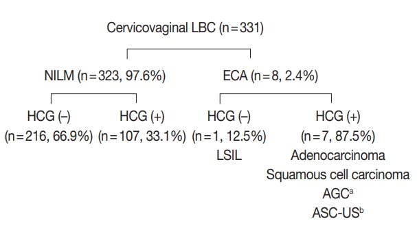
Hyperchromatic crowed groups (HCGs) are defined as three-dimensional aggregates of crowded cells with hyperchromatic nuclei, and are frequently encountered in cervicovaginal liquid-based cytology (LBC). Here, we aimed to examine the prevalence of HCGs in cervicovaginal LBC and the cytomorphological characteristics of various epithelial cell clusters presenting as HCGs.
Methods
We first examined the prevalence of HCGs in a “routine cohort” of LBC cytology (n=331), consisting of all cervicovaginal LBCs accessioned over 3 days from outpatient clinics (n=179) and the screening population (n=152). Then we examined a second “high-grade epithelial cell abnormalities (H-ECA) cohort” (n=69) of LBCs diagnosed as high-grade squamous intraepithelial lesion (HSIL), squamous cell carcinoma (SCC), or adenocarcinoma during 1 year.
Results
HCGs was observed in 34.4% of the routine cohort and were significantly more frequent in the epithelial cell abnormality category compared to the non-neoplastic category (p=.003). The majority of HCGs represented atrophy (70%). Of the 69 histologically confirmed H-ECA cases, all contained HCGs. The majority of cases were HSIL (62%), followed by SCC (16%). Individually scattered neoplastic cells outside the HCGs were significantly more frequent in SCCs compared to glandular neoplasia (p=.002). Despite the obscuring thick nature of the HCGs, examining the edges and the different focal planes of the HCGs and the background were helpful in defining the nature of the HCGs.
Conclusions
HCGs were frequently observed in cervicovaginal LBC and were mostly non-neoplastic; however, neoplastic HCGs were mostly high-grade lesions. Being aware of the cytomorphological features of different HCGs is important in order to avoid potential false-negative cytology interpretation.
-
Citations
Citations to this article as recorded by - Can Mitotic Figures in Hyperchromatic Crowded Groups be Cytodiagnostic Criteria for High-Grade Squamous Intra-epithelial Lesions?
Hisae Suzuki, Yumeno Kondo, Chihiro Oda, Takeshi Nishikawa, Mao Takeuchi, Shigenobu Tatsumi, Sho Hosokawa, Satoshi Irino, Tomoko Uchiyama, Tomomi Fujii, Yoshiaki Norimatsu
Journal of Cytology.2024; 41(2): 116. CrossRef - Quantitative Structural Analysis of Hyperchromatic Crowded Cell Groups in Cervical Cytology: Overcoming Diagnostic Pitfalls
Shinichi Tanaka, Tamami Yamamoto, Norihiro Teramoto
Cancers.2024; 16(24): 4258. CrossRef - Atypical glandular cells (AGC): Cytology of glandular lesions of the uterine cervix
Mir Yousufuddin Ali Khan, Sudeshna Bandyopadhyay, Ahmed Alrajjal, Moumita Saha Roy Choudhury, Rouba Ali-Fehmi, Vinod B. Shidham
Cytojournal.2022; 19: 31. CrossRef - Cytopathologic features of human papillomavirus–independent, gastric-type endocervical adenocarcinoma
Min-Kyung Yeo, Go Eun Bae, Dong-Hyun Kim, In-Ock Seong, Kwang-Sun Suh
Journal of Pathology and Translational Medicine.2022; 56(5): 260. CrossRef - The association of atypical squamous cells, cannot exclude a high grade squamous intraepithelial lesion, hyperchromatic crowded groups and high grade squamous intraepithelial lesions involving endocervical glands
Suzanne M. Selvaggi
Diagnostic Cytopathology.2021; 49(9): 1008. CrossRef
- High Cytoplasmic CXCR4 Expression Predicts Prolonged Survival in Triple-Negative Breast Cancer Patients Treated with Adjuvant Chemotherapy
-
Bobae Shim, Min‐Sun Jin, Ji Hye Moon, In Ae Park, Han Suk Ryu
-
J Pathol Transl Med. 2018;52(6):369-377. Published online October 1, 2018
-
DOI: https://doi.org/10.4132/jptm.2018.09.19
-
-
13,019
View
-
182
Download
-
10
Web of Science
-
12
Crossref
-
Abstract
PDF
- Background
Chemokine receptor CXC chemokine receptor type 4 (CXCR4) and its ligand CXC motif chemokine 12 (CXCL12; stromal cell-derived factor-1) are implicated in tumor growth, metastasis, and tumor cell-microenvironment interaction. A number of studies have reported that increased CXCR4 expression is associated with worse prognosis in triple-negative breast cancer (TNBC), but its prognostic significance has not been studied in TNBC patients treated with adjuvant chemotherapy.
Methods
Two hundred eighty-three TNBC patients who received adjuvant chemotherapy were retrospectively analyzed. Tissue microarray was constructed from formalinfixed, paraffin-embedded tumor tissue and immunohistochemistry for CXCR4 and CXCL12 was performed. Expression of each marker was compared with clinicopathologic characteristics and outcome.
Results
High cytoplasmic CXCR4 expression was associated with younger age (p = .008), higher histologic grade (p = .007) and lower pathologic stage (p = .045), while high CXCL12 expression was related to larger tumor size (p = .045), positive lymph node metastasis (p = .005), and higher pathologic stage (p = .017). The patients with high cytoplasmic CXCR4 experienced lower distant recurrence (p = .006) and better recurrence-free survival (RFS) (log-rank p = .020) after adjuvant chemotherapy. Cytoplasmic CXCR4 expression remained an independent factor of distant recurrence (p = .019) and RFS (p = .038) after multivariate analysis.
Conclusions
High cytoplasmic CXCR4 expression was associated with lower distant recurrence and better RFS in TNBC patients treated with adjuvant chemotherapy. This is the first study to correlate high CXCR4 expression to better TNBC prognosis, and the underlying mechanism needs to be elucidated in further studies.
-
Citations
Citations to this article as recorded by - Bisphenol A-induced cancer-associated adipocytes promotes breast carcinogenesis via CXCL12/AKT signaling
Zhiyuan Dong, Liping He, Jinyi Wu, Chunfeng Xie, Shanshan Geng, Jieshu Wu, Caiyun Zhong, Xiaoting Li
Molecular and Cellular Endocrinology.2025; 599: 112473. CrossRef - Distinct profiles of proliferating CD8+/TCF1+ T cells and CD163+/PD-L1+ macrophages predict risk of relapse differently among treatment-naïve breast cancer subtypes
Konstantinos Ntostoglou, Sofia D. P. Theodorou, Tanja Proctor, Ilias P. Nikas, Sinclair Awounvo, Athanasia Sepsa, Vassilis Georgoulias, Han Suk Ryu, Ioannis S. Pateras, Christos Kittas
Cancer Immunology, Immunotherapy.2024;[Epub] CrossRef - Unravelling the CXCL12/CXCR4 Axis in breast cancer: Insights into metastasis, microenvironment interactions, and therapeutic opportunities
Priyanka Garg, Venkateswara Rao Jallepalli, Sonali Verma
Human Gene.2024; 40: 201272. CrossRef - New Emerging Chemokine Receptors: CCR5 or CXCR5 on Tumor Is Associated with Poor Response to Chemotherapy and Poor Prognosis in Locally Advanced Triple-Negative Breast Cancer
Neslihan Cabioglu, Semen Onder, Hüseyin Karatay, Aysel Bayram, Gizem Oner, Mustafa Tukenmez, Mahmut Muslumanoglu, Abdullah Igci, Ahmet Dinccag, Vahit Ozmen, Adnan Aydiner, Pınar Saip, Ekrem Yavuz
Cancers.2024; 16(13): 2388. CrossRef - Cancer-Associated-Fibroblast-Mediated Paracrine and Autocrine SDF-1/CXCR4 Signaling Promotes Stemness and Aggressiveness of Colorectal Cancers
Chao-Yang Chen, Shih-Hsien Yang, Ping-Ying Chang, Su-Feng Chen, Shin Nieh, Wen-Yen Huang, Yu-Chun Lin, Oscar Kuang-Sheng Lee
Cells.2024; 13(16): 1334. CrossRef - Associations of CXCL12 polymorphisms with clinicopathological features in breast cancer: a case-control study
Shuai Lin, Yi Zheng, Meng Wang, Linghui Zhou, Yuyao Zhu, Yujiao Deng, Ying Wu, Dai Zhang, Na Li, Huafeng Kang, Zhijun Dai
Molecular Biology Reports.2022; 49(3): 2255. CrossRef - The clinicopathological and prognostic value of CXCR4 expression in patients with lung cancer: a meta-analysis
Liping Qiu, Yuanyuan Xu, Hui Xu, Biyun Yu
BMC Cancer.2022;[Epub] CrossRef - Demystifying the CXCR4 conundrum in cancer biology: Beyond the surface signaling paradigm
Mushtaq Ahmad Nengroo, Muqtada Ali Khan, Ayushi Verma, Dipak Datta
Biochimica et Biophysica Acta (BBA) - Reviews on Cancer.2022; 1877(5): 188790. CrossRef - Targeted dendrimers for antagonizing the migration and viability of NALM-6 lymphoblastic leukemia cells
Chuda Chittasupho, Chaiyawat Aonsri, Witcha Imaram
Bioorganic Chemistry.2021; 107: 104601. CrossRef - CXCR4 and RANK Combination as a Predictor of Breast Cancer Bone Metastasis in Indonesia
Yulian Erwin D
Journal of Surgery and Surgical Research.2021; : 020. CrossRef - CXCL12/CXCR4 axis in the microenvironment of solid tumors: A critical mediator of metastasis
Keywan Mortezaee
Life Sciences.2020; 249: 117534. CrossRef - Impact of the Chemokine Receptors CXCR4 and CXCR7 on Clinical Outcome in Adrenocortical Carcinoma
Irina Chifu, Britta Heinze, Carmina T. Fuss, Katharina Lang, Matthias Kroiss, Stefan Kircher, Cristina L. Ronchi, Barbara Altieri, Andreas Schirbel, Martin Fassnacht, Stefanie Hahner
Frontiers in Endocrinology.2020;[Epub] CrossRef
- Cytologic Diagnosis of Metastatic Alveolar Rhabdomyosarcoma in Cerebrospinal Fluid: A Case Report
-
Bobae Shim, Jiwon Koh, Ji Hye Moon, In Ae Park, Han Suk Ryu
-
J Pathol Transl Med. 2018;52(4):262-266. Published online June 14, 2018
-
DOI: https://doi.org/10.4132/jptm.2018.05.15
-
-
6,969
View
-
118
Download
-
3
Web of Science
-
3
Crossref
-
Abstract
PDF
- Rhabdomyosarcoma is a malignant soft tissue tumor which shows skeletal muscle differentiation. Leptomeningeal metastasis can occur as a late complication, but currently there are no reports that have documented the cytologic features in cerebrospinal fluid (CSF). We report a case of metastatic alveolar rhabdomyosarcoma diagnosed in the CSF of a 28-year-old male who was originally diagnosed with rhabdomyosarcoma on the neck, and that went through systemic therapy. The tumor was positive for anaplastic lymphoma kinase, but progressed despite additional therapy with crizotinib. The CSF specimen revealed small round cells, large atypical cells with abundant cytoplasm and eccentric nuclei, and cells with horseshoe-shaped nuclei. These cytologic findings were in agreement with previous literature and well-correlated with histopathology. This is the first report to document the cytologic feature of rhabdomyosarcoma in CSF. In many cases it is difficult to perform ancillary tests in a CSF specimen and cytopathologists should be aware of the cytomorphologic characteristics to avoid misdiagnosis.
-
Citations
Citations to this article as recorded by - A Review of Effusion Cytomorphology of Small Round Cell Tumors
Lucy M. Han, Christopher J. VandenBussche, Mads Abildtrup, Ashish Chandra, Poonam Vohra
Acta Cytologica.2022; 66(4): 336. CrossRef - Cytologic diagnosis of metastatic embryonal rhabdomyosarcoma in cerebrospinal fluid: A case report
Muxia Yan, Ying Wu, Jianqing Xia, Xiaohong Zhang, Yiqian Wang
Diagnostic Cytopathology.2021;[Epub] CrossRef - Effusion cytology of epithelioid rhabdomyosarcoma
Andrew A. Renshaw, Edwin W. Gould
Diagnostic Cytopathology.2019; 47(10): 1042. CrossRef
- Diagnostic Accuracy of Endoscopic Ultrasound-Guided Fine Needle Aspiration Cytology of Pancreatic Lesions
-
Hae Woon Baek, Min Jee Park, Ye-Young Rhee, Kyoung Bun Lee, Min A Kim, In Ae Park
-
J Pathol Transl Med. 2015;49(1):52-60. Published online January 15, 2015
-
DOI: https://doi.org/10.4132/jptm.2014.10.26
-
-
10,229
View
-
88
Download
-
33
Web of Science
-
32
Crossref
-
Abstract
PDF
- Background
Endoscopic ultrasound–guided fine needle aspiration cytology (EUS-FNAC) is currently the most commonly used procedure for obtaining cytologic specimens of the pancreas. It is accurate, minimally invasive, safe and cost-effective. However, there is discrepancy between cytological and surgical diagnoses. This study was aimed at evaluating the diagnostic accuracy of EUS-FNAC of the pancreas. Methods: We performed a retrospective review of 191 cases of pancreatic lesions initially diagnosed by EUS-FNAC with subsequent histological diagnosis between 2010 and 2012 in the Department of Pathology, Seoul National University Hospital. Cytologic and surgical diagnoses were categorized into five groups: negative, benign, atypical, malignant, and insufficient for diagnosis. Subsequently, 167 cases with satisfactory yield in both surgical and cytology specimens were statistically analyzed to determine correlations with diagnosis. Results: In comparison to surgical diagnoses, cytologic diagnoses were true-positive in 103 cases (61.7%), true-negative in 28 cases (16.8%), false-positive in 9 cases (5.4%), and false-negative in 27 cases (16.1%). The diagnostic accuracy was 78.4%, sensitivity was 79.2%, and specificity was 75.7%. The positive predictive value was 92.0%, and negative predictive value was 50.9%. Conclusions: EUS-FNAC has high accuracy, sensitivity, specificity and positive predictive value. Overcoming the limitations of EUS-FNAC will make it a useful and reliable diagnostic tool for accurate evaluation of pancreatic lesions.
-
Citations
Citations to this article as recorded by - miR-6855-5p Enhances Radioresistance and Promotes Migration of Pancreatic Cancer by Inducing Epithelial-Mesenchymal Transition via Suppressing FOXA1: Potential of Plasma Exosomal miR-6855-5p as an Indicator of Radiosensitivity in Patients with Pancreatic
Hiroki Ueda, Hidenori Takahashi, Shogo Kobayashi, Masahiko Kubo, Kazuki Sasaki, Yoshifumi Iwagami, Daisaku Yamada, Yoshito Tomimaru, Tadafumi Asaoka, Takehiro Noda, Junzo Shimizu, Yuichiro Doki, Hidetoshi Eguchi
Annals of Surgical Oncology.2025; 32(2): 720. CrossRef - Grading pancreatic adenocarcinomas on fine needle aspiration cytology. The outstanding issues
Mrinmay Kumar Mallik, Laila Rafiq Qadan, Asit Kumar Mohanty, Ali Alali, Kusum Kapila
Cytopathology.2024; 35(2): 256. CrossRef - Reporting Pancreatic FNAC using the Papanicolaou System: Still a Diagnostic Challenge
Parul Verma, Saloni Goyal, Ruchita Tyagi, Mehar Ghuman, Ramit Mahajan, Arshneet Kaur Selhi, Harpreet Kaur, Pavneet Kaur Selhi
Journal of Cytology.2024; 41(2): 123. CrossRef - Preoperative treatment response prediction for pancreatic cancer by multiple microRNAs in plasma exosomes: Optimization using machine learning and network analysis
Hiroki Ueda, Hidenori Takahashi, Ryoto Sakaniwa, Tetsuhisa Kitamura, Shogo Kobayashi, Yoshito Tomimaru, Masahiko Kubo, Kazuki Sasaki, Yoshifumi Iwagami, Daisaku Yamada, Tadafumi Asaoka, Takehiro Noda, Junzo Shimizu, Yuichiro Doki, Hidetoshi Eguchi
Pancreatology.2024; 24(7): 1097. CrossRef - Comparison of Endoscopic Ultrasound-Guided Fine-Needle Aspiration with Fine-Needle Biopsy for Solid Gastrointestinal Lesions: A Randomized Crossover Single-Center study
Shivaraj Afzalpurkar, Vijay Kumar Rai, Nikhil Sonthalia, Gajanan Rodge, Awanesh Tewary, Mahesh Goenka
Journal of Digestive Endoscopy.2023; 14(01): 014. CrossRef - Diagnosing and monitoring pancreatic cancer through cell-free DNA methylation: progress and prospects
María Victoria García-Ortiz, Pablo Cano-Ramírez, Marta Toledano-Fonseca, Enrique Aranda, Antonio Rodríguez-Ariza
Biomarker Research.2023;[Epub] CrossRef - The impact of preoperative EUS-FNA for distal resectable pancreatic cancer: Is it really effective enough to take risks?
Jin-Seok Park, Jae Hoon Lee, Tae Jun Song, Joune Seup Lee, Seok Jung Jo, Dong Wook Oh, Ki Byung Song, Dae Wook Hwang, Do Hyun Park, Sang Soo Lee, Song Cheol Kim, Dong Wan Seo, Sung Koo Lee, Myung-Hwan Kim
Surgical Endoscopy.2022; 36(5): 3192. CrossRef - Magnetic Resonance Imaging Radiomics‐Based Nomogram From Primary Tumor for Pretreatment Prediction of Peripancreatic Lymph Node Metastasis in Pancreatic Ductal Adenocarcinoma: A Multicenter Study
Zhenshan Shi, Chengle Ma, Xinming Huang, Dairong Cao
Journal of Magnetic Resonance Imaging.2022; 55(3): 823. CrossRef - Role of Pathologist in the Era of Image-Guided and EUS-Guided Aspirations: A 10-Year Study at a Single Tertiary Care Oncology Institute in North India
Gurudutt Gupta, Anila Sharma, Meenakshi Kamboj, Anurag Sharma, Sunil Pasricha, Garima Durga, Anurag Mehta, Avinash Rao
Acta Cytologica.2022; 66(3): 187. CrossRef - Predicting Factors for Pancreatic Malignancy with Computed Tomography and Endoscopic Ultrasonography in Chronic Pancreatitis
Jian-Han Lai, Keng-Han Lee, Chen-Wang Chang, Ming-Jen Chen, Ching-Chung Lin
Diagnostics.2022; 12(4): 1004. CrossRef - Phenotypic profiling of pancreatic ductal adenocarcinoma plasma-derived small extracellular vesicles for cancer diagnosis and cancer stage prediction: a proof-of-concept study
Wei Zhang, Ling Wang, Dan Li, Douglas H. Campbell, Bradley J. Walsh, Nicolle H. Packer, Qing Dong, Erkang Wang, Yuling Wang
Analytical Methods.2022; 14(23): 2255. CrossRef - Cancer-derived small extracellular vesicles: emerging biomarkers and therapies for pancreatic ductal adenocarcinoma diagnosis/prognosis and treatment
Wei Zhang, Douglas H. Campbell, Bradley J. Walsh, Nicolle H. Packer, Dingbin Liu, Yuling Wang
Journal of Nanobiotechnology.2022;[Epub] CrossRef - Single Cell RNA Sequencing: A New Frontier in Pancreatic Ductal Adenocarcinoma
Maroun Bou Zerdan, Malek Shatila, Dhruv Sarwal, Youssef Bouferraa, Morgan Bou Zerdan, Sabine Allam, Merima Ramovic, Stephen Graziano
Cancers.2022; 14(19): 4589. CrossRef - Molecular Subtyping and Precision Medicine for Pancreatic Cancer
Fieke Froeling, Raffaella Casolino, Antonio Pea, Andrew Biankin, David Chang
Journal of Clinical Medicine.2021; 10(1): 149. CrossRef - Reshaping preoperative treatment of pancreatic cancer in the era of precision medicine
R. Casolino, C. Braconi, G. Malleo, S. Paiella, C. Bassi, M. Milella, S.B. Dreyer, F.E.M. Froeling, D.K. Chang, A.V. Biankin, T. Golan
Annals of Oncology.2021; 32(2): 183. CrossRef - Endoscopic Ultrasound-Guided Fine Needle Aspiration Cytology of Pancreatic Adenocarcinomas Revisited. A Detailed Cytological Analysis
Mrinmay Kumar Mallik, Kusum Kapila, Asit Kumar Mohanty, Shafi Ahmed Inamdar, Ali AlAli, Abdullah Al Naseer
Journal of Cytology.2021; 38(1): 31. CrossRef - Diagnostic value of various liquid biopsy methods for pancreatic cancer
Yuzhou Zhu, Hao Zhang, Nan Chen, Jianqi Hao, Hongyu Jin, Xuelei Ma
Medicine.2020; 99(3): e18581. CrossRef - Factors affecting cytological results of endoscopic ultrasound guided-fine needle aspiration during learning
Jian-Han Lai, Hsiang-Hung Lin, Ching-Chung Lin
Diagnostic Pathology.2020;[Epub] CrossRef - Comparison between Conventional Smear and Liquid-Based Preparation in Endoscopic Ultrasonography-Fine Needle Aspiration Cytology of Pancreatic Lesions
Soo Hee Ko, Jung-Soo Pyo, Byoung Kwan Son, Hyo Young Lee, Il Whan Oh, Kwang Hyun Chung
Diagnostics.2020; 10(5): 293. CrossRef - Evaluation of Pancreatic Lesions With Endoscopic Ultrasound and Fine Needle Aspiration
Yan Luk, Wong Hoi She, Felix Che Lok Chow, Ka Wing Ma, Simon Hing Yin Tsang, Wing Chiu Dai, Tan To Cheung, Chung Mau Lo
Surgical Innovation.2020; 27(5): 431. CrossRef - The applicability of Papanicolaou Society of Cytopathology system on reporting endoscopic ultrasound‐guided fine needle aspiration cytology specimens of pancreatic lesions in situations with limited availability of ancillary tests. Experience at a single
Mrinmay Kumar Mallik, Laila Rafiq Qadan, Abdullah Al Naseer, Ali AlAli, Taiba Al Ansari, Shafi Ahmed Inamdar Naquib, Dilip Kumar Das, Kusum Kapila
Cytopathology.2020; 31(6): 564. CrossRef - Circulating Cell-Free DNA-Based Liquid Biopsy Markers for the Non-Invasive Prognosis and Monitoring of Metastatic Pancreatic Cancer
Marta Toledano-Fonseca, M. Teresa Cano, Elizabeth Inga, Rosa Rodríguez-Alonso, M. Auxiliadora Gómez-España, Silvia Guil-Luna, Rafael Mena-Osuna, Juan R. de la Haba-Rodríguez, Antonio Rodríguez-Ariza, Enrique Aranda
Cancers.2020; 12(7): 1754. CrossRef - Risk of malignancy in the categories of the Papanicolaou Society of Cytopathology system for reporting pancreaticobiliary cytology
Raza S. Hoda, Elizabeth B. Finer, Ronald N. Arpin, Matthew Rosenbaum, Martha B. Pitman
Journal of the American Society of Cytopathology.2019; 8(3): 120. CrossRef - Endoscopic ultrasound guided fine‐needle aspiration vs core needle biopsy for solid pancreatic lesions: Comparison of diagnostic accuracy and procedural efficiency
Aslam Syed, Olivia Babich, Bharat Rao, Shailendra Singh, Neil Carleton, Abhishek Gulati, Archana Kulkarni, Mrinal Garg, Katie Farah, Gursimran Kochhar, Suzanne Morrissey, Marcia Mitre, Abhijit Kulkarni, Manish Dhawan, Jan F. Silverman, Majed Pharaon, Shya
Diagnostic Cytopathology.2019; 47(11): 1138. CrossRef - Liquid Biopsy as Surrogate for Tissue for Molecular Profiling in Pancreatic Cancer: A Meta-Analysis Towards Precision Medicine
Claudio Luchini, Nicola Veronese, Alessia Nottegar, Vera Cappelletti, Maria G. Daidone, Lee Smith, Christopher Parris, Lodewijk A. A. Brosens, Maria G. Caruso, Liang Cheng, Christopher L. Wolfgang, Laura D. Wood, Michele Milella, Roberto Salvia, Aldo Scar
Cancers.2019; 11(8): 1152. CrossRef - It is necessary to exam bottom and top slide smears of EUS-FNA for pancreatic cancer
Jong-chan Lee, Haeryoung Kim, Hyoung Woo Kim, Jongchan Lee, Kyu-hyun Paik, Jingu Kang, Jin-Hyeok Hwang, Jaihwan Kim
Hepatobiliary & Pancreatic Diseases International.2018; 17(6): 553. CrossRef - Preoperative EUS-guided FNA: effects on peritoneal recurrence and survival in patients with pancreatic cancer
Sun Hwa Kim, Young Sik Woo, Kwang Hyuck Lee, Jong Kyun Lee, Kyu Taek Lee, Joo Kyung Park, Soo Hoon Kang, Ji Won Kim, Jae Keun Park, Sung-Wook Park
Gastrointestinal Endoscopy.2018; 88(6): 926. CrossRef - Cytological characteristics of atypical cells in endoscopic ultrasound-guided fine-needle aspiration specimens obtained from the pancreas
Shikine ESAKA, Yoko MATSUDA, Yuri HAMASHIMA, Masayuki IMAIZUMI, Hiroya KOJIMA, Yuri KISO, Hiroto SHIRAHATA, Mayumi KINOSHITA, Akemi SUZUKI, Tomio ARAI
The Journal of the Japanese Society of Clinical Cytology.2018; 57(4): 199. CrossRef - Performance measures for endoscopic retrograde cholangiopancreatography and endoscopic ultrasound: A European Society of Gastrointestinal Endoscopy (ESGE) Quality Improvement Initiative
Dirk Domagk, Kofi W Oppong, Lars Aabakken, Laszlo Czakó, Tibor Gyökeres, Gianpiero Manes, Peter Meier, Jan-Werner Poley, Thierry Ponchon, Andrea Tringali, Cristina Bellisario, Silvia Minozzi, Carlo Senore, Cathy Bennett, Michael Bretthauer, Cesare Hassan,
United European Gastroenterology Journal.2018; 6(10): 1448. CrossRef - Performance measures for ERCP and endoscopic ultrasound: a European Society of Gastrointestinal Endoscopy (ESGE) Quality Improvement Initiative
Dirk Domagk, Kofi W. Oppong, Lars Aabakken, Laszlo Czakó, Tibor Gyökeres, Gianpiero Manes, Peter Meier, Jan-Werner Poley, Thierry Ponchon, Andrea Tringali, Cristina Bellisario, Silvia Minozzi, Carlo Senore, Cathy Bennett, Michael Bretthauer, Cesare Hassan
Endoscopy.2018; 50(11): 1116. CrossRef - Imaging modalities for characterising focal pancreatic lesions
Lawrence MJ Best, Vishal Rawji, Stephen P Pereira, Brian R Davidson, Kurinchi Selvan Gurusamy
Cochrane Database of Systematic Reviews.2017;[Epub] CrossRef - Percutaneous ultrasound‐guided core needle biopsy of solid pancreatic masses: Results in 250 patients
Guven Kahriman, Nevzat Ozcan, Serap Dogan, Soner Ozmen, Kemal Deniz
Journal of Clinical Ultrasound.2016; 44(8): 470. CrossRef
- Simultaneous Occurrence of Ductal Carcinoma In Situ within Juvenile Fibroadenoma in Both Breasts: A Brief Case Report
-
Mi Jung Kwon, Hye-Rim Park, Jinwon Seo, Dong Hoon Kim, Kyoonsoon Jung, Young Ah Lim, Lee Su Kim, Hoonsik Bae, In Ae Park, Soo Kee Min
-
Korean J Pathol. 2014;48(2):164-166. Published online April 28, 2014
-
DOI: https://doi.org/10.4132/KoreanJPathol.2014.48.2.164
-
-
7,795
View
-
52
Download
-
1
Crossref
-
PDF
-
Citations
Citations to this article as recorded by - Breast Carcinoma within Fibroadenoma: A Systematic Review
Abdulwahid M. Salih, Lana R.A. Pshtiwan, Mohammed Gh. Hamasaeed, Sami S. Omar, Shaban Latif, Shadi H. Sidiq, Bushra O. Hussein, Hunar A. Hassan, Diyar A. Omar, Sarhang S. Abdalla, Hemn A. Hassan, Yousif M. Mahmood, Marwan N. Hassan, Dahat A.
Barw Medical Journal.2024;[Epub] CrossRef
- Evaluation of cytopathologic diagnosis of lung carcinoma.
-
In Ae Park, Eui Keun Ham
-
Korean J Cytopathol. 1991;2(1):20-27.
-
-
-
Abstract
PDF
- In order to evaluate the role of cytopathologic diagnosis of sputum, bronchial washing and bronchial brushing in the diagnosis of lung cancer, we performed this study. The patients included in this study had undergone sputum, bronchial washing and brushing cytology over the 20-month period of 1985 through 1987.
The total number of specimens was 5,495 of 2,242 patients, including 4,830 sputa and 665 bronchial washing and brushings. The average number of sputa and bronchial washings and brushings per case was 2.4 and 1.2 respectively. Among them, about 10% were unsatisfactory specimen, and three-fourths were negative specimens. In sputum cytology, the diagnosis of "atypical cells" was given to 3%, "suspicious for malignancy" was given to 1 %, and "malignancy" was given to 13%. In bronchial washing and brushing cytology, the diagnosis of "atypical cells", "suspicious for malignancy" and malignancy" was given to 6%, 3%, and 20% respectively. The cases diagnosed as "atypical cells" in cytology were actually malignancy in 95% and 84.8% of sputum and bronchial washing and brushings respectively, and the "suspicious for malignancy" were actually malignancy in 100% in both methods. The detection rates of malignancy were 50.4% and 55.2% in sputum and bronchial washing and brushing respectively, and the specificity was 100% in both methods. The accuracy of cell typing was 92% in sputum and 89.7% in bronchial washing and brushing.
- Carcinoma in situ of the urinary bladder in bladder washing cytology.
-
Doo Hyun Chung, In Ae Park, Eui Keun Ham
-
Korean J Cytopathol. 1991;2(1):51-55.
-
-
-
Abstract
PDF
- The diagnosis of carcinoma in situ of urinary bladder is difficult in that the symptoms and cystoscopic findings are nonspecific. The cytology of urine could be helpful for diagnosis of carcinoma in situ of urinary bladder.
We present a case of bladder washing cytology of carcinoma in situ.
A 54 year old man presented with dysuria for 1 year.
Cystoscopic findings revealed multifocal reddish trabeculated lesions. The bladder washing cytology revealed rather uniform tumor cells which were singly scattered or forming syncytium in the clean background. The nuclei were round to oval with inconspicious nucleoli. The cystoscopic biopsy revealed typical histologic features of carcinoma in situ of urinary bladder.
- Fine Needle Aspiration Cytology of the Mediastinal Lesions.
-
In Ae Park, Eui Keun Ham
-
Korean J Cytopathol. 1990;1(1):43-50.
-
-
-
Abstract
PDF
- The authors report 16 cases of mediastinal fine-needle aspiration cytology from Jan. 1985 to Mar. 1988 at the Seoul National University Hospital.
Among them, diagnostic materal were obtained in fifteen cases, establishing the diagnosis of 7 thymomas, 2 germinomas, 2 neurogenic tumosr, 1 lymphoma, and 3 meastatic carcinomas.
The 9 cytologic diagnoses could be confirmed by histologic examination in 8 patients and by another cytologic method in one patient, allowing concordance rate of 77%.
- WITHDRAWN:Uterine Arteriovenous Malformation Accompanied with a Uterine Stromomyoma
-
Yul Ri Chung, Dohee Kwon, Sehui Kim, Hee Young Na, Nayoung Han, Hyo Jin Kim, Min A Kim, In Ae Park
-
Received May 13, 2016 Accepted September 23, 2016 Published online August 4, 2017
-
DOI: https://doi.org/10.4132/jptm.2016.09.24
-
-
|
 E-submission