- Paediatric Primary Pachymeningeal Xanthogranuloma with Scattered Foci Displaying Reticulohistiocytoma-like Features
-
Miguel Fdo. Salazar, María del Rocío Estrada Hernández, Erick Gómez Apo, Laura G. Chávez Macías, Carlos Alfonso Rodríguez Álvarez
-
J Pathol Transl Med. 2015;49(5):403-408. Published online June 17, 2015
-
DOI: https://doi.org/10.4132/jptm.2015.05.28
-
-
9,338
View
-
51
Download
-
1
Web of Science
-
1
Crossref
-
 Abstract Abstract
 PDF PDF
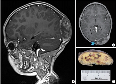
- We report a unique case of a 4-year-old girl with an intriguing fibrohistiocytic tumour. Magnetic resonance imaging scans showed a dural mass of variegated intensity compressing the left occipital pole and apparently extending toward the superior sagittal sinus. Grossly, the cut surface of the surgical specimen was yellow, pale, and soft with reddish kernel-like crusts. Histologically, the yellow areas resembled cholesterol granulomas with widespread coagulative necrosis, cholesterol clefts, powdery calcification, foreign body-type giant cells, and foamy macrophages, while the scattered red spots contained numerous multinucleated giant cells of foreign-body and Touton types, the former with amphophilic to slightly eosinophilic cytoplasm. Immunoperoxidase reactions confirmed the expression of histiocytic markers and vimentin. As far as we know, no tumour displaying these peculiar morphological features has yet been described.
-
Citations
Citations to this article as recorded by  - Reticulohistiocytoses: a revision of the full spectrum
A. Bonometti, E. Berti
Journal of the European Academy of Dermatology and Venereology.2020; 34(8): 1684. CrossRef
- WHO Grade IV Gliofibroma: A Grading Label Denoting Malignancy for an Otherwise Commonly Misinterpreted Neoplasm
-
Paola A. Escalante Abril, Miguel Fdo. Salazar, Nubia L. López García, Mónica N. Madrazo Moya, Yadir U. Zamora Guerra, Yadira Gandhi Mata Mendoza, Erick Gómez Apo, Laura G. Chávez Macías
-
J Pathol Transl Med. 2015;49(4):325-330. Published online June 17, 2015
-
DOI: https://doi.org/10.4132/jptm.2015.05.20
-
Correction in: J Pathol Transl Med 2015;49(6):538
-
9,167
View
-
78
Download
-
3
Web of Science
-
1
Crossref
-
Abstract
PDF
- We report a 50-year-old woman with no relevant clinical history who presented with headache and loss of memory. Magnetic resonance imaging showed a left parieto-temporal mass with annular enhancement after contrast media administration, rendering a radiological diagnosis of high-grade astrocytic neoplasm. Tumour sampling was performed but the patient ultimately died as a result of disease. Microscopically, the lesion had areas of glioblastoma mixed with a benign mesenchymal constituent; the former showed hypercellularity, endothelial proliferation, high mitotic activity and necrosis, while the latter showed fascicles of long spindle cells surrounded by collagen and reticulin fibers. With approximately 40 previously reported cases, gliofibroma is a rare neoplasm defined as either glio-desmoplastic or glial/benign mesenchymal. As shown in our case, its prognosis is apparently determined by the degree of anaplasia of the glial component.
-
Citations
Citations to this article as recorded by - Rare Pediatric Invasive Gliofibroma Has BRAFV600E Mutation and Transiently Responds to Targeted Therapy Before Progressive Clonal Evolution
Kristiyana Kaneva, Kee Kiat Yeo, Debra Hawes, Jianling Ji, Jaclyn A. Biegel, Marvin D. Nelson, Stefan Bluml, Mark D. Krieger, Anat Erdreich-Epstein
JCO Precision Oncology.2019; (3): 1. CrossRef
|
 E-submission