E-submission
Articles
- Page Path
- HOME > J Pathol Transl Med > Volume 48(6); 2014 > Article
-
Brief Case Report
Primary Leiomyosarcoma of Adrenal Gland with Tissue Eosinophilic Infiltration - Seungkoo Lee,, Gail Domecq C. Tanawit1, Rolando A. Lopez1, Jaime T. Zamuco1, Betsy Grace G. Cheng2, Menandro V. Siozon3
-
Korean Journal of Pathology 2014;48(6):423-425.
DOI: https://doi.org/10.4132/KoreanJPathol.2014.48.6.423
Published online: December 31, 2014
Department of Pathology, Kangwon National University Hospital, School of Medicine, Kangwon National University, Chuncheon, Korea
1Institute of Pathology, St. Luke’s Medical Center, Quezon City, Philippines
2Departments of Radiology, St. Luke’s Medical Center, Quezon City, Philippines
3Departments of Surgery, St. Luke’s Medical Center, Quezon City, Philippines
- Corresponding Author: Seungkoo Lee, M.D. Department of Anatomic Pathology, Kangwon National University Hospital, Kangwon National University School of Medicine, 1 Gangwondaehak-gil, Chuncheon 200-701, Korea Tel: +82-33-258-9172, Fax: +82-33-258-2475, E-mail: jsklee@kangwon.ac.kr
© 2014 The Korean Society of Pathologists/The Korean Society for Cytopathology
This is an Open Access article distributed under the terms of the Creative Commons Attribution Non-Commercial License (http://creativecommons.org/licenses/by-nc/3.0) which permits unrestricted non-commercial use, distribution, and reproduction in any medium, provided the original work is properly cited.
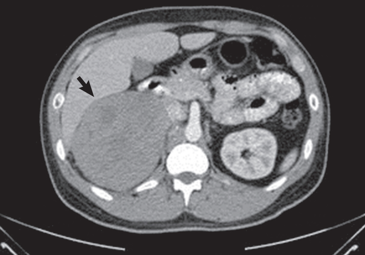
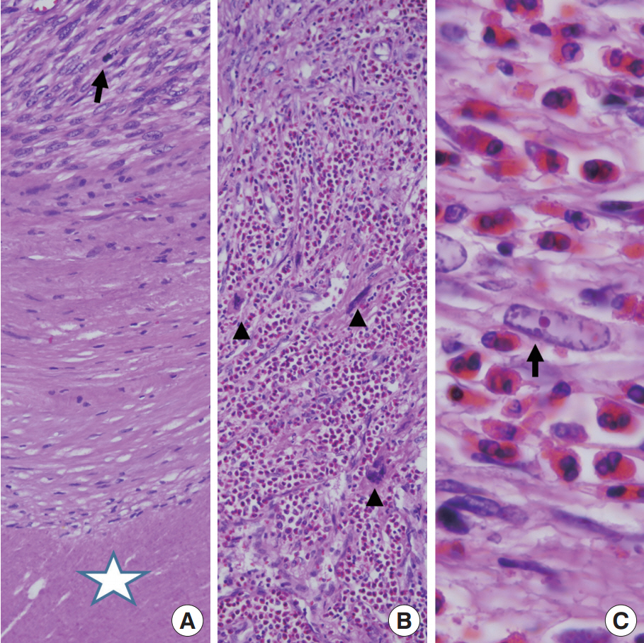
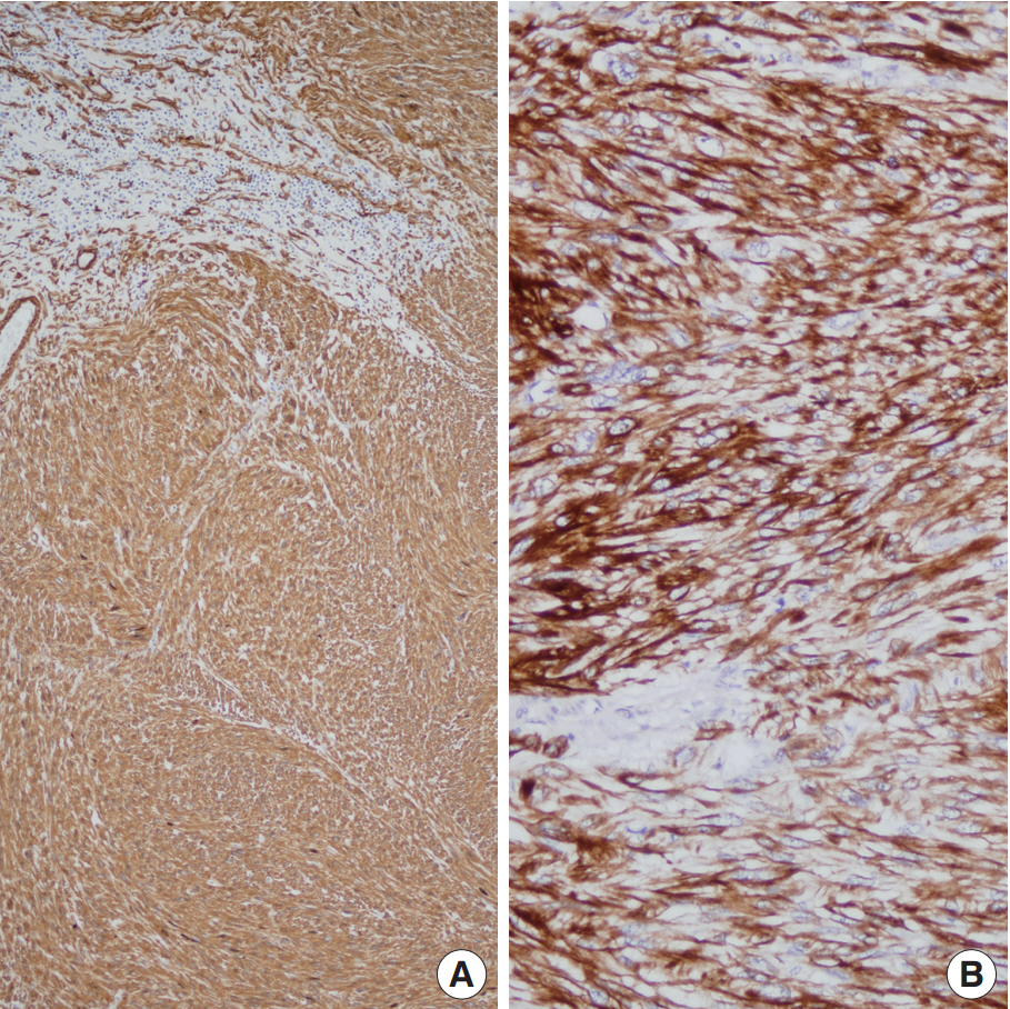
- A 28-year-old man presented with a three-month history of right flank pain, associated with persistent and stabbing pain and weight loss. Computed tomography showed a well-circumscribed heterogeneously enhancing 13.8-cm mass located at the right suprarenal area (Fig. 1). No metastatic lesion was noted. White blood cell count and eosinophil differential count were within the normal range. Biochemical examination revealed no functional tumor of the adrenal gland. The patient had no signs or symptoms of human immunodeficiency virus or Epstein-Barr virus infection. The patient underwent a right open adrenalectomy. The resected adrenal gland mass showed a large lobulated mass (15×13×6 cm, 792 g) almost replacing the adrenal gland. The mass was a well-circumscribed and partially encapsulated solid tumor. On histologic evaluation, the spindle cell tumor showed a rim of fibrous tissue with entrapped atrophic adrenal cortical cells, geographic coagulation necrosis with surrounding fibrosis and frequent mitotic figures (25/10 high-power field) (Fig. 2A). The surgical resection margin was free of tumor. We used Fédération Nationale des Centres de Lutte Contre le Cancer (FNCLCC) or the tumor grading and the tumor grade was 3. The tumor cells were strongly cytoplasmic positive for smooth muscle actin (Fig. 3A) and desmin (Fig. 3B). However, pan-cytokeratin, CD117, S100-protein, and human melanoma black 45 were all negative. On the bases of clinical data, histomorphological features and immunohistochemical studies, we diagnosed primary adrenal leiomyosarcoma. Interestingly, there was intense tissue eosinophilia (Fig. 2B). The eosinophilic infiltration was mainly present in the viable tumor cells. The center of the necrotic area did not show either eosinophils or other inflammatory cells (Fig. 2A). A few pleomorphic tumor cells with hyperchromatic nuclei were noted (Fig. 2B). Most of the tumor cells of the tissue eosinophilia revealed vesicular nuclei, cytoplasmic shrinkage, and occasional pink round nucleoli (Fig. 2C). The patient was alive without recurrent disease on 18-month follow-up.
CASE REPORT
- TATE has been observed in several cancers [1-4]. However, the mechanism by which eosinophils are recruited into tumor tissue is largely unknown. It is likely that stressed or necrotic tumor cells attract and activate eosinophils by expression of molecules such as damage-associated molecular patterns. TATE seems to have tumor destructive effector functions and immunoregulative and remodeling activities [2]. Although controversy remains, TATE are associated with good prognosis [3,4]. Our case was primary adrenal leiomyosarcoma with marked tissue eosinophilia. Although there were a few pleomorphic tumor cells, our case was considered to be the conventional type. Based on the French grading system (FNCLCC), the sub-scores of our case were two for tumor differentiation, three for mitotic count, and one for tumor necrosis, and the histologic grade was 3. The prognosis for primary adrenal leiomyosarcoma is generally poor. Among previously reported cases, only two cases were alive without metastasis after more than 15 months follow-up [5-7]. The distinct profile of the patient is high-grade sarcoma with marked tissue eosinophilia and 18 months survival without metastasis. Although TATE was found to be a good prognostic factor in a previous report [4], further study and longer patient follow-up are warranted to support the conclusion that TATE is a good prognostic factor for primary adrenal leiomyosarcoma.
DISCUSSION

- 1. Lowe D, Jorizzo J, Hutt MS. Tumour-associated eosinophilia: a review. J Clin Pathol 1981; 34: 1343-8. PubMedPMC
- 2. Lotfi R, Lee JJ, Lotze MT. Eosinophilic granulocytes and damage-associated molecular pattern molecules (DAMPs): role in the inflammatory response within tumors. J Immunother 2007; 30: 16-28. PubMed
- 3. Jain M, Kasetty S, Sudheendra US, Tijare M, Khan S, Desai A. Assessment of tissue eosinophilia as a prognosticator in oral epithelial dysplasia and oral squamous cell carcinoma-an image analysis study. Patholog Res Int 2014; 2014: ArticlePDF
- 4. Pal L, Parkash V, Chambers JT. Eosinophilia and uterine leiomyosarcoma. Obstet Gynecol 2003; 101(5 Pt 2):1130-2. PubMed
- 5. Azzouni F, Azabdaftari G, Safwat M, Schwaab T. Primary adrenal leiomyosarcoma: case report and review of literature. N Am J Med Sci 2012; 5: 58-63.
- 6. Lujan MG, Hoang MP. Pleomorphic leiomyosarcoma of the adrenal gland. Arch Pathol Lab Med 2003; 127: e32-5. PubMed
- 7. Lee H, Yoo J, Kang SJ, Kim BK. Primary leiomyosarcoma of adrenal gland: a case report. Korean J Pathol 2002; 36: 191-4.
REFERENCES
Figure & Data
References
Citations
- Outcomes and Follow-Up Trends in Adrenal Leiomyosarcoma: A Comprehensive Literature Review and Case Report
Federico Maria Mongardini, Maddalena Paolicelli, Antonio Catauro, Alessandra Conzo, Luigi Flagiello, Giusiana Nesta, Rosetta Esposito, Andrea Ronchi, Alessandro Romano, Renato Patrone, Ludovico Docimo, Giovanni Conzo
Journal of Clinical Medicine.2024; 13(12): 3499. CrossRef - Challenges in the diagnosis of the enigmatic primary adrenal leiomyosarcoma: two case reports and review of the literature
Sawako Suzuki, Naoya Takahashi, Masafumi Sugo, Kazuki Ishiwata, Akiko Ishida, Suzuka Watanabe, Katsushi Igarashi, Yutaro Ruike, Kumiko Naito, Masanori Fujimoto, Hisashi Koide, Yusuke Imamura, Shinichi Sakamoto, Tomohiko Ichikawa, Yoshihiro Kubota, Takeshi
BMC Endocrine Disorders.2023;[Epub] CrossRef - Primary adrenal leiomyosarcoma: clinical case and literature review
S. V. Lukyanov, K. M. Blikyan, S. S. Todorov, V. Y. Deribas, N. S. Lukyanov
Endocrine Surgery.2021; 15(1): 36. CrossRef Pleomorphic Leiomyosarcoma of the Adrenal Gland in a Young Woman: A Case Report and Review of the Literature
Yuanyuan Wang, Yongliang Teng, Shibo Na, Ye Yuan
OncoTargets and Therapy.2020; Volume 13: 4705. CrossRef- Primary Adrenal Leiomyosarcoma: Clinical, Radiological, and Histopathological Characteristics
Fatema Jabarkhel, Henri Puttonen, Lina Hansson, Andreas Muth, Oskar Ragnarsson
Journal of the Endocrine Society.2020;[Epub] CrossRef - Primary adrenal leiomyosarcoma with inferior vena cava extension in a 70-year-old man
Sai K Doppalapudi, Tejash Shah, Valerie A Fitzhugh, Vladislav Bargman
BMJ Case Reports.2019; 12(3): e227670. CrossRef - Primary Adrenal Leiomyosarcoma: An Extremely Rare Mesenchymal Tumor
D Lokanatha, Linu Abraham Jacob, MC Suresh Babu, KN Lokesh, Ram Krishna Sai, AH Rudresha, LK Rajeev, Smitha Saldanha, MN Suma, A Usha
Indian Journal of Medical and Paediatric Oncology.2019; 40(04): 559. CrossRef
 PubReader
 ePub Link
-
 Cite this Article
- Cite this Article
-
- Close
- Download Citation
- Close
- Figure
-