 E-submission
Search
- Page Path
- HOME > Search
Original Articles
- Correlation between HER2 gene copy number and immunohistochemistry categories in HER2-negative breast cancer: diagnostic utility for differentiating HER2-null, ultralow, and low tumors
- Min Chong Kim, Young Kyung Bae
- J Pathol Transl Med. 2026;60(2):193-201. Published online February 25, 2026
- DOI: https://doi.org/10.4132/jptm.2025.11.07

- 2,334 View
- 189 Download
-
 Abstract
 PDF
PDF - Background
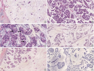
The recent recognition of human epidermal growth factor receptor 2 (HER2)–low and HER2-ultralow breast cancers (BCs) has expanded the therapeutic relevance of HER2 testing in the antibody-drug conjugate era. However, the biological continuum of HER2 expression measured by immunohistochemistry (IHC) and its relationship with the HER2 gene copy number remain unclear. Methods: We retrospectively analyzed 135 HER2-negative invasive BCs and reclassified them as HER2-null (IHC 0), HER2-ultralow (0+), or HER2-low (1+ or 2+ without amplification). HER2 gene copy number was determined using silver-enhanced in situ hybridization. Statistical analyses were performed to compare HER2 copy number among IHC categories and evaluate the discriminatory value of HER2 copy number for distinguishing IHC subgroups. Results: The mean HER2 copy number increased stepwise across IHC categories: 1.95 ± 0.54 (null), 2.03 ± 0.43 (ultralow), 2.25 ± 0.65 (low, 1+), and 3.29 ± 1.05 (low, 2+). Significant differences were observed between the ultralow and low groups (p = .003) and between the null and low groups (p < .001), but not between the null and ultralow groups or between the ultralow and 1+ groups. Conclusions: HER2 gene copy number was positively correlated with protein expression as reflected by IHC categories. Although HER2 gene copy number was statistically higher in HER2-low than in HER2-null tumors, the substantial overlap in copy number ranges likely limits its utility in distinguishing HER2-low from HER2- null BCs.
- Clinicopathologic characteristics of HER2-positive pure mucinous carcinoma of the breast
- Yunjeong Jang, Hera Jung, Han-Na Kim, Youjeong Seo, Emad Alsharif, Seok Jin Nam, Seok Won Kim, Jeong Eon Lee, Yeon Hee Park, Eun Yoon Cho, Soo Youn Cho
- J Pathol Transl Med. 2020;54(1):95-102. Published online November 13, 2019
- DOI: https://doi.org/10.4132/jptm.2019.10.24
- 12,470 View
- 301 Download
- 25 Web of Science
- 22 Crossref
-
Abstract
PDF
- Background
Pure mucinous carcinoma (PMC) is a rare type of breast cancer, estimated to represent 2% of invasive breast cancer. PMC is typically positive for estrogen receptors (ER) and progesterone receptors (PR) and negative for human epidermal growth factor receptor 2 (HER2). The clinicopathologic characteristics of HER2-positive PMC have not been investigated.
Methods
Pathology archives were searched for PMC diagnosed from January 1999 to April 2018. Clinicopathologic data and microscopic findings were reviewed and compared between HER2-positive PMC and HER2-negative PMC. We also analyzed the differences in disease-free survival (DFS) and overall survival according to clinicopathologic parameters including HER2 status in overall PMC cases.
Results
There were 21 HER2-positive cases (4.8%) in 438 PMCs. The average tumor size of HER2-positive PMC was 32.21 mm (± 26.55). Lymph node metastasis was present in seven cases. Compared to HER2-negative PMC, HER2-positive PMC presented with a more advanced T category (p < .001), more frequent lymph node metastasis (p = .009), and a higher nuclear and histologic grade (p < .001). Microscopically, signet ring cells were frequently observed in HER2-positive PMC (p < .001), whereas a micropapillary pattern was more frequent in HER2-negative PMC (p = .012). HER2-positive PMC was more frequently negative for ER (33.3% vs. 1.2%) and PR (28.6% vs. 7.2%) than HER2-negative PMC and showed a high Ki-67 labeling index. During follow-up, distant metastasis and recurrence developed in three HER2-positive PMC patients. Multivariate analysis revealed that only HER2-positivity and lymph node status were significantly associated with DFS.
Conclusions
Our results suggest that HER2-positive PMC is a more aggressive subgroup of PMC. HER2 positivity should be considered for adequate management of PMC. -
Citations
Citations to this article as recorded by
- Mucin‐producing breast lesions: a practical approach to diagnosis
Sunayana Misra, Mihir Gudi, Kimberly H Allison, Edi Brogi, Cecily Quinn, Hannah Y Wen, Puay Hoon Tan
Histopathology.2026; 88(5): 939. CrossRef - Signet-ring cell cytomorphology in breast cancer: Unveiling the overlooked
Shitong Su, Zijian Liu, Bifeng Yao, Qiuyang Jing, Ruijie Liu, Kuansong Wang
Critical Reviews in Oncology/Hematology.2026; 220: 105170. CrossRef - Predictive factors and prognostic significance of HER2-low early breast cancer with long-term follow-up
Yuka Niwa, Mitsuo Terada, Yumi Wanifuchi-Endo, Takashi Fujita, Tomoko Asano, Hidetoshi Kawaguchi, Kazuki Nozawa, Nana Matsumoto, Ayaka Isogai, Hikaru Kawahara, Marie Mizumoto, Tatsuya Toyama
Surgical Oncology.2026; 64: 102360. CrossRef - Mucinous carcinoma of the breast: morphological spectrum, diagnostic pitfalls and classification challenges
Emad A Rakha, Bara Wazwaz, Stephen B Fox
Journal of Clinical Pathology.2026; 79(7): 433. CrossRef - Clinicopathological characteristics of mucinous breast cancer: a retrospective analysis of a 6-years study from national cancer center in Vietnam
Thi Huyen Phung, Thanh Tung Pham, Huu Thang Nguyen, Dinh Thach Nguyen, Thanh Long Nguyen, Thi Hoai Hoang
Breast Cancer Research and Treatment.2025; 209(3): 667. CrossRef - Poor response of HER2-positive mucinous carcinomas of breast to neoadjuvant HER2-targeted therapy: A study of four cases
Min Han, Daniel Schmolze, Javier A. Arias-Stella, Christina H. Wei, Joanne Mortimer, Fang Fan
Annals of Diagnostic Pathology.2025; 74: 152396. CrossRef - Comprehensive Immunohistochemical Analysis of Mesonephric Marker Expression in Low-grade Endometrial Endometrioid Carcinoma
Yurimi Lee, Sangjoon Choi, Hyun-Soo Kim
International Journal of Gynecological Pathology.2024; 43(3): 221. CrossRef - Clinicopathological features and prognosis of mucinous breast carcinoma with a micropapillary structure
Beibei Yang, Menglu Shen, Bo Sun, Jing Zhao, Meng Wang
Thoracic Cancer.2024; 15(36): 2530. CrossRef - Pure Mucinous Carcinoma of the Breast: Radiologic-Pathologic Correlation
Cherie M Kuzmiak, Benjamin C Calhoun
Journal of Breast Imaging.2023;[Epub] CrossRef - Role of circ-FOXO3 and miR-23a in radiosensitivity of breast cancer
Elahe Abdollahi, Hossein Mozdarani, Behrooz Z. Alizadeh
Breast Cancer.2023; 30(5): 714. CrossRef - On Ultrasonographic Features of Mucinous Carcinoma with Micropapillary Pattern
Wei-Sen Yang, Yang Li, Ya Gao
Breast Cancer: Targets and Therapy.2023; Volume 15: 473. CrossRef - Spectrum of Mucin-containing Lesions of the Breast: Multimodality Imaging Review with Pathologic Correlation
Janice N. Thai, Melinda F. Lerwill, Shinn-Huey S. Chou
RadioGraphics.2023;[Epub] CrossRef - Mesonephric-like Adenocarcinoma of the Ovary: Clinicopathological and Molecular Characteristics
Hyun Hee Koh, Eunhyang Park, Hyun-Soo Kim
Diagnostics.2022; 12(2): 326. CrossRef - Alveolar Soft Part Sarcoma of the Uterus: Clinicopathological and Molecular Characteristics
Yurimi Lee, Kiyong Na, Ha Young Woo, Hyun-Soo Kim
Diagnostics.2022; 12(5): 1102. CrossRef - Metastasis of the Mucionous adenocarcinoma of breast to the mandibular gingiva: Rare case report
Ivana Mijatov, Aleksandra Fejsa Levakov, Aleksandar Spasić, Jelena Nikolić, Saša Mijatov
Medicine.2022; 101(38): e30732. CrossRef - Endometrioid Carcinomas of the Ovaries and Endometrium Involving Endocervical Polyps: Comprehensive Clinicopathological Analyses
Jihee Sohn, Yurimi Lee, Hyun-Soo Kim
Diagnostics.2022; 12(10): 2339. CrossRef - Serous Carcinoma of the Endometrium with Mesonephric-Like Differentiation Initially Misdiagnosed as Uterine Mesonephric-Like Adenocarcinoma: A Case Report with Emphasis on the Immunostaining and the Identification of Splice Site TP53 Mutation
Sangjoon Choi, Yoon Yang Jung, Hyun-Soo Kim
Diagnostics.2021; 11(4): 717. CrossRef - HER2 positive mucinous carcinoma of breast with micropapillary features: Report of a case and review of literature
Dinesh Chandra Doval, Rupal Tripathi, Sunil Pasricha, Pankaj Goyal, Chaturbhuj Agrawal, Anurag Mehta
Human Pathology: Case Reports.2021; 25: 200531. CrossRef - Carcinoma mucosecretor de mama HER2-positivo, un caso clínico
A.M. González Aranda, E. Martínez Gómez, A. Santana Costa, F. Arnanz Velasco, M.H. González de Diego, A. Zapico Goñi
Clínica e Investigación en Ginecología y Obstetricia.2021; 48(4): 100685. CrossRef - Clinicopathologic features of unexpectedly HER2 positive breast carcinomas: An institutional experience
Carissa LaBoy, Kalliopi P. Siziopikou, Lauren Rosen, Luis Z. Blanco, Jennifer L. Pincus
Pathology - Research and Practice.2021; 222: 153441. CrossRef - Mesonephric-like Differentiation of Endometrial Endometrioid Carcinoma: Clinicopathological and Molecular Characteristics Distinct from Those of Uterine Mesonephric-like Adenocarcinoma
Sujin Park, Go Eun Bae, Jiyoung Kim, Hyun-Soo Kim
Diagnostics.2021; 11(8): 1450. CrossRef - Mesonephric-like Adenocarcinoma of the Uterine Corpus: Comprehensive Immunohistochemical Analyses Using Markers for Mesonephric, Endometrioid and Serous Tumors
Hyunjin Kim, Kiyong Na, Go Eun Bae, Hyun-Soo Kim
Diagnostics.2021; 11(11): 2042. CrossRef
- Mucin‐producing breast lesions: a practical approach to diagnosis
- The Expression of p53, c-erbB-2 and nm23 Proteins in Breast Cancer.
- Kyo Young Lee, Yong Goo Kim, Young Shin Kim, Kyung Ja Han, Chang Suk Kang, Jean A Kim, Won Il Kim, Sang In Shim
- Korean J Pathol. 1999;33(2):88-95.
- 2,052 View
- 10 Download
-
Abstract
- Recently, p53, c-erbB-2 and nm23 proteins have been studied in breast cancer. The expression of p53 protein indicates the mutation of p53 gene known as a tumor supressor gene, and c-erbB-2 gene amplification has been considered an indicator of poor prognosis and nm23 a metastsis suppressor gene. In order to elucidate the roles and relations of these proteins in the develpoment, progression and metastasis in breast cancer, we studied 89 cases of invasive breast cancer and 32 cases of lymph node metastasis for the expression of p53, c-erbB-2 and nm23 proteins using an immunohistochemical method. The results were as follows: 1) The expression rates of p53, c-erbB-2, and nm23 proteins in breast cancer were 40.4%, 34.8% and 55.1%, respectively. Co-expression of p53 protein and c-erbB-2 protein was found in 20.2% of cases, showing the highest incidence in poorly differentiated type (40%). 2) p53 protein expression was increased in poorly differentiated type but was not statistically significant. On the other hand, the expression of nm23 protein was decreased in poorly differentiated type, which was statistically significant (p<0.05). 3) The correlation of p53 protein expression with c-erbB-2 protein expression was statistically significant (p<0.05) but that with nm23 protein was not. 4) In the cases with lymph node metastasis, discordant expression of p53, c-erbB-2 and nm23 proteins between primary tumor and the lymph node metastatic tumor was found in 9.4%, 3.1% and 18.8% of cases, respectively. The above results suggest that overexpression of p53 and c-erbB-2 proteins and downregulation of nm23 protein are associated with the tumor progression in the breast cancer.
- The Expression of c-erbB-2, EGFR, p53 and Ki-67 in Ovarian Borderline Tumors and Carcinomas of the Ovary.
- Kyueng Whan Min, Moon Hyang Park
- Korean J Pathol. 2007;41(5):296-306.
- 2,865 View
- 63 Download
-
Abstract
PDF
- BACKGROUND
An ovarian surface epithelial tumor is a heterogenous disease, and various biological and molecular factors are important for its development and progression. Several findings support EGFR or c-erbB-2 as adverse prognostic indicators for an ovarian carcinoma.
METHODS
We reviewed the histological and clinical findings of 52 carcinomas (17 endometrioid, 16 serous, 13 mucinous and 6 clear cell tumors), and 26 borderline (10 serous and 16 mucinous) tumors. Expression of c-erbB-2, EGFR, p53, and Ki-67 was evaluated on paraffinembedded tissue from a primary ovarian tumor by immunohistochemical methods.
RESULTS
Expression of c-erbB-2 was found in 7.6% of tumors and expression of EGFR was found in 9.6% of tumors by immunohistochemical analysis. No significance was found between cerbB- 2 and EGFR expression as indicators of a poor prognosis. The expression of p53 and Ki-67 (>50%) correlated with the grade and type of tumor in the ovarian cancers. p53 and Ki- 67 overexpression (>50%) was absent in the borderline ovarian tumors, whereas ovarian carcinomas showed expression of both p53 and Ki-67.
CONCLUSION
Expression of c-erbB- 2, EGFR, p53, and Ki-67 as determined by immunohistochemical analysis did not correlate with prognostic significance. However, p53 and Ki-67 expression may be used as markers to predict aggressive behavior, and to differentiate between malignant and borderline epithelial ovarian tumors. Further large-scale studies are required to clarify the significance of c-erbB-2 and EGFR expression in ovarian tumors.
- Rarity of EGFR and c-ErbB-2 Overexpressions in Hepatocellular Carcinoma: An Immunohistochemical Study.
- Woo Sung Moon, Hyun Jin Son, Ho Sung Park, Min Young Park
- Korean J Pathol. 2004;38(4):244-248.
- 2,514 View
- 13 Download
-
Abstract
PDF
- BACKGROUND
The overexpression of epidermal growth factor receptor (EGFR) and c-erbB-2 oncogenes has been implicated in the development of many types of cancer. However, the role of EGFR and c-erbB-2 overexpression in hepatocellular carcinoma (HCC) has not been fully elucidated.
METHODS
The aim of this study was to evaluate the immunohistochemical expression of EGFR and c-erbB-2 oncoprotein in a series of 52 HCCs.
RESULTS
All but one of the HCC tumor tissues were negative for EGFR monoclonal antibody, clone H11. All of the HCC tumor tissue samples were negative for EGFR monoclonal antibody, clone 29.1.1. However, strong EGFR immunoreactivity was detected in sinusoidal endothelial cells of HCC in 25 tumors (48%) using EGFR 29.1.1 antibody. The expression of c-erbB-2 was observed in 6% (3/52) of the HCCs. No significant correlation was found between p53 mutation and the expression of c-erbB-2.
CONCLUSION
Our results suggest that both EGFR and c-erbB-2 oncoprotein overexpressions in tumor cells are rare and do not seem to predominantly contribute to the malignant phenotype in HCC.
- Correlation between Expression of c-erbB-2 Oncogene and Various Prognostic Factors in the Colorectal Carcinoma.
- Wan Kim, Hong Ran Choi, Ji Shin Lee, Jong Tae Park, Chang Soo Park, Kyu Hyuk Cho
- Korean J Pathol. 1993;27(3):217-225.
- 2,214 View
- 12 Download
-
Abstract
PDF
- The c-erbB-2 oncogene, which is a new human proto-oncogene similar to EGFR structurally, generates a glycoprotein of tyrosine kinase family with a molecular weight of 185,000 To evaluate the prognostic significance of c-erbB-2 oncogene expression in colorectal carcinoma, We analysed 73 colorectal carcinomas in paraffin sections immunohistochemically, using the monoclonal antibody specific for the c-erbB-2 oncogene product and correlated with clinicopathological data. The results were as follows 1) The immunoreactivity for c-erbB-2 oncogene was localized to cell membrane of the tumor cells and occasionally observed within the cytoplasm. 2) The positivity of c-erbB-2 oncogene expression was 71.2%(52/73) of the colorectal carcinomas overall. According to the histological types, the positivity of c-erbB-2 oncogene in adenocarcinoma(77.4%) was higher than that in mucinous carcinoma(36.4%)(p<0.05). 3) Expression of c-erbB-2 oncogene was significantly correlated with lymph node metastasis or distant metastasis(p=0.0117), Dukes stage(p=0.0432), and TNM classification(p=0.0102). These results suggest that c-erbB-2 oncogene expression may be used as a prognostic factor of colorectal carcinoma because of its correlation with other clinicopathological prognostic factors.
- Expression of p53 Protein and c-erbB-2 Oncoprotein in Breast Carcinoma.
- Eun Hee Lee, Dong Sug Kim, Tae Sook Lee, Soo Jung Lee
- Korean J Pathol. 1995;29(5):596-606.
- 2,098 View
- 10 Download
-
Abstract
- This study was conducted to evaluate the expression of p53 and c-erbB-2 using immuno-histochemical methods in 145 primary breast carcinomas and to correlate it with other histo-pathological prognostic factors. Invasive ductal carcinoma represented 129 of the cases. Expression of p53 protein and c-erbB-2 oncoprotein was present in 48% (62/129) and 30% (39/129) of invasive ductal carcinomas, respectively. The expression of p53 protein was stongly associated with a high score of degree of differentiation (p<0.05), nuclear pleomorphism (p<0.05), mitotic index (p<0.05), SBR grade (p<0.05) and MSBR grade (p<0.05), but it was not associated with patient's age, size of tumor or axillary node metastasis. The overexpression of c-erbB-2 C-erbB-2 oncoprotein was strongly associated with a high score of nuclear pleomorphism and a high SBR grade (p<0.05), but not associated with patient's age, size of tumor, axillary node metastasis, degree of differentiation, mitotic index or MSBR grade. An inverse relationship between the expression of p53 protein and estrogen receptor status was found, but the expression of c-erbB-2 was not associated with estrogen receptor status. It is concluded that p53 protein and c-erbB-2 oncoprotein are important prognostic factors in breast cancers, and that the aberrant expression of p53 protein is the most useful prognostic factor becausd of strong association of known histopathological prognostic factors and negative estrogen receptor status.
- The Expressions of Tyrosine Kinase Receptors, EphA2, c-met and c-erbB-2 in the Human Breast.
- Soo Kee Min, Hyun Deuk Cho, Seong Jin Cho, Hye Rim Park, Hyung Sik Shin, Young Euy Park, Bom Woo Yeom
- Korean J Pathol. 2005;39(1):15-22.
- 2,298 View
- 23 Download
-
Abstract
PDF
- BACKGROUND
Tyrosine kinase receptor (TKR) is an important protein for normal-development, growth and tumorigenesis in human tissues. The purpose of this study was to evaluate the effect of TKR in the progression of breast cancer.
METHODS
The expressions of EphA2, c-met and c-erbB-2 were examined, by using immunohistochemical methods and RT-PCR, in samples of breast tissue that included 111 samples of normal epithelium, 34 samples of ductal carcinoma in situ (DCIS), and 109 samples of invasive ductal carcinomas (IDC). The results were compared with the prognostic parameters of breast cancer including the tumor grade, growth pattern, lymph node metastasis and the expressions of ER, PR, p53 and Ki-67.
RESULTS
The protein expressions of the three TKRs were higher in DCIS and IDC than in normal epithelium. The protein expression of EphA2 was correlated with a tumor grade, a labeling index of Ki-67, and the protein expression of c-met. Overexpression of c-erbB-2 was correlated with lymph node metastasis. The mRNA levels of the three TKRs were correlated with each other in normal tissue and IDC. The level of c-met mRNA was higher in the low grade tumors.
CONCLUSIONS
The three TKRs may play roles in the tumorigenesis of human breast cancer. The overexpressions of EphA2 and c-erbB-2 may be a poor prognostic parameter in breast cancers.
- c-erbB-2 Oncoprotein Overexpression in Breast Cancer.
- Tae Sook Hwang, Kyung Ja Cho, Young Bae Kim, Joo Ryung Huh, Ja June Jang
- Korean J Pathol. 1994;28(1):1-7.
- 2,394 View
- 15 Download
-
Abstract
PDF
- c-erbB-2 oncogene is a normal cellular proto-oncogene coding transmembrane glycoprotein structurally similar to the epidermal growth factor receptor. Amplification of this oncogene in a variety of human adenocarcinomas has been reported and is particularly well documented in breast carcinoma. It has been suggested that amplification of this oncogene is indicative of poor prognosis and is valuable only second to the lymph node status. Using immunohistochemical staining for the c-erbB-2 protein, overexpression of this protein was analysed in 228 primary breast cancer specimens and the frequency of overexpression and the relationship between overexpression and the other established prognostic variables are evaluated. Ninty three cases out of 228 cases(40.8%) show postive oncoprotein overexpression and using the chi-squared test for a trend, a significant correlation was found between c-erbB-2 protein staining and the histological grade, lymph node status, and estrogen receptor status(P<0.05). No significant association was found between staining and the patient's age and tumor size. Most of the tumors with histological types known to have good prognosis showed negative expression. Above findings strongly suggest that expression of c-erbB-2 oncogene is another independent indicator of poor prognosis in breast carcinoma.
- Expression of Epidermal Growth Factor Related Peptides, EGF-R, and c-erbB-2 and Their Relationship with the Prognostic Factors in Gastric Carcinoma.
- Joo Heon Kim, Jin Wook Lee, Woo Sung Moon, Myoung Jae Kang, Dong Geun Lee
- Korean J Pathol. 1999;33(11):1039-1046.
- 2,196 View
- 11 Download
-
Abstract
PDF
- Recent investigations have revealed that autocrine growth factors and their receptors are closely related and play an important role in controlling cancer cell growth. We performed an immunohistochemical study on the expression of epidermal growth factor (EGF), transforming growth factor-alpha (TGF-alpha), epidermal growth factor receptor (EGF-R), c-erbB-2, and PCNA labelling index in 60 cases of human gastric carcinomas. TGF-alpha was detected in 38 cases (63.3%), EGF in 26 cases (43.3%), EGF-R in 44 cases (73.3%), and c-erbB-2 in 18 cases (30%). These growth factors, EGF-R and c-erbB-2, were found more often in advanced gastric cancers. The PCNA labeling index was significantly higher in tumors with the expression of EGF-R or c-erbB-2. Tumors with simultaneous expression of EGF, TGF-alpha, EGF-R and c-erbB-2 was associated with a high PCNA labeling index. A correlation was observed between the synchronous expression of growth factors and its receptors and histological differentiation. The results suggest that the expression of EGF, TGF-alpha, EGF-R and c-erbB-2 are closely related and plays an important role in the growth and progression of human gastric carcinoma.
- Comparing Fluorescence In Situ Hybridization and Immunohistochemistry to Determine the HER-2/neu Status in Breast Carcinoma.
- Kyeongmee Park, Jungyoen Kim, Sungjig Lim
- Korean J Pathol. 2002;36(4):243-248.
- 2,250 View
- 13 Download
-
Abstract
PDF
- BACKGROUND
Identification of HER-2/neu status is important in predicting the response to specific chemotherapy in breast carcinoma patients and HER-2/neu status is associated with poor clinical outcome even with systemic chemotherapy. Introduction of fluorescence in situ hybridization (FISH) allows an accurate assessment of the level of gene amplification with information about distribution of gene copies in histologic sections.
METHODS
HER-2/neu status was performed on paraffin sections of 176 primary breast carcinomas by FISH, using PathVysion and by immunohistochemistry (IHC), using HercepTest. The results of HER-2/neu amplification was compared with clinical and pathological prognostic factors.
RESULTS
HER-2/neu amplification and overexpression were detected in 51 tumors (29.0%) by FISH and 32 tumors (18.2%) by IHC. The results of each method agreed with each other in 157 tumors (concordance: 89.2%, kappa=0.783). HER-2/neu amplification was associated with poor nuclear grade, marked nuclear pleomorphism, and presence of the combined ductal carcinoma in situ in the invasive ductal carcinomas as well as Van Nuys grade of the ductal carcinoma in situ component (p<0.05).
CONCLUSIONS
The comparison of FISH and IHC demonstrated an excellent correlation of HER-2/neu overexpression 2+ and 3+ with gene amplification. However, FISH may be a more accurate and reliable method for negative and 1+ cases. HER-2/neu amplification proves to be of prognostic relevance.
- Altered Expression of p53, p21WAF1, p16, Rb, Smad4 and c-erbB-2 in Resected Pancreatic Ductal Adenocarcinoma.
- Yun Kyung Kang, Woo Ho Kim
- Korean J Pathol. 2002;36(6):382-388.
- 2,129 View
- 20 Download
-
Abstract
PDF
- BACKGROUND
Our aim was to undertake a comprehensive analysis of the expression of key molecular markers in a series of pancreatic ductal adenocarcinomas and to determine their association with clinicopathologic variables.
METHODS
By using immunohistochemical staining, we examined the expressions of five tumor suppressor genes (p53, p21WAF1, p16, Rb, Smad4) and a growth factor receptor (c-erbB-2) in 52 surgically resected pancreatic ductal adenocarcinomas.
RESULTS
Abnormal nuclear overexpression of p53 was noted in 28/52 (53.8%) cases. Total loss of p21WAF1, p16, Rb, and Smad4 was detected in 15/52 (28.8%), 33/52 (63.5%), 4/52 (7.7%), and 26/52 (50%) cases, respectively. Overexpression of c-erbB-2 was noted in 21/52 (40.4%) cases. Forty-nine (94.2%) cases revealed aberration of at least one of the markers examined. There was a positive correlation between p53 and c-erbB-2 overexpression (p<0.05). Among the examined genes, overexpression of c-erbB-2 was found to have a positive relationship with the tumor stage (p<0.05). There was also a significant correlation between the histologic grade and the number of abnormally expressed genes per tumor (p<0.05).
CONCLUSION
Among the various tumor-associated proteins evaluated in this study, c-erbB-2 could have the most likely clinical implication.
- Fascin-1 Protein Expression in Gastric Carcinoma.
- Seoung Wan Chae, Jin Hee Sohn
- Korean J Pathol. 2006;40(2):112-117.
- 2,448 View
- 27 Download
-
Abstract
PDF
- BACKGROUND
Fascin-1 is a globular cross-linking and actin bundling protein that provides mechanical support to cellular protrusions and cell motility. The expression of fascin in epithelial neoplasms has been recently reported, but its exact mechanism in cancer is unknown. The purpose of this study was to assess the expression of fascin and its relationship with the clinicopathologic parameters and the other tumor markers in gastric carcinoma.
METHODS
Immunohistochemical stainings for fascin, c-erbB-2, p53 and Ki-67 labeling index were performed in 62 gastric carcinoma specimens.
RESULTS
Fascin-1 protein was not expressed in the normal gastric glandular epithelial cells. It had an expression in 35.5% of the gastric adenocarcinomas. The fascin-1 expression in carcinoma was slightly increased in the well to moderately differentiated tumors compared with the poorly differentiated tumors. The fascin-1 expression was correlated with the c-erbB-2 protein expression. There was no significant correlation with the clinicopathologic factors such as tumor size, nodal metastasis, pathologic stage, p53 protein expression and Ki-67 labeling index.
CONCLUSIONS
This study reveals the possibility that the fascin-1 protein expression in gastric carcinoma may be closely linked with the c-erbB-2 protein expression. However, further study on fascin-1 and c-erbB-2 protein at the cellular level and their clinical relevance is needed.
- A Comparative Study of Immunohistochemical Expression of p53, bcl-2, c-erbB-2, and MIB-1 in Polypoid and Infiltrative Colorectal Carcinomas.
- Jeong Seok Moon, Seong Hwan Park, Bong Kyong Shin, Ju Han Lee, Joon Ho Shin, Bom Woo Yeom
- Korean J Pathol. 1998;32(8):581-589.
- 2,147 View
- 10 Download
-
Abstract
- Almost all colorectal carcinomas have been thought to develop from pre-existing adenomas. However, some colorectal carcinomas can arise directly from normal flat mucosa, and usually form infiltrative mass at the early stage. The carcinogenesis of this infiltrative carcinoma may be different from the well-known adenoma-carcinoma sequence, which usually forms a polypoid mass. The purpose of this study is to investigate the different expression of various oncogenes in polypoid carcinoma and infiltrative carcinoma. We performed immunohistochemical staining on p53, bcl-2, c-erbB-2 and MIB-1 in 29 polypoid carcinomas arised from adenomas, and 21 infiltrative carcinomas. The average tumor size of infiltrative carcinomas (5.5 cm) was larger than that of polypoid carcinomas (3.1 cm), and the polypoid carcinomas were differentiated more than the infiltrative carcinomas. The results of p53, bcl-2, c-erbB-2, and MIB-1 antisera immunoreactivity in the polypoid carcinoma were 79%, 17%, 21%, and 100%, and those in the infiltrative carcinoma were 71%, 29%, 29%, and 100%, respectively. However the diffuse positivities of p53 and MIB-1 antisera were slightly higher in the infiltraive carcinomas (62%, 76%) than in the polypoid carcinomas (55%, 41%) (p=0.63, 0.01). And the results of p53 and c-erbB-2 immunoreactivity in the adenomas were 52% and 17%, respectively, which is significantly lower than that in the polypoid carcinoma(p=0.03, 0.74). The immunoreactivty of bcl-2 in the adenoma was 72%, which was significantly higher than that in the polypoid carcinoma (17%) (p<0.01). In summary, we did not show the significant difference in expression of p53, bcl-2, c-erbB-2, and MIB-1 proteins between polypoid and infiltrative carcinomas. However, the tendency of infiltrative carcinomas having a more aggressive nature suggests another carcinogenetic mechanism is involved in the colorectal carcinogenesis.
- c-erbB-2 Oncoprotein Expression in Ductal Carcinoma in situ and Paget's Disease of the Breast.
- Jung Yeon Kim, Kyung Ja Cho, Seung Sook Lee, Shin Kwang Khang, Nam Sun Paik
- Korean J Pathol. 1996;30(11):972-980.
- 2,324 View
- 21 Download
-
Abstract
PDF
- A clinico-pathologic study with an immunohistochemical examination for c-erbB-2 expression in 54 cases of ductal carcinoma in situ and 16 cases of Paget's disease of the breast was performed. c-erbB-2 oncoprotein overexpression was observed in 45% (24/54) and 88% (14/16) of ductal carcinoma in situ and Paget's disease, respectively. The overexpression of c-erbB-2 oncoprotein was significantly correlated with the nuclear grade of tumors and inversely with the status of the estrogen receptor. c-erbB-2 was positive in 4 out of 5 patients with metastasis to axillary lymph nodes and 3 out of 4 patients who died of the disease. Prognostic significance of c-erbB-2 oncoprotein in ductal carcinoma in situ was highly suggested. The expression of c-erbB-2 oncoprotein in Paget's disease was well correlated with coexisting infiltrating or in situ ductal carcinoma. The high positive rate of c-erbB-2 oncoprotein in ductal carcinoma with Paget's disease could be understood with a recent hypothesis that c-erbB-2 oncoprotein is involved in promotion of cell motility and the spread of carcinoma cells.
- Immunohistochemical Characteristics of Biliary Tract Carcinoma and Its Precancerous Lesions.
- Jiyoung Kim, Youngnyun Park, Hogeun Kim
- Korean J Pathol. 1998;32(11):985-992.
- 2,581 View
- 10 Download
-
Abstract
- Carcinomas of the biliary tract are known to be more common in East Asia than in Western countries, but their exact histopathological characteristics and tumorigenesis are not well elucidated. To examine the histological and immunohistochemical characteristics of the biliary tract carcinomas according to their anatomical sites and to elucidate their tumorigenesis, we performed histological review and immunohistochemical study in a total of 135 cases of biliary tract carcinomas; 24 intrahepatic bile duct, 34 gallbladder, 51 common bile duct, and 26 periampullary carcinomas. Precancerous lesions were associated with 5 (20.8%) cases of intrahepatic duct carcinomas (dysplasia 5), 7 (20.6%) cases of gallbladder carcinomas (adenoma 5, dysplasia 2), 10 (19.6%) cases of common bile duct carcinomas (adenoma 7, dysplasia 3), and 2(7.7%)cases of periampullary carcinomas (adenoma 2). Immunohistochemically, c-erbB-2 expression in gallbladder carcinoma (21/34, 62%) was significantly higher than that of intrahepatic (8/24, 33%). Ki-67 indices were higher in common bile duct carcinomas (19%) than those of intrahepatic bile duct (14%) or periampullary carcinomas (12%). Overexpression of p53 gene product in the periampullary carcinomas (20/22, 77%) was higher than that of intrahepatic (12/24, 50%) or common bile duct carcinoma (26/51, 51%). In the precancerous lesions the c-erbB-2 expression was present in 29% of the gallbladder, 20% of the intrahepatic, 10% of the common bile duct precancerous lesions and none of the 2 cases of adenomas in the periampullary region. The p53 overexpression in the precancerous lesions was frequent, ranging from 43% to 60%. These results suggest that a mechanism involving p53 gene mutation and c-erbB-2 gene activation is present in the tumorigenesis in a significant number of the biliary tract carcinomas and they may be the early events in the tumorigenesis of the biliary tract carcinomas.
- A Study of the Correlation between Expression of c-erbB-2 Oncoprotein and Various Clinicopathological Prognostic Factors in Breast Carcinoma.
- Jong Hee Nam, Kyung Soo Kim, Chang Soo Park, Sang Woo Juhng
- Korean J Pathol. 1995;29(2):136-144.
- 1,911 View
- 12 Download
-
Abstract
PDF
- Immunohistochemical study for c-erbB-2 oncoprotein was performed on paraffin sections of 76 primary breast carcinomas to determine the relationship between expression of c-erbB-2 and various clinicopathological prognostic indicators, including the expression of epidermal growth factor receptor (EGFR). Positive reaction for c-erbB-2 oncoprotein revealed an intense red granular staining predominantly located at the tumor cell membrane, with some cells exhibiting a weak cytoplasmic staining as well. The epithelial cells of the normal lobule and duct showed a negative reaction. Positive reaction for EGFR revealed a granular staining in the cytoplasm and the cell membrane of the tumor cells. Some tumors showed a positive EGFR staining in the epithelial cells of normal duct and lobule. Twenty six of 76 cases (34.2%) of primary breast carcinomas revealed a positive reaction for c-erbB-2 oncoprotein, and 28 cases (36.8%) were positive for EGFR. Expression of c-erbB-2 oncoprotein and EGFR was evident in 37.7% and 40.6% of 69 classic invasive ductal carcinomas, respectively. None of the other histological types showed a positive reaction. Expression of c-erbB-2 oncoprotein was strongly associated with tumor size(p=0.0015), histologic grade(.p=0.0175), vascular invasion(p=0.0043), and lymph node metastasis(p=0.0024), but not with age at diagnosis(p=0.1836). No significant association was found between expression of c-erbB-2 oncoprotein and EGFR. Co-expression of c-erbB-2 oncoprotein and EGFR was also strongly associated with tumor size (p=0.0029). These results suggest that c-erbB-2 oncoprotein is biologically distinct from EGFR, and may be used as a prognostic indicator of breast carcinoma due to its strong association with various clinicopathological prognostic factors.
- A Study on the DNA Ploidy and Expression of c-erbB-2 Oncogen in the Ovarian Carcinomas.
- Jong Jae Jung, Chang Soo Park, Sang Woo Juhng
- Korean J Pathol. 1997;31(1):15-22.
- 1,989 View
- 15 Download
-
Abstract
PDF
- To evaluate the relationships among the c-erbB-2 oncogene expression, DNA ploidy and other prognostic factors, an immunohistochemical study of the c-erbB-2 oncogene product and flow cytometric analysis of DNA ploidy were performed in paraffin sections of 42 cases of ovarian carcinomas. The results were as follows: 1) The positive reaction for c-erbB-2 oncogene product was observed mainly along the cytoplasmic membrane, and occasionally within the cytoplasm of the tumor cells. 2) Overall the positivity of c-erbB-2 oncogene expression was 45.2% of the ovarian carcinomas. By the histological types, the positivity was 35.7% in serous carcinoma, 80.0% in mucinous carcinoma, and 45.2% in endometrioid carcinoma; by the degree of differentiation, 57.1% in well differentiated carcinoma, 40.0% in moderately differentiated, and 27.3% in poorly differentiated; by the nuclear grading, 58.3% in grade I, 52.6% in grade II, and 18.2 % in grade III; and by the clinical staging, 57.1% in stage I, 42.8% in stage II, and 35.0% in stage III. The expression of the c-erbB-2 oncogene in the ovarian carcinomas was higher in the tumors of good differentiation, of the lower nuclear grade and of the lower clinical stage. 3) The incidence of DNA aneuploidy in the cases positive for the c-erbB-2 oncogene expression(47.3%) was higher than that in the negative cases(31.4%). From the above results, therefore, it is suggested that the c-erbB-2 oncogene may be involved in the early stage of ovarian carcinogenesis. Also suggested is that ovarian carcinomas positive for the c-erbB-2 oncogene in the early stages may have higher probability of having a DNA aneuploid cell line during the progress of the tumors.
- Expressions of Epidermal Growth Factor Receptor, c-erbB-2 and p53 Protein as Useful Markers of Malignant Potential in a Transitional Cell Carcinoma of the Urinary Bladder.
- Gu Kong, Ki Yong Shin, Sun Jin Kim, Young Hyeh Ko, Hae Young Park, Young Nam Woo, Jung Dal Lee
- Korean J Pathol. 1997;31(1):51-58.
- 2,175 View
- 11 Download
-
Abstract
PDF
- Transitional cell carcinoma(TCC) of the urinary bladder shows marked heterogeneity in biological behaviors. Evidence has accumulated that biological markers may provide significant information to predict the potential aggressiveness of TCC. We have assessed the expression of the epidermal growth factor receptor (EGF-R), c-erbB-2 and p53 proteins in 56 cases of TCC to investigate the prognostic significance of differential expression of these oncoproteins using an immunohistochemical method. We analysed the expression patterns of these oncoproteins according to tumor stage and grade. And we assessed the probability of progression-free survival in stage T1 tumors according to their expressions. Positive rates of EGF-R (>+3 staining intensity), c-erbB-2 (intense membrane staining) and p53 proteins (>20% positive cells) were 73.2%, 37.5% and 42.9%, respectively. Invasive tumors had significantly higher positive rates of all three factors than did superficial tumors (p<0.005 for EGF-R and c-erbB-2, p<0.05 for p53). High grade tumors had significantly higher positive rates of c-erbB-2 and p53 proteins (p<0.005). In superficial tumors, T1 tumors had higher positive rate of p53 protein compared with Ta tumors (p<0.05). Twelve cases of superficial tumors (34.3%) were positive for EGF-R and negative for c-erbB-2 and p53 proteins. Nine cases of superficial tumors(25.7%) were negative for all three factors. In invasive tumors, however, 42.5% of the cases were positive for all three factors. The overexpression of p53 protein was the only useful marker to predict the rapid progression in stage T1 tumors (p<0.05, log-rank test). These results suggest that the differential overexpression of EGF-R, c-erbB-2 and p53 proteins could be useful to depict tumor aggressiveness of TCC of the urinary bladder. And, the overexpression of a p53 protein may be a useful marker to predict the possibility of rapid progression in stage T1 tumors.
- Expression of c-erbB-2 and Cyclooxygenase-2 in Pancreatic Ductal Adenocarcinoma.
- Hye Jeong Choi, Hong Jin Kim, Sung Soo Yun, Joon Hyuck Choi
- Korean J Pathol. 2007;41(3):171-175.
- 2,458 View
- 20 Download
-
Abstract
PDF
- Background
: Carcinoma of the pancreas is a fatal malignant disease with limited therapeutic options. Cyclooxygenase-2 (COX-2) and c-erbB-2 are known to be involved in the carcinogenesis, differentiation and invasiveness of various neoplasms. We studied the immunohistochemical expressions of c-erbB-2 and COX-2 and the correlation between these expressions and the clinicopathologic parameters and the relation between the expressions.
Methods
: Immunohistochemical staining for c-erbB-2 and COX-2 were performed on the paraffin embedded sections of 36 cases of surgically resected ductal adenocarcinoma of the pancreas and 10 cases of non-neoplastic pancreas tissue.
Results
: The non-neoplastic control group showed a c-erbB-2 expression in the acini (8/10) and ducts (2/10), and a COX-2 expression in the acini (6/10) and ducts (3/10). The overexpression of c-erbB-2 was observed in 58% (21/36) of the carcinoma specimens. No significant correlation was found between c-erbB-2 and age, gender, tumor size, gross type, histologic grade, vascular invasion, perineural invasion, lymph node metastasis, and the TNM stage. The overexpression of COX-2 was observed in 41.7% (15/36) of the carcinoma specimens. The COX-2 expression was significantly high in the lymph node metastasis group (p<0.05), but it was not correlated with the other clinicopathologic parameters. Also there was no significant correlation between the c-erbB-2 and COX-2 expressions.
Conclusions
: In pancreatic ductal adenocarcinomas, c-erbB-2 and COX-2 were frequently overexpressed, and COX-2 overexpression was correlated with lymph node metastasis.
 First
First Prev
Prev


