 E-submission
Search
- Page Path
- HOME > Search
- Single-center study on clinicopathological and typical molecular pathologic features of metastatic brain tumor
- Su Hwa Kim, Young Suk Lee, Sung Hak Lee, Yeoun Eun Sung, Ahwon Lee, Jun Kang, Jae-Sung Park, Sin Soo Jeun, Youn Soo Lee
- J Pathol Transl Med. 2023;57(4):217-231. Published online July 11, 2023
- DOI: https://doi.org/10.4132/jptm.2023.06.10
- 7,199 View
- 176 Download
- 1 Crossref
-
 Abstract
 PDF
PDF - Background
The metastatic brain tumor is the most common brain tumor. The aim of this study was to demonstrate the clinicopathological and molecular pathologic features of brain metastases (BM).
Methods
A total of 269 patients were diagnosed with BM through surgical resection at Seoul St. Mary’s Hospital from January 2010 to March 2020. We reviewed the clinicopathological features and molecular status of primary and metastatic brain tissues using immunohistochemistry and molecular pathology results.
Results
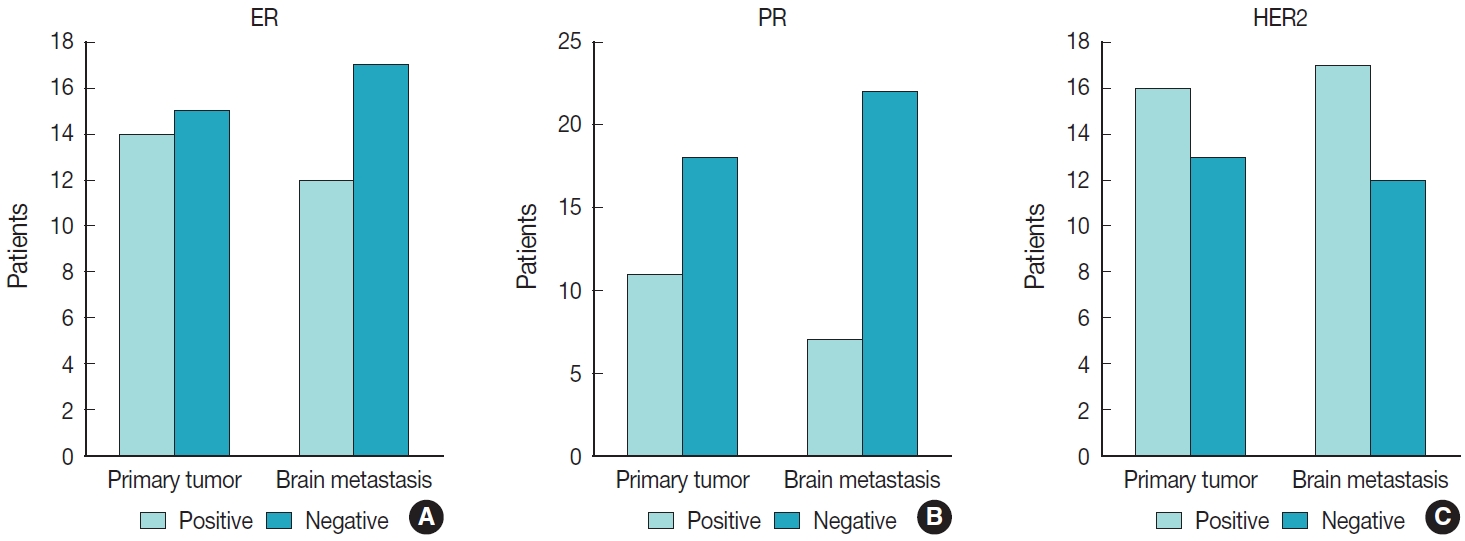
Among 269 patients, 139 males and 130 females were included. The median age of primary tumor was 58 years (range, 13 to 87 years) and 86 patients (32.0%) had BM at initial presentation. Median BM free interval was 28.0 months (range, 1 to 286 months). The most frequent primary site was lung 46.5% (125/269), and followed by breast 15.6% (42/269), colorectum 10.0% (27/269). Epidermal growth factor receptor (EGFR) mutation was found in 50.8% (32/63) and 58.0% (40/69) of lung primary and BM, respectively. In both breast primary and breast cancer with BM, luminal B was the most frequent subtype at 37.9% (11/29) and 42.9% (18/42), respectively, followed by human epidermal growth factor receptor 2 with 31.0% (9/29) and 33.3% (14/42). Triple-negative was 20.7% (6/29) and 16.7% (7/42), and luminal A was 10.3% (3/29) and 7.1% (3/42) of breast primary and BM, respectively. In colorectal primary and colorectal cancer with BM, KRAS mutation was found in 76.9% (10/13) and 66.7% (2/3), respectively.
Conclusions
We report the clinicopathological and molecular pathologic features of BM that can provide useful information for understanding the pathogenesis of metastasis and for clinical trials based on the tumor’s molecular pathology. -
Citations
Citations to this article as recorded by
- Colorectal cancer metastasis to the brain: A scoping review of incidence, treatment, and outcomes
Hunter J Hutchinson, Melanie Gonzalez, Diana Feier, Colin E Welch, Brandon Lucke-Wold
World Journal of Gastrointestinal Pathophysiology.2025;[Epub] CrossRef
- Colorectal cancer metastasis to the brain: A scoping review of incidence, treatment, and outcomes
- Clinicopathologic characterization of cervical metastasis from an unknown primary tumor: a multicenter study in Korea
- Miseon Lee, Uiree Jo, Joon Seon Song, Youn Soo Lee, Chang Gok Woo, Dong-Hoon Kim, Jung Yeon Kim, Sun Och Yoon, Kyung-Ja Cho
- J Pathol Transl Med. 2023;57(3):166-177. Published online May 10, 2023
- DOI: https://doi.org/10.4132/jptm.2023.04.12
- 8,337 View
- 179 Download
- 6 Web of Science
- 5 Crossref
-
Abstract
PDF
 Supplementary Material
 - Background
Research regarding cervical metastasis from an unknown primary tumor (CUP) according to human papillomavirus (HPV) and Epstein-Barr virus (EBV) status in Korea has been sporadic and small-scale. This study aims to analyze and understand the characteristics of CUP in Korea according to viral and p16 and p53 status through a multicenter study.
Methods
Ninety-five cases of CUP retrieved from six hospitals in Korea between January 2006 and December 2016 were subjected to high-risk HPV detection (DNA in situ hybridization [ISH] or real-time polymerase chain reaction), EBV detection (ISH), and immunohistochemistry for p16 and p53.
Results
CUP was HPV-related in 37 cases (38.9%), EBV-related in five cases (5.3%), and unrelated to HPV or EBV in 46 cases (48.4%). HPV-related CUP cases had the best overall survival (OS) (p = .004). According to the multivariate analysis, virus-unrelated disease (p = .023) and longer smoking duration (p < .005) were prognostic factors for poor OS. Cystic change (p = .016) and basaloid pattern (p < .001) were more frequent in HPV-related cases, and lymphoepithelial lesion was frequent in EBV-related cases (p = .010). There was no significant association between viral status and p53 positivity (p = .341), smoking status (p = .728), or smoking duration (p = .187). Korean data differ from Western data in the absence of an association among HPV, p53 positivity, and smoking history.
Conclusions
Virus-unrelated CUP in Korea had the highest frequency among all CUP cases. HPV-related CUP is similar to HPV-mediated oropharyngeal cancer and EBVrelated CUP is similar to nasopharyngeal cancer in terms of characteristics, respectively. -
Citations
Citations to this article as recorded by- Management of squamous cell carcinoma of unknown primary in the head and neck: current evidence-based diagnostic and treatment strategies
Marcel Kloppenburg, Matthias Santer, Lukas Schmutzler, Felix Johnson, Benedikt Hofauer, Teresa Steinbichler
memo - Magazine of European Medical Oncology.2026; 19(1): 45. CrossRef - Differenzierung von benignen und malignen Halszysten – eine diagnostische Herausforderung
Christina Sauter, Matthias Sand, Karim Plath, Michaela Maria Plath
Laryngo-Rhino-Otologie.2025; 104(05): 296. CrossRef - Unlocking the Hidden: Advancing Imaging Techniques in Diagnosing Cancers of Unknown Primary in the Head and Neck Region
Daniela Messineo, Filippo Valentini, Giovanni Francesco Niccolini, Federica Zoccali, Francesca Ripari, Enrico Marotta, Marcello Caratozzolo, Pasquale Frisina
Applied Sciences.2025; 15(4): 2194. CrossRef - Characterization of undifferentiated carcinoma of the salivary gland: clinicopathological and immunohistochemical analyses in comparison with lymphoepithelial carcinoma
Sangjoon Choi, Gyuheon Choi, Hee Jin Lee, Joon Seon Song, Yoon Se Lee, Seung-Ho Choi, Kyung-Ja Cho
Journal of Pathology and Translational Medicine.2025; 59(6): 361. CrossRef - Expansion of tumor-infiltrating lymphocytes from head and neck squamous cell carcinoma to assess the potential of adoptive cell therapy
Sangjoon Choi, Mofazzal Hossain, Hyun Lee, Jina Baek, Hye Seon Park, Chae-Lyul Lim, DoYeon Han, Taehyun Park, Jong Hyeok Kim, Gyungyub Gong, Mi-Na Kweon, Hee Jin Lee
Cancer Immunology, Immunotherapy.2024;[Epub] CrossRef
- Management of squamous cell carcinoma of unknown primary in the head and neck: current evidence-based diagnostic and treatment strategies
- Prevalence of high-risk human papillomavirus and its genotype distribution in head and neck squamous cell carcinomas
- Yuil Kim, Young-Hoon Joo, Min-Sik Kim, Youn Soo Lee
- J Pathol Transl Med. 2020;54(5):411-418. Published online July 21, 2020
- DOI: https://doi.org/10.4132/jptm.2020.06.22
- 13,970 View
- 191 Download
- 23 Web of Science
- 26 Crossref
-
Abstract
PDF
- Background
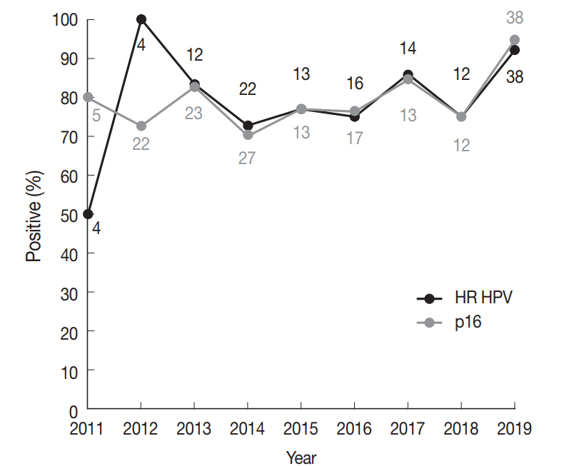
High-risk (HR) human papillomavirus (HPV) is found in a subset of head and neck (HN) squamous cell carcinomas (SCCs). For oropharyngeal SCCs, HR HPV positivity is known to be associated with good prognosis, and a separate staging system for HPV-associated carcinomas using p16 immunohistochemistry (IHC) as a surrogate test has been adopted in the 8th American Joint Committee on Cancer staging system. We examined the HR HPV status and the genotype distribution in five HN subsites.
Methods
Formalin-fixed paraffin-embedded tissue sections were used for p16 IHC and DNA extraction. HPV DNA detection and genotyping were done employing either a DNA chip-based or real-time polymerase chain reaction–based method.
Results
During 2011–2019, a total of 466 SCCs were tested for HPV DNA with 34.1% positivity for HR HPV. Among HN subsites, the oropharynx showed the highest HR HPV prevalence (149/205, 75.1%), followed by the sinonasal tract (3/14, 21.4%), larynx (5/43, 11.6%), hypopharynx (1/38, 2.6%), and oral cavity (1/166, 0.6%). The most common HPV genotype was HPV16 (84.3%) followed by HPV35 (6.9%) and HPV33 (4.4%). Compared with HR HPV status, the sensitivity and specificity of p16 IHC were 98.6% and 94.3% for the oropharynx, and 99.2% and 93.8% for the tonsil, respectively.
Conclusions
Using a Korean dataset, we confirmed that HR HPV is most frequently detected in oropharyngeal SCCs. p16 positivity showed a good concordance with HR HPV DNA for oropharyngeal and especially tonsillar carcinomas. The use of p16 IHC may further be extended to predict HR HPV positivity in sinonasal tract SCCs. -
Citations
Citations to this article as recorded by- Prevalence of Human Papillomavirus in Head and Neck Cancers in South Korea: A Targeted Literature Review
Aneesha Fathima Syed Mohamed, Ruixuan Wang, Seyoung Oh, Ying Hui Wu, Gyongseon Yang, Isaya Sukarom, Sei Young Lee, Wei Wang
Korean Journal of Otorhinolaryngology-Head and Neck Surgery.2026; 69(3): 115. CrossRef - Impact of histopathological parameters in prognosis of oral squamous cell carcinoma
R. P. Ekanayaka, W. M. Tilakaratne
Oral Diseases.2025; 31(5): 1420. CrossRef - Prevalence of human papilloma virus in head and neck mucous squamous cell carcinoma and genotypes by location: an observational study
Emilie Uhlrich, Jerzy Klijanienko, Joey Martin, Emmanuelle Jeannot, Anne Vincent-Salomon, Paul Freneaux, Christophe Le Tourneau, Olivier Choussy, Antoine Dubray-Vautrin
European Journal of Cancer Prevention.2025; 34(5): 426. CrossRef - Risk factors for cervical lymph node metastasis in oropharyngeal cancer and its impact on prognosis
Li Zhang, Zhilin Li, Jing Wang, Chen Wang, Shuxin Wen
Brazilian Journal of Otorhinolaryngology.2025; 91(2): 101520. CrossRef - Co-infection of human papillomavirus genotypes and Epstein-Barr virus in tumors of the oral cavity and oropharynx: a retrospective study in Northeastern Mexico
Gerardo del Carmen Palacios-Saucedo, Jose Manuel Vazquez-Guillen, Alondra Yamileth Alanis-Valdez, Leticia Lizeth Valdez-Treviño, Luis Roberto Galindo-Mendez, Angel Zavala-Pompa, Lydia Guadalupe Rivera-Morales, Ana Carolina Martinez-Torres, Roberto Lopez-V
IJID Regions.2025; 14: 100555. CrossRef - Rates of p16 and p53 expression in head and neck cutaneous squamous cell carcinoma vary according to human papillomavirus status
Rachid Ait Addi
World Journal of Clinical Cases.2025;[Epub] CrossRef - The epidemiological trends and survival of HPV-related oropharyngeal cancer other than tonsils and base of tongue − a systematic review and meta-analysis
Anas Mohammad Al Fadel, Kathrine Kronberg Jakobsen, Lasse Holmgaard Jensen, Amanda-Louise Fenger Carlander, Christian Grønhøj, Christian von Buchwald
Oral Oncology.2025; 165: 107311. CrossRef - Oropharyngeal Helicobacter pylori colonization increases risk and worsens prognosis of head and neck squamous cell carcinoma
Xianyao Jiang, Yongjin Huang, Changwu Li, Hongyan Jiang
Scientific Reports.2025;[Epub] CrossRef - Characteristics of human papillomavirus infection among oropharyngeal cancer patients: A systematic review and meta-analysis
Meimei Cui, Jinling Cheng, Huijuan Cheng, Ming Zhao, Dan Zhou, Min Zhang, Jingjing Jia, Limei Luo
Archives of Oral Biology.2024; 157: 105830. CrossRef - Longitudinal Screening for Oral High-Risk Non-HPV16 and Non-HPV18 Strains of Human Papillomavirus Reveals Increasing Prevalence among Adult and Pediatric Biorepository Samples: A Pilot Study
Jordan Jacobs, Eugene Chon, Karl Kingsley
Vaccines.2024; 12(8): 895. CrossRef - Position Statement about Gender-Neutral HPV Vaccination in Korea
Kyung-Jin Min, Yung-Taek Ouh, Sangrak Bae, Yong-Bae Ji, Jae-Kwan Lee, Jae-Weon Kim, Kwang-Jae Cho, Dong-Hun Im
Vaccines.2024; 12(10): 1110. CrossRef - High-risk HPV Does not Appear to be an Important Risk Factor for Sinonasal Carcinomas in Turkish Population: A Tertiary Center Experience
Evsen Apaydin Arikan, Levent Aydemir, Murat Ulusan, Dilek Yilmazbayhan, Yasemin Ozluk
International Journal of Surgical Pathology.2023; 31(2): 124. CrossRef - Practical Application of Circulating Tumor-Related DNA of Human Papillomavirus in Liquid Biopsy to Evaluate the Molecular Response in Patients with Oropharyngeal Cancer
Agnieszka M. Mazurek, Tomasz W. Rutkowski
Cancers.2023; 15(4): 1047. CrossRef - The Prevalence of HPV in Oral Cavity Squamous Cell Carcinoma
Seyed Keybud Katirachi, Mathias Peter Grønlund, Kathrine Kronberg Jakobsen, Christian Grønhøj, Christian von Buchwald
Viruses.2023; 15(2): 451. CrossRef - The Protective Role of Cranberries and Blueberries in Oral Cancer
César Esquivel-Chirino, Mario Augusto Bolaños-Carrillo, Daniela Carmona-Ruiz, Ambar Lopéz-Macay, Fernando Hernández-Sánchez, Delina Montés-Sánchez, Montserrat Escuadra-Landeros, Luis Alberto Gaitán-Cepeda, Silvia Maldonado-Frías, Beatriz Raquel Yáñez-Ocam
Plants.2023; 12(12): 2330. CrossRef - Unusual cases of sinonasal malignancies: a letter to the editor on HPV-positive sinonasal squamous cell carcinomas
Benedicte Bitsch Lauritzen, Sannia Sjöstedt, Jakob Myllerup Jensen, Katalin Kiss, Christian von Buchwald
Acta Oncologica.2023; 62(6): 608. CrossRef - Prevalence of human Papillomavirus associated oropharyngeal and oral squamous cell carcinoma in Asian countries: A systematic review and large-scale meta-analysis
Yy Jean Tan, Ken Wong Siong Hou, Galvin Sim Siang Lin, Jasmine Lim Suk Wun, Wan Nor Amira Wan Ahmad Abdul Nasir, Lynn Wei Linn Ko
Acta Marisiensis - Seria Medica.2023; 69(2): 77. CrossRef - Top 100 most cited articles on human papillomavirus-induced head and neck squamous cell carcinoma: A bibliographic review
Rahul Mohandas, Subhashree Mohapatra, Mary Oshin, ShubhangiSambhaji Hajare
Journal of International Oral Health.2023; 15(3): 219. CrossRef - Intracellular Toll-Like Receptors Modulate Adaptive Immune Responses in Head and Neck Cancer
Sangeetha K. Nayanar, Deepak Roshan V.G., Shruthi Surendran, Göran Kjeller, Bengt Hasséus, Daniel Giglio
Viral Immunology.2023; 36(10): 659. CrossRef - Positive Rate of Human Papillomavirus and Its Trend in Head and Neck Cancer in South Korea
Hyun Woong Jun, Yong Bae Ji, Chang Myeon Song, Jae Kyung Myung, Hae Jin Park, Kyung Tae
Frontiers in Surgery.2022;[Epub] CrossRef - Transcriptionally active HPV in OPMD and OSCC: A systematic review following the CAP/ASCO guidelines
Laura Borges Kirschnick, Lauren Frenzel Schuch, Maria Eduarda Pérez‐de‐Oliveira, Ana Gabriela Costa Normando, Bruno Augusto Linhares Almeida Mariz, Eliete Neves Silva Guerra, Felipe Martins Silveira, Ana Carolina Uchoa Vasconcelos, Luciana Estevam Simonat
Oral Diseases.2022; 28(8): 2309. CrossRef - Effect of National Oral Health Screening Program on the Risk of Head and Neck Cancer: A Korean National Population-Based
Chan Woo Wee, Hyo-Jung Lee, Jae-Ryun Lee, Hyejin Lee, Min-Jeong Kwoen, Woo-Jin Jeong, Keun-Yong Eom
Cancer Research and Treatment.2022; 54(3): 709. CrossRef - Expression of p16, p53, and TLR9 in HPV-Associated Head and Neck Squamous Cell Carcinoma: Clinicopathological Correlations and Potential Prognostic Significance
Shu Wang, Xibing Zhuang, Caixia Gao, Tiankui Qiao
OncoTargets and Therapy.2021; Volume 14: 867. CrossRef - The Role of Human Papilloma Virus in Dictating Outcomes in Head and Neck Squamous Cell Carcinoma
Shane Brennan, Anne-Marie Baird, Esther O’Regan, Orla Sheils
Frontiers in Molecular Biosciences.2021;[Epub] CrossRef - A Contemporary Systematic Review on Repartition of HPV-Positivity in Oropharyngeal Cancer Worldwide
Amanda F. Carlander, Kathrine K. Jakobsen, Simone K. Bendtsen, Martin Garset-Zamani, Charlotte D. Lynggaard, Jakob Schmidt Jensen, Christian Grønhøj, Christian von Buchwald
Viruses.2021; 13(7): 1326. CrossRef - The Prevalence of High- and Low-Risk Types of HPV in Patients with Squamous Cell Carcinoma of the Head and Neck, Patients with Chronic Tonsillitis, and Healthy Individuals Living in Poland
Joanna Katarzyna Strzelczyk, Krzysztof Biernacki, Jadwiga Gaździcka, Elżbieta Chełmecka, Katarzyna Miśkiewicz-Orczyk, Natalia Zięba, Janusz Strzelczyk, Maciej Misiołek
Diagnostics.2021; 11(12): 2180. CrossRef
- Prevalence of Human Papillomavirus in Head and Neck Cancers in South Korea: A Targeted Literature Review
- Molecular characteristics of meningiomas
- Young Suk Lee, Youn Soo Lee
- J Pathol Transl Med. 2020;54(1):45-63. Published online January 15, 2020
- DOI: https://doi.org/10.4132/jptm.2019.11.05
- 25,697 View
- 705 Download
- 54 Web of Science
- 59 Crossref
-
Abstract
PDF
- Meningioma is the most common primary intracranial tumor in adults. The grading of meningioma is based on World Health Organization criteria, which rely on histopathological features alone. This grading system is unable to conclusively predict the clinical behavior of these tumors (i.e., recurrence or prognosis in benign or atypical grades). Advances in molecular techniques over the last decade that include genomic and epigenomic data associated with meningiomas have been used to identify genetic biomarkers that can predict tumor behavior. This review summarizes the molecular characteristics of meningioma using genetic and epigenetic biomarkers. Molecular alterations that can predict meningioma behavior may be integrated into the upcoming World Health Organization grading system.
-
Citations
Citations to this article as recorded by- Multi-Institutional Modified Delphi For Genomics in Expert Consensus Survey of Genomic Testing for Anterior Skull Base Malignancies
Anirudh Saraswathula, Shreya Sriram, Corinna Levine, Nyall R. London, Shirley Y. Su, Mathew Geltzeiler, Sanjeet V. Rangarajan, Ian Witterick, Brian Thorp, Kathleen Kelly Gallagher, Kenneth Byrd, Ricardo Carrau, Waleed Abuzeid, Eric Wang, Carl Snyderman, E
Journal of Neurological Surgery Part B: Skull Base.2026; 87(01): 014. CrossRef - Long-Term Prognostic Impact of the Dural Tail in the Local Control of Intracranial Meningiomas
Franco Rubino, Romulo A. de Almeida, Komal Shah, Shaan M. Raza, Franco DeMonte
Journal of Neurological Surgery Part B: Skull Base.2026;[Epub] CrossRef - Multidisciplinary management of meningiomas in the era of precision oncology
Majid Assadi, Malik E. Juweid, Philipp Lohmann, Giuseppe Minniti, Felix Sahm, Jana Ivanidze, Philipp Karschnia, Alexander Landry, Felix M. Mottaghy, Gelareh Zadeh
Nature Reviews Clinical Oncology.2026; 23(7): 539. CrossRef - From imaging to molecular pathways: a comprehensive narrative review of the distinctive landscape of secretory meningioma
Erfan Shahabinejad, Mahsa Kamali, Amirreza Shakoeizadeh, Zeinab Falakian, Aynaz Foroughi Eghbal, Seyyed Mohammad Alipour, Maria Krywyj, Jason P. Sheehan, Qais Alrashidi, Hamid Borghei-Razavi
International Journal of Neuroscience.2026; : 1. CrossRef - Recurrence of Resected Skull Base Meningiomas during Long-term Follow-up: Incidence and Predisposing Factors
Joshua Ian Macarthur, Cathal John Hannan, Callum Howard, Jane Halliday, Omar Nathan Pathmanaban, Charlotte Hammerbeck-Ward, Scott A. Rutherford, Andrew T. King
Journal of Neurological Surgery Part B: Skull Base.2025; 86(03): 245. CrossRef - Role of H3K27me3 and Ki-67 Labeling Index in Assessing the Biological Behavior of Meningiomas
Shalaka Deshpande, Bhavna Nayal, Rajesh Nair, Deepak Nayak, Padmapriya J, Geetha V
World Neurosurgery.2025; 194: 123514. CrossRef - Context aware machine learning techniques for brain tumor classification and detection – A review
Usman Amjad, Asif Raza, Muhammad Fahad, Doaa Farid, Adnan Akhunzada, Muhammad Abubakar, Hira Beenish
Heliyon.2025; 11(2): e41835. CrossRef - Case report: Clonal evolution analysis of a rare case of meningioma lung metastases identifies actionable alterations in matched longitudinal tumour samples
Nicola Cosgrove, Orla M. Fitzpatrick, Liam Grogan, Bryan T. Hennessy, Simon J. Furney, Sinead Toomey
Frontiers in Oncology.2025;[Epub] CrossRef - Post-operative Hemorrhage After Tumor Removal of Multiple Meningiomas
Bob Irfan Syahputra, Akhmad Imron, Dhany Febriantara, Helza Efriani
International Journal of Recent Surgical and Medical Sciences.2025; 11: e005. CrossRef - Diarylpentanoid, a curcumin analog, inhibits malignant meningioma growth in both in vitro and in vivo models
Anna Terasawa, Kazuhiro Shimazu, Hiroshi Nanjo, Masatomo Miura, Hiroyuki Shibata
World Journal of Experimental Medicine.2025;[Epub] CrossRef - Tumour-associated macrophages in human meningiomas
Rahmina Meta, Henrik Sahlin Pettersen, Sofie Eline Tollefsen, Borgny Ytterhus, Øyvind Olav Salvesen, Wenche Sjursen, Sverre Helge Torp, Jianhong Zhou
PLOS One.2025; 20(5): e0319960. CrossRef - Binary Classification of Meningioma Grades Using CNN and VGG16 + XGBoost Deep Learning Models
L. Priya, D. Saraswathi, M. Bhuvaneshwari, K. Dhanya, K. Krishna Kousalya, Deepali
Applied Computational Intelligence and Soft Computing.2025;[Epub] CrossRef - Unusual adrenal metastasis of anaplastic meningioma: A case report
M. Inouss, C. Rhoul, A. Kharkhach, T. Bouhout, B. Serji
International Journal of Surgery Case Reports.2025;[Epub] CrossRef - TERT promoter mutations in meningiomas: associations with clinicopathological features and insights into spatial and temporal heterogeneity in a 165-case cohort
Ahmet Boduroğlu, Mualla Özcan, Güzide Ayşe Ocak
Pathology - Research and Practice.2025; 274: 156191. CrossRef - The Gut–Brain Axis in Brain Tumors: Insights into Tumor Development, Progression, and Therapy
Sarah Adriana Scuderi, Alessio Ardizzone, Elsa Calcaterra, Nicoletta Palermo, Fabiola De Luca, Antonio Catalfamo, Emanuela Esposito, Anna Paola Capra
Biomedicines.2025; 13(9): 2172. CrossRef - The Natural History and Treatment of Meningiomas: An Update
Arsene Daniel Nyalundja, Fabrice Mugisha, Claire Karekezi
Seminars in Neurology.2024; 44(01): 001. CrossRef - Epidemiology, Genetics, and DNA Methylation Grouping of Hyperostotic Meningiomas
Gray Umbach, Edwina B. Tran, Charlotte D. Eaton, Abrar Choudhury, Ramin Morshed, Javier E. Villanueva-Meyer, Philip V. Theodosopoulos, Stephen T. Magill, Michael W. McDermott, David R. Raleigh, Ezequiel Goldschmidt
Operative Neurosurgery.2024; 26(6): 662. CrossRef - The Evolving Classification of Meningiomas: Integration of Molecular Discoveries to Inform Patient Care
S. Joy Trybula, Mark W. Youngblood, Constantine L. Karras, Nikhil K. Murthy, Amy B. Heimberger, Rimas V. Lukas, Sean Sachdev, John A. Kalapurakal, James P. Chandler, Daniel J. Brat, Craig M. Horbinski, Stephen T. Magill
Cancers.2024; 16(9): 1753. CrossRef - Minimally Invasive Approaches in the Surgical Treatment of Intracranial Meningiomas: An Analysis of 54 Cases
Guenther C. Feigl, Daniel Staribacher, Gavin Britz, Dzmitry Kuzmin
Brain Tumor Research and Treatment.2024; 12(2): 93. CrossRef - Related mechanisms, current treatments, and new perspectives in meningioma
Gizem Inetas‐Yengin, Omer Faruk Bayrak
Genes, Chromosomes and Cancer.2024;[Epub] CrossRef - Clinical application of intraoperative ultrasound superb microvascular imaging in brain tumors resections: contributing to the achievement of total tumoral resection
Siman Cai, Hao Xing, Yuekun Wang, Yu Wang, Wenbin Ma, Yuxin Jiang, Jianchu Li, Hongyan Wang
BMC Medical Imaging.2024;[Epub] CrossRef - WHO CNS 5 and meningiomas: What’s new?
Indranil Chakrabarti, Sujaya Mazumder
IP Archives of Cytology and Histopathology Research.2024; 9(2): 67. CrossRef - Molecular Developments in Parasellar Tumors and Potential Therapeutic Implications
Paraskevi Xekouki, Vasiliki Venetsanaki, Georgios Kyriakopoulos, Krystallenia Alexandraki, Anna Angelousi, Gregory Kaltsas
Endocrine Reviews.2024; 45(6): 880. CrossRef - The Impact of Molecular and Genetic Analysis on the Treatment of Patients with Atypical Meningiomas
Janez Ravnik, Hojka Rowbottom
Diagnostics.2024; 14(16): 1782. CrossRef - Differential Expression of Proteins and Genes at the Tumor‐Brain Interface in Invasive Meningioma
Kornwika Senglek, Chinachote Teerapakpinyo, Nutchawan Jittapiromsak, Pakrit Jittapiromsak, Irin Lertparinyaphorn, Paul Scott Thorner, Shanop Shuangshoti
Genes, Chromosomes and Cancer.2024;[Epub] CrossRef - Protein expression of CD44 in patients with meningioma tumors: association with clinicopathological parameters and survival
Trupti Trivedi, Neha Bhalala, Kirti Dialani, Priti Trivedi
Journal of the Egyptian National Cancer Institute.2024;[Epub] CrossRef - DNA methylation profiling of meningiomas highlights clinically distinct molecular subgroups
Jyotsna Singh, Ravi Sharma, Nidhi Shukla, Priya Narwal, Amit Katiyar, Swati Mahajan, Saumya Sahu, Ajay Garg, Mehar C. Sharma, Ashish Suri, Chitra sarkar, Vaishali Suri
Journal of Neuro-Oncology.2023; 161(2): 339. CrossRef - Spinal meningiomas, from biology to management - A literature review
Nicolas Serratrice, Imène Lameche, Christian Attieh, Moussa A Chalah, Joe Faddoul, Bilal Tarabay, Rabih Bou-Nassif, Youssef Ali, Joseph G Mattar, François Nataf, Samar S Ayache, Georges N Abi Lahoud
Frontiers in Oncology.2023;[Epub] CrossRef - Somatic mutation landscape in a cohort of meningiomas that have undergone grade progression
Sarah A Cain, Bernard Pope, Stefano Mangiola, Theo Mantamadiotis, Katharine J Drummond
BMC Cancer.2023;[Epub] CrossRef - Actualización sobre el meningioma: correlación clínico-radiológica y radio-patológica
A. Navarro-Ballester, M. Aleixandre-Barrachina, S.F. Marco-Doménech
Radiología.2023; 65(5): 458. CrossRef - SMARCE1-related meningiomas: A clear example of cancer predisposing syndrome
Erika Fiorentini, Laura Giunti, Andrea Di Rita, Simone Peraio, Carla Fonte, Chiara Caporalini, Anna Maria Buccoliero, Maria Luigia Censullo, Giulia Gori, Alice Noris, Rosa Pasquariello, Roberta Battini, Rossana Pavone, Flavio Giordano, Sabrina Giglio, Ber
European Journal of Medical Genetics.2023; 66(7): 104784. CrossRef - Grade scoring system reveals distinct molecular subtypes and identifies KIF20A as a novel biomarker for predicting temozolomide treatment efficiency in gliomas
Liguo Ye, Shi’ao Tong, Yaning Wang, Yu Wang, Wenbin Ma
Journal of Cancer Research and Clinical Oncology.2023; 149(12): 9857. CrossRef - Integrated clinical genomic analysis reveals xenobiotic metabolic genes are downregulated in meningiomas of current smokers
A. Basit Khan, Rajan Patel, Malcolm F. McDonald, Eric Goethe, Collin English, Ron Gadot, Arya Shetty, Shervin Hosseingholi Nouri, Arif O. Harmanci, Akdes S. Harmanci, Tiemo J. Klisch, Akash J. Patel
Journal of Neuro-Oncology.2023; 163(2): 397. CrossRef - DNA methylation meningioma biomarkers: attributes and limitations
Zhaohui Li, Yufei Gao, Jinnan Zhang, Liang Han, Hang Zhao
Frontiers in Molecular Neuroscience.2023;[Epub] CrossRef - Meningioma: A Biography—Tumor Forever Tied to the Origins and “Soul of Neurosurgery”
Nolan J. Brown, Zach Pennington, Cathleen C. Kuo, Julian Gendreau, Sachiv Chakravarti, Rohin Singh, Dontré M. Douse, Jamie J. Van Gompel
World Neurosurgery.2023; 178: 191. CrossRef - Molecular genetic features of meningiomas
E.S. Makashova, N.V. Lasunin, M.V. Galkin, S.V. Zolotova, K.O. Karandasheva, A.V. Golanov
Burdenko's Journal of Neurosurgery.2023; 87(4): 101. CrossRef - Novel Advances in Treatment of Meningiomas: Prognostic and Therapeutic Implications
Gerardo Caruso, Rosamaria Ferrarotto, Antonello Curcio, Luisa Metro, Francesco Pasqualetti, Paola Gaviani, Valeria Barresi, Filippo Flavio Angileri, Maria Caffo
Cancers.2023; 15(18): 4521. CrossRef - Update on meningioma: Clinical-radiological and radio-pathological correlation
A. Navarro-Ballester, M. Aleixandre-Barrachina, S.F. Marco-Doménech
Radiología (English Edition).2023; 65(5): 458. CrossRef - Early Preventive Strategies and CNS Meningioma – Is This Feasible? A Comprehensive Review of the Literature
Daniel Sescu, Aminta Chansiriwongs, Katarzyna Julia Minta, Jyothi Vasudevan, Chandrasekaran Kaliaperumal
World Neurosurgery.2023; 180: 123. CrossRef - Domestic Animal Models of Central Nervous System Tumors: Focus on Meningiomas
Michele Tomanelli, Tullio Florio, Gabriela Vargas, Aldo Pagano, Paola Modesto
Life.2023; 13(12): 2284. CrossRef - Assessment of parameters of the acid-base state among patients with meningiomas and gliomas in the postoperative period
E. S. Orlova, I. O. Ishchenko, K. K. Kukanov, N. E. Voinov, A. P. Gerasimov, N. E. Ivanova
Russian Neurosurgical Journal named after Professor A. L. Polenov.2023; 15(2): 21. CrossRef - The integrated multiomic diagnosis of sporadic meningiomas: a review of its clinical implications
Stephanie M. Robert, Shaurey Vetsa, Arushii Nadar, Sagar Vasandani, Mark W. Youngblood, Evan Gorelick, Lan Jin, Neelan Marianayagam, E Zeynep Erson-Omay, Murat Günel, Jennifer Moliterno
Journal of Neuro-Oncology.2022; 156(2): 205. CrossRef - Clinical presentation, diagnostic findings and outcome of dogs undergoing surgical resection for intracranial meningioma: 101 dogs
Alexander K. Forward, Holger Andreas Volk, Giunio Bruto Cherubini, Tom Harcourt-Brown, Ioannis N. Plessas, Laurent Garosi, Steven De Decker
BMC Veterinary Research.2022;[Epub] CrossRef - Sphenoid wing meningiomas: peritumoral brain edema as a prognostic factor in surgical outcome
Abdalrahman Nassar, Volodymyr Smolanka, Andriy Smolanka, Dipak Chaulagain, Oleg Devinyak
Neurosurgical Review.2022; 45(4): 2951. CrossRef - Potential Molecular Mechanisms of Recurrent and Progressive Meningiomas: A Review of the Latest Literature
Wenjie Peng, Pei Wu, Minghao Yuan, Bo Yuan, Lian Zhu, Jiesong Zhou, Qian Li
Frontiers in Oncology.2022;[Epub] CrossRef - Case Report: Upper Thoracic Purely Extradural Spinal Meningioma With Nerve Root Attachment: A Case Report and Literature Review
Zhao-Lin Wang, Jian-Hui Mou, Dong Sun, Peng Liu
Frontiers in Surgery.2022;[Epub] CrossRef - Molecular diagnosis and treatment of meningiomas: an expert consensus (2022)
Jiaojiao Deng, Lingyang Hua, Liuguan Bian, Hong Chen, Ligang Chen, Hongwei Cheng, Changwu Dou, Dangmurenjiapu Geng, Tao Hong, Hongming Ji, Yugang Jiang, Qing Lan, Gang Li, Zhixiong Liu, Songtao Qi, Yan Qu, Songsheng Shi, Xiaochuan Sun, Haijun Wang, Yongpi
Chinese Medical Journal.2022; 135(16): 1894. CrossRef - Оновлена інформація про менінгіоми крила клиноподібної кістки
Abdalrahman Nassar, Volodymyr Smolanka
INTERNATIONAL NEUROLOGICAL JOURNAL.2022; 18(1): 43. CrossRef - The Prognostic Value of Methylation Signatures and NF2 Mutations in Atypical Meningiomas
Rahmina Meta, Henning B. Boldt, Bjarne W. Kristensen, Felix Sahm, Wenche Sjursen, Sverre H. Torp
Cancers.2021; 13(6): 1262. CrossRef - Neurofibromatosis Type 2 (NF2) and the Implications for Vestibular Schwannoma and Meningioma Pathogenesis
Suha Bachir, Sanjit Shah, Scott Shapiro, Abigail Koehler, Abdelkader Mahammedi, Ravi N. Samy, Mario Zuccarello, Elizabeth Schorry, Soma Sengupta
International Journal of Molecular Sciences.2021; 22(2): 690. CrossRef - Meningioma: A Review of Epidemiology, Pathology, Diagnosis, Treatment, and Future Directions
Christian Ogasawara, Brandon D. Philbrick, D. Cory Adamson
Biomedicines.2021; 9(3): 319. CrossRef - The substantial loss of H3K27me3 can stratify risk in grade 2, but not in grade 3 meningioma
Minsun Jung, Seong-Ik Kim, Ka Young Lim, Jeongmo Bae, Chul-Kee Park, Seung Hong Choi, Sung-Hye Park, Jae-Kyung Won
Human Pathology.2021; 115: 96. CrossRef - Papillary Meningioma: Case Presentation with Emphasis on Surgical and Medical Therapy of a Rare Variant of Meningioma
Gerardo Cazzato, Valeria Internò, Antonietta Cimmino, Raffaella Messina, Marco Tucci, Teresa Lettini, Leonardo Resta, Giuseppe Ingravallo
Diseases.2021; 9(3): 63. CrossRef - An Overview of Managements in Meningiomas
Lianhua Zhao, Wei Zhao, Yanwei Hou, Cuixia Wen, Jing Wang, Pei Wu, Zaiyu Guo
Frontiers in Oncology.2020;[Epub] CrossRef - Multi-Omics Analysis in Initiation and Progression of Meningiomas: From Pathogenesis to Diagnosis
Jiachen Liu, Congcong Xia, Gaiqing Wang
Frontiers in Oncology.2020;[Epub] CrossRef - Molecular Mechanism and Approach in Progression of Meningioma
Zhiwei Shao, Lihong Liu, Yanghao Zheng, Sheng Tu, Yuanbo Pan, Sheng Yan, Qichun Wei, Anwen Shao, Jianmin Zhang
Frontiers in Oncology.2020;[Epub] CrossRef - Multiple meningiomas: does quantity matter? a population-based survival analysis with underlined age and sex differences
Andres Ramos-Fresnedo, Ricardo A. Domingo, Tito Vivas-Buitrago, Larry Lundy, Daniel M. Trifiletti, Mark E. Jentoft, Amit B. Desai, Alfredo Quiñones-Hinojosa
Journal of Neuro-Oncology.2020; 149(3): 413. CrossRef - Meningioma: A Review of Clinicopathological and Molecular Aspects
Kristin Huntoon, Angus Martin Shaw Toland, Sonika Dahiya
Frontiers in Oncology.2020;[Epub] CrossRef - Neues zur Einteilung und Therapie von Meningeomen
Corinna Seliger, Wolfgang Wick
Neurologie up2date.2020; 3(04): 343. CrossRef
- Multi-Institutional Modified Delphi For Genomics in Expert Consensus Survey of Genomic Testing for Anterior Skull Base Malignancies
- PD-L1 Expression and Combined Status of PD-L1/PD-1–Positive Tumor Infiltrating Mononuclear Cell Density Predict Prognosis in Glioblastoma Patients
- Jiheun Han, Yongkil Hong, Youn Soo Lee
- J Pathol Transl Med. 2017;51(1):40-48. Published online December 15, 2016
- DOI: https://doi.org/10.4132/jptm.2016.08.31
- 18,329 View
- 281 Download
- 35 Web of Science
- 40 Crossref
-
Abstract
PDF
- Background
Programmed death ligand 1 (PD-L1) in tumor cells is known to promote immune escape of cancer by interacting with programmed cell death 1 (PD-1) in tumor infiltrating immune cells. Immunotherapy targeting these molecules is emerging as a new strategy for the treatment of glioblastoma (GBM). Understanding the relationship between the PD-L1/PD-1 axis and prognosis in GBM patients may be helpful to predict the effects of immunotherapy.
Methods
PD-L1 expression and PD-1–positive tumor infiltrating mononuclear cell (PD-1+tumor infiltrating mononuclear cell [TIMC]) density were evaluated using tissue microarray containing 54 GBM cases by immunohistochemical analysis; the associations with patient clinical outcomes were evaluated.
Results
PD-L1 expression and high PD-1+TIMC density were observed in 31.5% and 50% of GBM cases, respectively. High expression of PD-L1 in tumor cells was an independent and significant predictive factor for worse overall survival (OS; hazard ratio, 4.958; p = .007) but was not a significant factor in disease-free survival (DFS). PD-1+TIMC density was not correlated with OS or DFS. When patients were classified based on PD-1 expression and PD-1+TIMC density, patients with PD-L1+/PD-1+TIMC low status had the shortest OS (13 months, p = .009) and DFS (7 months, p = .053).
Conclusions
PD-L1 expression in GBM was an independent prognostic factor for poor OS. In addition, combined status of PD-L1 expression and PD-1+TIMC density also predicted patient outcomes, suggesting that the therapeutic role of the PD-1/PD-L1 axis should be considered in the context of GBM immunity. -
Citations
Citations to this article as recorded by- Dual biomarker role of PD-L1 and LC3B in glioblastoma: prognostic and therapeutic potential
Rana Fathy Torky, Rania Makboul, Dalia M. Badary, Wael M. A. El-Ghani, Ahmed El-Hakeem, Rabab M. H. El Ghorori
Neurosurgical Review.2026;[Epub] CrossRef - Expression features of targets for anti-glioma CAR-T cell immunotherapy
Peng Zhang, Chunzhao Li, Yi Wang, Xiaohan Chi, Tai Sun, Qianhe Zhang, Yang Zhang, Nan Ji
Journal of Neuro-Oncology.2025; 171(1): 179. CrossRef - Expression of Programmed Cell Death-Ligand 1 (PD-L1) in Astrocytic Tumors and Its Correlation With Histopathological Grade and Proliferative Index (Ki-67): A Cross-Sectional Study
Namita Singh, Ranjana Giri, Prita Pradhan, Diptiranjan Satapathy, Ipsita Debata
Cureus.2025;[Epub] CrossRef - Prognostic significance of PD-L1 and CD45RO+ cells in glioblastoma: The modulating role of MMR status
Yousef Mohammadi, Elina Kaviani, Simin Ahmadvand, Amirreza Dehghanian, Abbas Ghaderi
Journal of Neuroimmunology.2025; 406: 578669. CrossRef - PD-L1 Clones and Their Relevance in Glioblastoma, IDH-Wildtype: A Comparative Analysis
Michal Hendrych, Frantisek Vana, Marketa Hermanova, Radek Lakomy, Tomas Kazda, Kvetoslava Matulova, Alena Kopkova, Martina Jelinkova, Radim Jancalek, Martin Smrcka, Vaclav Vybihal, Jiri Sana
Bratislava Medical Journal.2025; 126(9): 2233. CrossRef - Tumor-associated microenvironment, PD-L1 expression and their relationship with immunotherapy in glioblastoma, IDH-wild type: A comprehensive review with emphasis on the implications for neuropathologists
Giuseppe Broggi, Giuseppe Angelico, Jessica Farina, Giordana Tinnirello, Valeria Barresi, Magda Zanelli, Andrea Palicelli, Francesco Certo, Giuseppe Barbagallo, Gaetano Magro, Rosario Caltabiano
Pathology - Research and Practice.2024; 254: 155144. CrossRef - Treatment advances in high-grade gliomas
Xi Chen, Yi Cui, Liqun Zou
Frontiers in Oncology.2024;[Epub] CrossRef - TRP-2 / gp100 DNA vaccine and PD-1 checkpoint blockade combination for the treatment of intracranial tumors
Joshua R. D. Pearson, Carles Puig-Saenz, Jubini E. Thomas, Lydia D. Hardowar, Murrium Ahmad, Louise C. Wainwright, Adam M. McVicar, Victoria A. Brentville, Chris J. Tinsley, A. Graham Pockley, Lindy G. Durrant, Stephanie E. B. McArdle
Cancer Immunology, Immunotherapy.2024;[Epub] CrossRef - Advanced immunotherapies for glioblastoma: tumor neoantigen vaccines in combination with immunomodulators
Berta Segura-Collar, Sara Hiller-Vallina, Olaya de Dios, Marta Caamaño-Moreno, Lucia Mondejar-Ruescas, Juan M. Sepulveda-Sanchez, Ricardo Gargini
Acta Neuropathologica Communications.2023;[Epub] CrossRef - Immunohistochemical Analysis of PD-1 and FOXP3 in Tumor-Infiltrating Lymphocytes in Human Gliomas
Priyanka Kanagaraj, Archana Balasubramanian, Raveena Suresh, Bhargavi Somasundaram, Sandhya Sundaram, Priyathersini Nagarajan
Cureus.2023;[Epub] CrossRef - Expression, prognostic significance and therapeutic implications of PD‐L1 in gliomas
Gayaththri Vimalathas, Bjarne Winther Kristensen
Neuropathology and Applied Neurobiology.2022;[Epub] CrossRef - PD-L1 tumor expression is associated with poor prognosis and systemic immunosuppression in glioblastoma
Carolina Noronha, Ana Sofia Ribeiro, Ricardo Taipa, Dina Leitão, Fernando Schmitt, Joaquim Reis, Cláudia Faria, Joana Paredes
Journal of Neuro-Oncology.2022; 156(3): 453. CrossRef - Assessment of radiographic and prognostic characteristics of programmed death-ligand 1 expression in high-grade gliomas
Makoto Ohno, Shigehisa Kitano, Kaishi Satomi, Akihiko Yoshida, Yasuji Miyakita, Masamichi Takahashi, Shunsuke Yanagisawa, Yukie Tamura, Koichi Ichimura, Yoshitaka Narita
Journal of Neuro-Oncology.2022; 160(2): 463. CrossRef - The prognostic significance of PD-L1 expression in patients with glioblastoma: A meta-analysis
Xin Guo, Yuelin Zhang, Hengxing Jiao, Xingyu Miao
Frontiers in Oncology.2022;[Epub] CrossRef - LncRNA UCA1 attenuated the killing effect of cytotoxic CD8 + T cells on anaplastic thyroid carcinoma via miR-148a/PD-L1 pathway
Xiaoming Wang, Yan Zhang, Jian Zheng, Cuixian Yao, Xiubo Lu
Cancer Immunology, Immunotherapy.2021; 70(8): 2235. CrossRef - Low tumour-infiltrating lymphocyte density in primary and recurrent glioblastoma
Kelsey Maddison, Moira C. Graves, Nikola A. Bowden, Michael Fay, Ricardo E. Vilain, Sam Faulkner, Paul A. Tooney
Oncotarget.2021; 12(21): 2177. CrossRef - A Systematic Review of the Tumor-Infiltrating CD8+ T-Cells/PD-L1 Axis in High-Grade Glial Tumors: Toward Personalized Immuno-Oncology
Mahdi Abdoli Shadbad, Zahra Asadzadeh, Negar Hosseinkhani, Afshin Derakhshani, Nazila Alizadeh, Oronzo Brunetti, Nicola Silvestris, Behzad Baradaran
Frontiers in Immunology.2021;[Epub] CrossRef - Prognostic value of programmed death ligand 1 (PD-L1) in glioblastoma: a systematic review, meta-analysis and validation based on dataset
Huan Wang, Youchao Xiao, Xingguang Ren, Dahai Wan
Bioengineered.2021; 12(2): 10366. CrossRef - Expression of Programmed Cell Death Ligand 1 and Associated Lymphocyte Infiltration in Olfactory Neuroblastoma
Nyall R. London, Lisa M. Rooper, Justin A. Bishop, Haiying Xu, Lydia J. Bernhardt, Masaru Ishii, Christine L. Hann, Janis M. Taube, Evgeny Izumchenko, Daria A. Gaykalova, Gary L. Gallia
World Neurosurgery.2020; 135: e187. CrossRef - CCR2 inhibition reduces tumor myeloid cells and unmasks a checkpoint inhibitor effect to slow progression of resistant murine gliomas
Joseph A. Flores-Toro, Defang Luo, Adithya Gopinath, Matthew R. Sarkisian, James J. Campbell, Israel F. Charo, Rajinder Singh, Thomas J. Schall, Meenal Datta, Rakesh K. Jain, Duane A. Mitchell, Jeffrey K. Harrison
Proceedings of the National Academy of Sciences.2020; 117(2): 1129. CrossRef - Treatment Results for Recurrent Glioblastoma and Alteration of Programmed Death-Ligand 1 Expression After Recurrence
Kyoung Su Sung, Tae Hoon Roh, Ju Hyung Moon, Eui Hyun Kim, Seok-Gu Kang, Se Hoon Kim, Jong Hee Chang
World Neurosurgery.2020; 135: e459. CrossRef - Current advances in PD-1/PD-L1 axis-related tumour-infiltrating immune cells and therapeutic regimens in glioblastoma
Chang Shu, Qingguo Li
Critical Reviews in Oncology/Hematology.2020; 151: 102965. CrossRef - PD-L1 Expression in Glioblastoma, the Clinical and Prognostic Significance: A Systematic Literature Review and Meta-Analysis
Chengcheng Hao, Gang Chen, Huishan Zhao, Yan Li, Jianxin Chen, Hongmei Zhang, Shan Li, Yuze Zhao, Feng Chen, Wenbin Li, Wen G. Jiang
Frontiers in Oncology.2020;[Epub] CrossRef - Checkpoint inhibitor immunotherapy for glioblastoma: current progress, challenges and future outlook
Patrick C. Gedeon, Cosette D. Champion, Kristen E. Rhodin, Karolina Woroniecka, Hanna R. Kemeny, Alexa N. Bramall, Joshua D. Bernstock, Bryan D. Choi, John H. Sampson
Expert Review of Clinical Pharmacology.2020; 13(10): 1147. CrossRef - Current clinical management of elderly patients with glioma
Alessia Pellerino, Francesco Bruno, Valeria Internò, Roberta Rudà, Riccardo Soffietti
Expert Review of Anticancer Therapy.2020; 20(12): 1037. CrossRef - The Prognostic and Therapeutic Value of PD-L1 in Glioma
Ruo Qiao Chen, Feng Liu, Xin Yao Qiu, Xiao Qian Chen
Frontiers in Pharmacology.2019;[Epub] CrossRef - Challenges and potential of PD-1/PD-L1 checkpoint blockade immunotherapy for glioblastoma
Xin Wang, Gaochao Guo, Hui Guan, Yang Yu, Jie Lu, Jinming Yu
Journal of Experimental & Clinical Cancer Research.2019;[Epub] CrossRef - Association Between Programmed Death-Ligand 1 Expression and Clinicopathological Characteristics, Structural Recurrence, and Biochemical Recurrence/Persistent Disease in Medullary Thyroid Carcinoma
Xiao Shi, Peng-Cheng Yu, Bo-Wen Lei, Cui-Wei Li, Yan Zhang, Li-Cheng Tan, Rong-Liang Shi, Jie Wang, Ben Ma, Wei-Bo Xu, Xiao Wang, Jia-Qian Hu, Nai-Si Huang, Wen-Jun Wei, Yu Wang, Tong-Zhen Chen, Yu-Long Wang, Qing-Hai Ji
Thyroid®.2019; 29(9): 1269. CrossRef - The Binding of PD-L1 and Akt Facilitates Glioma Cell Invasion Upon Starvation via Akt/Autophagy/F-Actin Signaling
Ruo Qiao Chen, Xiao Hong Xu, Feng Liu, Chun Yang Li, Yuan Jun Li, Xiang Rui Li, Guo Yong Jiang, Feng Hu, Di Liu, Feng Pan, Xin Yao Qiu, Xiao Qian Chen
Frontiers in Oncology.2019;[Epub] CrossRef - Analysis of PD-L1 expression in salivary duct carcinoma with its efficacy as a tumor marker
Yong Ju Lee, Yoon Woo Koh, Sun Och Yoon, Hyang Joo Ryu, Hye Ryun Kim, Hyang Ae Shin
Korean Society for Head and Neck Oncology.2019; 35(1): 13. CrossRef - Prognostic relevance of programmed cell death ligand 1 expression in glioblastoma
Kyu Sang Lee, Kyoungyul Lee, Sumi Yun, Seyoung Moon, Yujun Park, Jung Ho Han, Chae-Yong Kim, Hye Seung Lee, Gheeyoung Choe
Journal of Neuro-Oncology.2018; 136(3): 453. CrossRef - Programmed Death-Ligand 1 Expression and Its Correlation with Lymph Node Metastasis in Papillary Thyroid Carcinoma
Hyo Jung An, Gyung Hyuck Ko, Jeong-Hee Lee, Jong Sil Lee, Dong Chul Kim, Jung Wook Yang, Min Hye Kim, Jin Pyeong Kim, Eun Jung Jung, Dae Hyun Song
Journal of Pathology and Translational Medicine.2018; 52(1): 9. CrossRef - Radiological evaluation of response to immunotherapy in brain tumors: Where are we now and where are we going?
Michele Porcu, Cinzia Solinas, Paolo Garofalo, Evandro de Azambuja, Mario Scartozzi, Karen Willard-Gallo, Matthias Preusser, Luca Saba
Critical Reviews in Oncology/Hematology.2018; 126: 135. CrossRef - The expression of programed death ligand‐1 could be related with unfavorable prognosis in salivary duct carcinoma
Fumihiko Sato, Jun Akiba, Akihiko Kawahara, Yoshiki Naito, Takeharu Ono, Yorihiko Takase, Kazuya Murata, Hideyuki Abe, Tomohiko Yamaguchi, Hiroaki Miyoshi, Yushi Abe, Yutaro Mihara, Masahiko Tanikawa, Momoko Akashi, Hirofumi Kurose, Hirohito Umeno, Hirohi
Journal of Oral Pathology & Medicine.2018; 47(7): 683. CrossRef - Expression Patterns, Prognostic Value, and Intratumoral Heterogeneity of PD-L1 and PD-1 in Thymoma and Thymic Carcinoma
Dwight Owen, Benjamin Chu, Amy M. Lehman, Lakshmanan Annamalai, Jennifer H. Yearley, Konstantin Shilo, Gregory A. Otterson
Journal of Thoracic Oncology.2018; 13(8): 1204. CrossRef - Immuohistochemical expression and prognostic significance of PD-L1 and P53 expression in astrocytoma
Marwa S. Abd Allah, Rasha M. Abd Raboo, Shaimaa N. Elzamly
Egyptian Journal of Pathology.2018; 38(2): 319. CrossRef - PD-L1 and immune escape: insights from melanoma and other lineage-unrelated malignancies
Noah Frydenlund, Meera Mahalingam
Human Pathology.2017; 66: 13. CrossRef - Clinical Trials Investigating Immune Checkpoint Blockade in Glioblastoma
Russell Maxwell, Christopher M. Jackson, Michael Lim
Current Treatment Options in Oncology.2017;[Epub] CrossRef - Topotecan Decreases the Expression of Programmed Death-Ligand 1 in Glioblastoma Cell Lines; Implications for Immunotherapy
Joshua Bernstock, Daniel Ye, Florian Gessler, Luca Peruzzotti-Jametti, Mark Gilbert, Yves Pommier, Stefano Pluchino, Ichiro Nakano, John Hallenbeck
Matters.2017;[Epub] CrossRef - Relationship between expression of PD-L1 and tumor angiogenesis, proliferation, and invasion in glioma
Song Xue, Man Hu, Peifeng Li, Ji Ma, Li Xie, Feifei Teng, Yufang Zhu, Bingjie Fan, Dianbin Mu, Jinming Yu
Oncotarget.2017; 8(30): 49702. CrossRef
- Dual biomarker role of PD-L1 and LC3B in glioblastoma: prognostic and therapeutic potential
- Human Papillomavirus Infection–Associated Adenoid Cystic Carcinoma of the Hard Palate
- Arthur Minwoo Chung, Dong Il Sun, Eun Sun Jung, Youn Soo Lee
- J Pathol Transl Med. 2017;51(3):329-331. Published online December 5, 2016
- DOI: https://doi.org/10.4132/jptm.2016.07.07
- 10,424 View
- 106 Download
- 1 Web of Science
- 1 Crossref
-
PDF
-
Citations
Citations to this article as recorded by- Adenoid Cystic Carcinoma of the Skull Base: Response to Radiation Therapy and Outcomes in a Retrospective Case Series
Shekhar K. Gadkaree, Anuraag S. Parikh, Alejandro I. Rodarte, Ashton Lehmann, Stacey T. Gray, Derrick T. Lin
Journal of Neurological Surgery Part B: Skull Base.2020; 81(05): 505. CrossRef
- Adenoid Cystic Carcinoma of the Skull Base: Response to Radiation Therapy and Outcomes in a Retrospective Case Series
- The Predictive Value of Pathologic Features in Pituitary Adenoma and Correlation with Pituitary Adenoma Recurrence
- Jee Soon Kim, Youn Soo Lee, Min Jung Jung, Yong Kil Hong
- J Pathol Transl Med. 2016;50(6):419-425. Published online October 6, 2016
- DOI: https://doi.org/10.4132/jptm.2016.06.30
- 11,616 View
- 239 Download
- 18 Web of Science
- 19 Crossref
-
Abstract
PDF
- Background
The 2004 World Health Organization classification introduced atypical pituitary adenoma (aPA), which was equivocally defined as invasion with increased mitotic activity that had a Ki-67 labeling index (LI) greater than 3%, and extensive p53 immunoreactivity. However, aPAs that exhibit all of these features are rare and the predictive value for recurrence in pituitary adenomas (PAs) remains uncertain. Thus, we sought to characterize pathological features of PAs that correlated with recurrence.
Methods
One hundred and sixty-seven cases of surgically resected PA or aPA were retrieved from 2011 to 2013 in Seoul St. Mary’s Hospital. Among them, 28 cases were confirmed to be recurrent, based on pathologic or radiologic examination. The pathologic characteristics including mitosis, invasion, Ki-67 LI and p53 immunoreactivity were analyzed in relation to recurrence.
Results
Analysis of the pathologic features indicated that only Ki-67 LI over 3% was significantly associated with tumor recurrence (p = .02). The cases with at least one pathologic feature showed significantly higher recurrence rates (p < .01). Analysis indicated that cases with two pathologic features, Ki-67 LI over 3% and extensive p53 immunoreactivity 20% or more, were significantly associated with tumor recurrence (p < .01).
Conclusions
Based on these results, PA tumor recurrence can be predicted by using mitosis, invasion, Ki-67 LI (3%), or extensive p53 immunoreactivity (≥ 20%). Assessment of these features is recommended for PA diagnosis for more accurate prediction of recurrence. -
Citations
Citations to this article as recorded by- Long-term outcomes of adjuvant proton radiotherapy (PRT) for residual pituitary adenoma (PA) in adults – a retrospective, single institute experience
Fabian J. K. Allmendinger, Maximilian Deng, Sebastian Regnery, Lars Wessel, Katharina Kozyra, Felix Englert, Ricarda Wickert, Jannik Walter, Lucas Mose, Thomas Tessonnier, Sandro M. Krieg, Jürgen Debus, Laila König, Tanja Adena-Eichkorn
Journal of Neuro-Oncology.2026;[Epub] CrossRef - Experience using temozolomide in the treatment of aggressive pituitary adenomas
P. L. Kalinin, L. I. Astafyeva, I. V. Chernov, G. L. Kobyakov, D. V. Fomichev, Yu. Yu. Trunin
Russian journal of neurosurgery.2025; 26(4): 54. CrossRef - The Value of ER∝ in the Prognosis of GH- and PRL-Secreting PitNETs: Clinicopathological Correlations
Roxana-Ioana Dumitriu-Stan, Iulia-Florentina Burcea, Valeria Nicoleta Nastase, Raluca Amalia Ceaușu, Anda Dumitrascu, Laurentiu Catalin Cocosila, Alexandra Bastian, Sabina Zurac, Marius Raica, Catalina Poiana
International Journal of Molecular Sciences.2023; 24(22): 16162. CrossRef - Ki-67/MIB-1 and Recurrence in Pituitary Adenoma
Kent Tadokoro, Colten Wolf, Joseph Toth, Cara Joyce, Meharvan Singh, Anand Germanwala, Chirag Patel
Journal of Neurological Surgery Part B: Skull Base.2022; 83(S 02): e580. CrossRef - Association of PTTG1 expression with invasiveness of non-functioning pituitary adenomas
Su Jung Kum, Hye Won Lee, Soon Gu Kim, Hyungsik Park, Ilseon Hwang, Sang Pyo Kim
Journal of Pathology and Translational Medicine.2022; 56(1): 22. CrossRef - A Preoperative MRI-Based Radiomics-Clinicopathological Classifier to Predict the Recurrence of Pituitary Macroadenoma Within 5 Years
Yu Zhang, Yuqi Luo, Xin Kong, Tao Wan, Yunling Long, Jun Ma
Frontiers in Neurology.2022;[Epub] CrossRef - Endoscopic Endonasal Pituitary Surgery For Nonfunctioning Pituitary Adenomas: Long-Term Outcomes and Management of Recurrent Tumors
Anne-Laure Bernat, Pénélope Troude, Stefano Maria Priola, Ahmad Elsawy, Faisal Farrash, Ozgur Mete, Shereen Ezzat, Sylvia L. Asa, John De Almeida, Allan Vescan, Eric Monteiro, Joao Paulo Almeida, Gelareh Mohammed Zadeh, Fred Gentili
World Neurosurgery.2021; 146: e341. CrossRef - A Nomogram for Preoperatively Predicting the Ki-67 Index of a Pituitary Tumor: A Retrospective Cohort Study
Xiangming Cai, Junhao Zhu, Jin Yang, Chao Tang, Feng Yuan, Zixiang Cong, Chiyuan Ma
Frontiers in Oncology.2021;[Epub] CrossRef - Comparative Proteomic Study Shows the Expression of Hint-1 in Pituitary Adenomas
Carolina Carrillo-Najar, Daniel Rembao-Bojórquez, Martha L. Tena-Suck, Sergio Zavala-Vega, Noemí Gelista-Herrera, Miguel A. Ramos-Peek, Juan L. Gómez-Amador, Febe Cazares-Raga, Fidel de la Cruz Hernández-Hernández, Alma Ortiz-Plata
Diagnostics.2021; 11(2): 330. CrossRef - Prediction of recurrence in solid nonfunctioning pituitary macroadenomas: additional benefits of diffusion-weighted MR imaging
Ching-Chung Ko, Tai-Yuan Chen, Sher-Wei Lim, Yu-Ting Kuo, Te-Chang Wu, Jeon-Hor Chen
Journal of Neurosurgery.2020; 132(2): 351. CrossRef - Pituitary tumors: epidemiology and clinical presentation spectrum
Marta Araujo-Castro, Víctor Rodríguez Berrocal, Eider Pascual-Corrales
Hormones.2020; 19(2): 145. CrossRef - Ki67 in endocrine neoplasms: to count or not to count, this is the question! A systematic review from the English language literature
E. Guadagno, E. D’Avella, P. Cappabianca, A. Colao, M. Del Basso De Caro
Journal of Endocrinological Investigation.2020; 43(10): 1429. CrossRef - Study of Simple Immunohistochemical Cytocolorimetric Assay Application for More Accurate Assessment of Prognosis in Patients with Pituitary Adenomas
Pavel V. Nikitin, Marina V. Ryzhova, Lyudmila V. Shishkina, Svetlana V. Shugay, Irina V. Zubova
World Neurosurgery.2019; 122: e1047. CrossRef - The Prognostic Roles of the Ki-67 Proliferation Index, P53 Expression, Mitotic Index, and Radiological Tumor Invasion in Pituitary Adenomas
Rovshan Hasanov, Berna İmge Aydoğan, Saba Kiremitçi, Esra Erden, Sevim Güllü
Endocrine Pathology.2019; 30(1): 49. CrossRef - Residual Tumor Confers a 10-Fold Increased Risk of Regrowth in Clinically Nonfunctioning Pituitary Tumors
Jelena Maletkovic, Asmaa Dabbagh, Dongyun Zhang, Abdul Zahid, Marvin Bergsneider, Marilene B Wang, Michael Linetsky, Noriko Salamon, William H Yong, Harry V Vinters, Anthony P Heaney
Journal of the Endocrine Society.2019; 3(10): 1931. CrossRef - Atypical pituitary adenoma: a clinicopathologic case series
Martin J. Rutkowski, Ryan M. Alward, Rebecca Chen, Jeffrey Wagner, Arman Jahangiri, Derek G. Southwell, Sandeep Kunwar, Lewis Blevins, Han Lee, Manish K. Aghi
Journal of Neurosurgery.2018; 128(4): 1058. CrossRef - Both invasiveness and proliferation criteria predict recurrence of non-functioning pituitary macroadenomas after surgery: a retrospective analysis of a monocentric cohort of 120 patients
Julie Lelotte, Anne Mourin, Edward Fomekong, Alex Michotte, Christian Raftopoulos, Dominique Maiter
European Journal of Endocrinology.2018; 178(3): 237. CrossRef - Letter to the Editor. Atypical pituitary adenoma
Lauren E. Rotman, T. Brooks Vaughan, James R. Hackney, Kristen O. Riley
Journal of Neurosurgery.2018; 129(6): 1657. CrossRef - Molecular targeted therapies in adrenal, pituitary and parathyroid malignancies
Anna Angelousi, Georgios K Dimitriadis, Georgios Zografos, Svenja Nölting, Gregory Kaltsas, Ashley Grossman
Endocrine-Related Cancer.2017; 24(6): R239. CrossRef
- Long-term outcomes of adjuvant proton radiotherapy (PRT) for residual pituitary adenoma (PA) in adults – a retrospective, single institute experience
- Histologic Disorderliness in the Arrangement of Tumor Cells as an Objective Measure of Tumor Differentiation
- Sungwook Suh, Gyeongsin Park, Young Sub Lee, Yosep Chong, Youn Soo Lee, Yeong Jin Choi
- Korean J Pathol. 2014;48(5):339-345. Published online October 27, 2014
- DOI: https://doi.org/10.4132/KoreanJPathol.2014.48.5.339
- 8,248 View
- 68 Download
-
Abstract
PDF
- Background: Inter-observer and intra-observer variation in histologic tumor grading are well documented. To determine whether histologic disorderliness in the arrangement of tumor cells may serve as an objective criterion for grading, we tested the hypothesis the degree of disorderliness is related to the degree of tumor differentiation on which tumor grading is primarily based. Methods: Borrowing from the statistical thermodynamic definition of entropy, we defined a novel mathematical formula to compute the relative degree of histologic disorderliness of tumor cells. We then analyzed a total of 51 photomicrographs of normal colorectal mucosa and colorectal adenocarcinoma with varying degrees of differentiation using our formula. Results: A one-way analysis of variance followed by post hoc pairwise comparisons using Bonferroni correction indicated that the mean disorderliness score was the lowest for the normal colorectal mucosa and increased with decreasing tumor differentiation. Conclusions: Disorderliness, a pathologic feature of malignant tumors that originate from highly organized structures is useful as an objective tumor grading proxy in the field of digital pathology.
- Classic Papillary Thyroid Carcinoma with Tall Cell Features and Tall Cell Variant Have Similar Clinicopathologic Features
- Woo Jin Oh, Young Sub Lee, Uiju Cho, Ja Seong Bae, Sohee Lee, Min Hee Kim, Dong Jun Lim, Gyeong Sin Park, Youn Soo Lee, Chan Kwon Jung
- Korean J Pathol. 2014;48(3):201-208. Published online June 26, 2014
- DOI: https://doi.org/10.4132/KoreanJPathol.2014.48.3.201
- 23,104 View
- 179 Download
- 40 Crossref
-
Abstract
PDF
Background The tall cell variant of papillary thyroid carcinoma (TCVPTC) is more aggressive than classic papillary thyroid carcinoma (PTC), but the percentage of tall cells needed to diagnose TCVPTC remains controversial. In addition, little is known about the clinicopathologic features of classic PTC with tall cell features (TCF).
Methods We retrospectively selected and reviewed the clinicopathologic features and presence of the
BRAF mutation in 203 cases of classic PTC, 149 cases of classic PTC with TCF, and 95 cases of TCVPTCs, which were defined as PTCs having <10%, 10-50%, and ≥50% tall cells, respectively.Results TCVPTCs and classic PTCs with TCF did not vary significantly in clinicopathologic characteristics such as pathologic (p) T stage, extrathyroidal extension, pN stage, lateral lymph node metastasis, or
BRAF mutations; however, these features differed significantly in TCVPTCs and classic PTCs with TCF in comparison to classic PTCs. Similar results were obtained in a subanalysis of patients with microcarcinomas (≤1.0 cm in size).Conclusions Classic PTCs with TCF showed a similar
BRAF mutation rate and clinicopathologic features to TCVPTCs, but more aggressive characteristics than classic PTCs.-
Citations
Citations to this article as recorded by- Clinicopathologic characteristics of papillary thyroid carcinoma, tall cell subtype and subtype with tall cell features, an institutional experience
Xueting Jin, Shunsuke Koga, Xiao Zhou, Niaz Z. Khan, Zubair W. Baloch
Human Pathology.2025; 161: 105867. CrossRef - Association Between BRAF V600E Allele Frequency and Aggressive Behavior in Papillary Thyroid Microcarcinoma
Luiza Tatar, Saruchi Bandargal, Marc P. Pusztaszeri, Véronique-Isabelle Forest, Michael P. Hier, Jasmine Kouz, Raisa Chowdhury, Richard J. Payne
Cancers.2025; 17(15): 2553. CrossRef - Papillary Thyroid Carcinoma and Body Mass Index: The Role of Immune System in Tumor Microenvironment
Rebecca Sparavelli, Riccardo Giannini, Francesca Signorini, Gabriele Materazzi, Alessio Basolo, Ferruccio Santini, Clara Ugolini
International Journal of Molecular Sciences.2025; 26(17): 8290. CrossRef - External validation of a deep learning-based algorithm for detection of tall cells in papillary thyroid carcinoma: A multicenter study
Sebastian Stenman, Sylvain Bétrisey, Paula Vainio, Jutta Huvila, Mikael Lundin, Nina Linder, Anja Schmitt, Aurel Perren, Matthias S. Dettmer, Caj Haglund, Johanna Arola, Johan Lundin
Journal of Pathology Informatics.2024; 15: 100366. CrossRef - Focal Tall Cell Change in Papillary Thyroid Carcinoma: Lessons Learned from Practices Adopting Rigid Criteria (Height to Width Ratio of 3)
Esther Diana Rossi, Liron Pantanowitz
Endocrine Pathology.2024; 35(1): 80. CrossRef - Predicting tall-cell subtype of papillary thyroid carcinomas independently with preoperative multimodal ultrasound
Bei-Bei Ye, Yun-Yun Liu, Ying Zhang, Bo-Ji Liu, Le-Hang Guo, Qing Wei, Yi-Feng Zhang, Hui-Xiong Xu
British Journal of Radiology.2024; 97(1159): 1311. CrossRef - TERT mutations and aggressive histopathologic characteristics of radioiodine-refractory papillary thyroid cancer
Ju Yeon Pyo, Yoon Jin Cha, SoonWon Hong
Journal of Pathology and Translational Medicine.2024; 58(6): 310. CrossRef - Papillary Thyroid Carcinomas with Tall Cell Features: An Intermediate Entity Between Classic and Tall Cell Subtypes
Athanasios Bikas, Kristine Wong, Theodora Pappa, Sara Ahmadi, Craig B. Wakefield, Ellen Marqusee, Pingping Xiang, Benjamin Altshuler, Jacob Haase, Justine A. Barletta, Iñigo Landa, Erik K. Alexander
Thyroid.2023; 33(6): 697. CrossRef - A novel nomogram for identifying high-risk patients among active surveillance candidates with papillary thyroid microcarcinoma
Li Zhang, Peisong Wang, Kaixuan Li, Shuai Xue
Frontiers in Endocrinology.2023;[Epub] CrossRef - The Impact of BRAF V600E Mutation Allele Frequency on the Histopathological Characteristics of Thyroid Cancer
Mawaddah Abdulhaleem, Saruchi Bandargal, Marc Philippe Pusztaszeri, Mohannad Rajab, Hannah Greenspoon, Joshua Ross Krasner, Sabrina Daniela Da Silva, Véronique-Isabelle Forest, Richard J. Payne
Cancers.2023; 16(1): 113. CrossRef - Protruding Huge Thyroid Mass Concurrent Hemorrhage and Skin Necrosis: A Case Report
Solji An, Joonseon Park, Kwangsoon Kim, Ja Seong Bae, Jeong Soo Kim
Journal of Endocrine Surgery.2023; 23(4): 143. CrossRef - CD56 Expression in Papillary Thyroid Carcinoma Is Highly Dependent on the Histologic Subtype: A Potential Diagnostic Pitfall
Uiju Cho, Yourha Kim, Sora Jeon, Chan Kwon Jung
Applied Immunohistochemistry & Molecular Morphology.2022; 30(5): 389. CrossRef - Aggressive histopathological variants of papillary thyroid carcinoma, diagnostic challenge, and clinical significance—A case series
PK Pravanya, KR Anila, Shaji Thomas, A Sreekumar, K Jayasree
Medical Journal of Dr. D.Y. Patil Vidyapeeth.2022; 15(6): 922. CrossRef - Tall cell variant papillary thyroid carcinoma impacts disease-free survival at the 10 % cut-point on multivariate analysis
Shabnam Samankan, Leah Militello, Gabriella Seo, Sedef Everest, Quinn O'Malley, Sarah L. Spaulding, Monica Xing, Ammar Matloob, John Beute, Raymond Chai, Scott Doyle, Mark L. Urken, Margaret Brandwein-Weber
Pathology - Research and Practice.2022; 236: 154012. CrossRef - A population-based study of the three major variants of papillary thyroid carcinoma
Junming Xu, Yingying Zhang, Jun Liu, Shenglong Qiu, Min Wang
Journal of International Medical Research.2021;[Epub] CrossRef - Tall cell percentage alone in PTC without aggressive features should not guide patients’ clinical management
Anello Marcello Poma, David Viola, Elisabetta Macerola, Agnese Proietti, Eleonora Molinaro, Dario De Vietro, Rossella Elisei, Gabriele Materazzi, Paolo Miccoli, Fulvio Basolo, Clara Ugolini
The Journal of Clinical Endocrinology & Metabolism.2021; 106(10): e4109. CrossRef - Molecular Pathology of Non-familial Follicular Epithelial–Derived Thyroid Cancer in Adults: From RAS/BRAF-like Tumor Designations to Molecular Risk Stratification
Paula Soares, Antónia Afonso Póvoa, Miguel Melo, João Vinagre, Valdemar Máximo, Catarina Eloy, José Manuel Cameselle-Teijeiro, Manuel Sobrinho-Simões
Endocrine Pathology.2021; 32(1): 44. CrossRef - Deep Neck Infection: Atypical Presentation of Papillary Thyroid Cancer
Apichana Mahattanapreut, Rangsima Aroonroch, Chalermchai Chintrakarn, Chutintorn Sriphrapradang, Dinesh K. Chhetri
Case Reports in Otolaryngology.2021; 2021: 1. CrossRef - The evolving concept of aggressive histological variants of differentiated thyroid cancer
Juan C. Hernandez-Prera
Seminars in Diagnostic Pathology.2020; 37(5): 228. CrossRef - Papillary Thyroid Cancer—Aggressive Variants and Impact on Management: A Narrative Review
Andrés Coca-Pelaz, Jatin P. Shah, Juan C. Hernandez-Prera, Ronald A. Ghossein, Juan P. Rodrigo, Dana M. Hartl, Kerry D. Olsen, Ashok R. Shaha, Mark Zafereo, Carlos Suarez, Iain J. Nixon, Gregory W. Randolph, Antti A. Mäkitie, Luiz P. Kowalski, Vincent Van
Advances in Therapy.2020; 37(7): 3112. CrossRef - Contemporary evaluation and management of tall cell variant of papillary thyroid carcinoma
Sara Cartwright, Abbey Fingeret
Current Opinion in Endocrinology, Diabetes & Obesity.2020; 27(5): 351. CrossRef - Le carcinome papillaire de la thyroïde avec contingent à cellules hautes : facteurs pronostiques
I. Riahi, H. Jaafoura, H. Saibi, E. Chebil, I. Ben Nacef, M. Ksentini, T. Ben Ghachem, R. Lahiani, M. Ben Salah
Annales d'Endocrinologie.2020; 81(4): 345. CrossRef - Updates in the Pathologic Classification of Thyroid Neoplasms: A Review of the World Health Organization Classification
Yanhua Bai, Kennichi Kakudo, Chan Kwon Jung
Endocrinology and Metabolism.2020; 35(4): 696. CrossRef - Tall Cell Variant of Papillary Thyroid Carcinoma: Impact of Change in WHO Definition and Molecular Analysis
Kristine S. Wong, Sara E. Higgins, Ellen Marqusee, Matthew A. Nehs, Trevor Angell, Justine A. Barletta
Endocrine Pathology.2019; 30(1): 43. CrossRef - Histopatological and molecular genetic characteristics of clinically aggressive variants of papillary thyroid carcinoma
A. V. Bogolyubova, A. Yu. Abrosimov, L. S. Selivanova, P. V. Belousov
Arkhiv patologii.2019; 81(1): 46. CrossRef - Papillary Thyroid Cancers with Focal Tall Cell Change are as Aggressive as Tall Cell Variants and Should Not be Considered as Low-Risk Disease
Pim J. Bongers, Wouter P. Kluijfhout, Raoul Verzijl, Mattan Lustgarten, Marloes Vermeer, David P. Goldstein, Karen Devon, Lorne E. Rotstein, Sylvia L. Asa, James D. Brierley, Richard W. Tsang, Shereen Ezzat, Menno R. Vriens, Ozgur Mete, Jesse D. Pasternak
Annals of Surgical Oncology.2019; 26(8): 2533. CrossRef - A case-based approach to aggressive variants of papillary thyroid carcinoma with literature review
JosephAntoine Flordelis Chatto, AnnetteLaurente Salillas
Thyroid Research and Practice.2019; 16(3): 128. CrossRef - Clinically Relevant Prognostic Parameters in Differentiated Thyroid Carcinoma
Tyler Janovitz, Justine A. Barletta
Endocrine Pathology.2018; 29(4): 357. CrossRef - Prediction of novel target genes and pathways involved in tall cell variant papillary thyroid carcinoma
Fada Xia, Bo Jiang, Yong Chen, Xin Du, Yao Peng, Wenlong Wang, Zhuolu Wang, Xinying Li
Medicine.2018; 97(51): e13802. CrossRef - Papillary thyroid carcinoma with tall cell features is as aggressive as tall cell variant: a meta-analysis
Huy Gia Vuong, Nguyen Phuoc Long, Nguyen Hoang Anh, Tran Diem Nghi, Mai Van Hieu, Le Phi Hung, Tadao Nakazawa, Ryohei Katoh, Tetsuo Kondo
Endocrine Connections.2018; 7(12): R286. CrossRef - TERT Promoter Mutation in an Aggressive Cribriform Morular Variant of Papillary Thyroid Carcinoma
Eun Ji Oh, Sohee Lee, Ja Seong Bae, Yourha Kim, Sora Jeon, Chan Kwon Jung
Endocrine Pathology.2017; 28(1): 49. CrossRef - Update on the cytologic features of papillary thyroid carcinoma variants
Marc Pusztaszeri, Manon Auger
Diagnostic Cytopathology.2017; 45(8): 714. CrossRef - Molecular correlates and rate of lymph node metastasis of non-invasive follicular thyroid neoplasm with papillary-like nuclear features and invasive follicular variant papillary thyroid carcinoma: the impact of rigid criteria to distinguish non-invasive f
Uiju Cho, Ozgur Mete, Min-Hee Kim, Ja Seong Bae, Chan Kwon Jung
Modern Pathology.2017; 30(6): 810. CrossRef - BRAF-positive paucicellular variant of anaplastic carcinoma in the presence of tall cell variant papillary thyroid cancer
O. V. Dolzhansky, E. M. Paltseva, D. N. Khmelkova, F. A. Konovalov, I. V. Kanivets, A. V. Lavrov, D. V. Pyankov, S. A. Korostelev, O. A. Levendyuk, V. M. Pominalnaya, D. N. Fedorov
Arkhiv patologii.2017; 79(3): 27. CrossRef - A comparison of the clinicopathological features and prognoses of the classical and the tall cell variant of papillary thyroid cancer: a meta-analysis
Zeming Liu, Wen Zeng, Tianwen Chen, Yawen Guo, Chao Zhang, Chunping Liu, Tao Huang
Oncotarget.2017; 8(4): 6222. CrossRef - Clinical utility of TERT promoter mutations and ALK rearrangement in thyroid cancer patients with a high prevalence of the BRAF V600E mutation
Ja Seong Bae, Yourha Kim, Sora Jeon, Se Hee Kim, Tae Jung Kim, Sohee Lee, Min-Hee Kim, Dong Jun Lim, Youn Soo Lee, Chan Kwon Jung
Diagnostic Pathology.2016;[Epub] CrossRef - Tall cell variant of papillary thyroid carcinoma: current evidence on clinicopathologic features and molecular biology
Xiaofei Wang, Wenli Cheng, Chongqing Liu, Jingdong Li
Oncotarget.2016; 7(26): 40792. CrossRef - The Warthin-Like Variant of Papillary Thyroid Carcinoma: A Comparison with Classic Type in the Patients with Coexisting Hashimoto’s Thyroiditis
Min-kyung Yeo, Ja Seong Bae, Sohee Lee, Min-Hee Kim, Dong-Jun Lim, Youn Soo Lee, Chan Kwon Jung
International Journal of Endocrinology.2015; 2015: 1. CrossRef - BRAF Immunohistochemistry Using Clone VE1 is Strongly Concordant with BRAFV600E Mutation Test in Papillary Thyroid Carcinoma
Jung-Soo Pyo, Jin Hee Sohn, Guhyun Kang
Endocrine Pathology.2015; 26(3): 211. CrossRef - Pathologie de la thyroïde. Cas no 3. Carcinome papillaire de la thyroïde, variante à cellules hautes
Emmanuelle Leteurtre
Annales de Pathologie.2015; 35(5): 402. CrossRef
- Clinicopathologic characteristics of papillary thyroid carcinoma, tall cell subtype and subtype with tall cell features, an institutional experience
- Esophageal Squamous Cell Carcinoma
In Situ Overlying Leiomyoma Mimicking Invasive Cancer: A Brief Case Report - Woo Jin Oh, Eun Jung Lee, Youn Soo Lee, Tae-Jung Kim
- Korean J Pathol. 2014;48(2):162-163. Published online April 28, 2014
- DOI: https://doi.org/10.4132/KoreanJPathol.2014.48.2.162
- 8,293 View
- 43 Download
- 2 Crossref
-
PDF
-
Citations
Citations to this article as recorded by- Esophageal squamous cell carcinoma or high-grade dysplasia overlying leiomyoma, rare but not to be neglected
Changyuan Guo, Dan Liu, Yong Liu, Lei Guo, Lulu Rong, Guiqi Wang, Ning Lu, Liyan Xue
Esophagus.2021; 18(1): 125. CrossRef - Esophageal leiomyoma and simultaneous overlying squamous cell carcinoma: a case report and review of the literature
Saadat Mehrabi, Mohammad Javad Yavari Barhaghtalab, Safoora Hejazinia, Hossein Saedi
BMC Surgery.2021;[Epub] CrossRef
- Esophageal squamous cell carcinoma or high-grade dysplasia overlying leiomyoma, rare but not to be neglected
- Microtubule-Associated Protein Tau, α-Tubulin and βIII-Tubulin Expression in Breast Cancer
- Soyoung Im, Changyoung Yoo, Ji-Han Jung, Ye-Won Jeon, Young Jin Suh, Youn Soo Lee, Hyun Joo Choi
- Korean J Pathol. 2013;47(6):534-540. Published online December 24, 2013
- DOI: https://doi.org/10.4132/KoreanJPathol.2013.47.6.534
- 10,159 View
- 62 Download
- 11 Crossref
-
Abstract
PDF
Background The microtubule-associated protein Tau binds to both inner and outer surfaces of microtubules, leading to tubulin assembly and microtubule stabilization. The aim of this study was to evaluate the significance of Tau, α-tubulin, and βIII-tubulin expression in breast carcinoma and to assess their relationships with disease progression in the context of taxane treatment.
Methods Immunohistochemical expressions of Tau, α-tubulin, and βIII-tubulin were assessed in 183 breast cancer cases. Expression was correlated with clinicopathologic parameters, disease progression and overall survival.
Results Tau expression was correlated with lymph node metastasis and estrogen receptor (ER) positivity (p=.003 and p<.001, respectively). Loss of α-tubulin was significantly correlated with distant metastasis (p=.034). Loss of βIII-tubulin was correlated with lymph node metastasis and ER positivity (p=.004 and p<.001, respectively). In taxane-treated cases, Tau expression and loss of α-tubulin and βIII-tubulin expression were related to disease progression (p=.001, p=.028, and p=.030, respectively). Tau expression was associated with a worse survival rate in taxane-treated patients (p=.049).
Conclusions Tau expression and loss of α-tubulin and βIII-tubulin expression were correlated with aggressive behavior in taxane-treated breast cancer. Further evaluation of Tau, α-tubulin and βIII-tubulin may be useful in predicting clinical behavior and seeking therapeutic measures in taxane-based chemotherapy for breast cancer.
-
Citations
Citations to this article as recorded by- Cytoskeletal dynamics in breast cancer: mechanistic insights and therapeutic opportunities
RamaRao Malla, Anshu Tumbali, Pavani Chode, Krithika Manda, Anuveda Sree Samudrala, Yerusha Nuthalapati, Charanteja Mangam, Priyamvada Bhamidipati, Mundla Srilatha, Ganji Purnachandra Nagaraju
Journal of the National Cancer Center.2025;[Epub] CrossRef - Genes Related to Motility in an Ionizing Radiation and Estrogen Breast Cancer Model
Tania Koning, Gloria M. Calaf
Biology.2024; 13(11): 849. CrossRef - Tubulin Isotypes: Emerging Roles in Defining Cancer Stem Cell Niche
Tessy Thomas Maliekal, Dhrishya Dharmapal, Suparna Sengupta
Frontiers in Immunology.2022;[Epub] CrossRef - RAD6 inhibition enhances paclitaxel sensitivity of triple negative breast cancer cells by aggravating mitotic spindle damage
Brittany M. Haynes, Kristen Cunningham, Malathy P. V. Shekhar
BMC Cancer.2022;[Epub] CrossRef - Influence of Paclitaxel and Doxorubicin Therapy of ßIII-Tubulin, Carbonic Anhydrase IX, and Survivin in Chemically Induced Breast Cancer in Female Rat
Alena Pastornická, Silvia Rybárová, Slávka Drahošová, Jozef Mihalik, Andrea Kreheľová, Andriana Pavliuk-Karachevtseva, Ingrid Hodorová
International Journal of Molecular Sciences.2021; 22(12): 6363. CrossRef - Intelligently thermoresponsive flower-like hollow nano-ruthenium system for sustained release of nerve growth factor to inhibit hyperphosphorylation of tau and neuronal damage for the treatment of Alzheimer's disease
Hui Zhou, Youcong Gong, Yanan Liu, Anlian Huang, Xufeng Zhu, Jiawei Liu, Guanglong Yuan, Li Zhang, Ji-an Wei, Jie Liu
Biomaterials.2020; 237: 119822. CrossRef - HE4 promotes collateral resistance to cisplatin and paclitaxel in ovarian cancer cells
J. R. Ribeiro, C. Schorl, N. Yano, N. Romano, K. K. Kim, R. K. Singh, R. G. Moore
Journal of Ovarian Research.2016;[Epub] CrossRef - A strategy to identify housekeeping genes suitable for analysis in breast cancer diseases
Tatiana M. Tilli, Cláudio da Silva Castro, Jack A. Tuszynski, Nicolas Carels
BMC Genomics.2016;[Epub] CrossRef - Increased expression of αTubulin is associated with poor prognosis in patients with pancreatic cancer after surgical resection
Chao Lin, Guo-chao Zhao, Ya-dong Xu, Dan-song Wang, Da-yong Jin, Yuan Ji, Wen-hui Lou, Wen-chuan Wu
Oncotarget.2016; 7(37): 60657. CrossRef - Oblongifolin C inhibits metastasis by up-regulating keratin 18 and tubulins
Xiaoyu Wang, Yuanzhi Lao, Naihan Xu, Zhichao Xi, Man Wu, Hua Wang, Xiyi Li, Hongsheng Tan, Menghong Sun, Hongxi Xu
Scientific Reports.2015;[Epub] CrossRef - Regulation of human MAPT gene expression
Marie-Laure Caillet-Boudin, Luc Buée, Nicolas Sergeant, Bruno Lefebvre
Molecular Neurodegeneration.2015;[Epub] CrossRef
- Cytoskeletal dynamics in breast cancer: mechanistic insights and therapeutic opportunities
- Cytological Evaluation and REBA HPV-ID HPV Testing of Newly Developed Liquid-Based Cytology, EASYPREP: Comparison with SurePath
- Youn Soo Lee, Gyungyub Gong, Jin Hee Sohn, Ki Sung Ryu, Jung Hun Lee, Shin Kwang Khang, Kyung-Ja Cho, Yong-Man Kim, Chang Suk Kang
- Korean J Pathol. 2013;47(3):265-274. Published online June 25, 2013
- DOI: https://doi.org/10.4132/KoreanJPathol.2013.47.3.265
- 12,854 View
- 99 Download
- 4 Crossref
-
Abstract
PDF
Background The objective of this study was to evaluate a newly-developed EASYPREP liquid-based cytology method in cervicovaginal specimens and compare it with SurePath.
Methods Cervicovaginal specimens were prospectively collected from 1,000 patients with EASYPREP and SurePath. The specimens were first collected by brushing for SurePath and second for EASYPREP. The specimens of both methods were diagnosed according to the Bethesda System. Additionally, we performed to REBA HPV-ID genotyping and sequencing analysis for human papillomavirus (HPV) on 249 specimens.
Results EASYPREP and SurePath showed even distribution of cells and were equal in cellularity and staining quality. The diagnostic agreement between the two methods was 96.5%. Based on the standard of SurePath, the sensitivity, specificity, positive predictive value, and negative predictive value of EASYPREP were 90.7%, 99.2%, 94.8%, and 98.5%, respectively. The positivity of REBA HPV-ID was 49.4% and 95.1% in normal and abnormal cytological samples, respectively. The result of REBA HPV-ID had high concordance with sequencing analysis.
Conclusions EASYPREP provided comparable results to SurePath in the diagnosis and staining quality of cytology examinations and in HPV testing with REBA HPV-ID. EASYPREP could be another LBC method choice for the cervicovaginal specimens. Additionally, REBA HPV-ID may be a useful method for HPV genotyping.
-
Citations
Citations to this article as recorded by- Virome capture sequencing for comprehensive HPV genotyping in cervical samples
Thanayod Sasivimolrattana, Sasiprapa Liewchalermwong, Wasun Chantratita, Insee Sensorn, Arkom Chaiwongkot, Parvapan Bhattarakosol
Science Progress.2025;[Epub] CrossRef - High-Risk Human Papillomavirus Detection via Cobas® 4800 and REBA HPV-ID® Assays
Sasiprapa Liewchalermwong, Shina Oranratanaphan, Wichai Termrungruanglert, Surang Triratanachat, Patou Tantbirojn, Nakarin Kitkumthorn, Parvapan Bhattarakosol, Arkom Chaiwongkot
Viruses.2022; 14(12): 2713. CrossRef - Evaluation of nuclear chromatin using grayscale intensity and thresholded percentage area in liquid‐based cervical cytology
Hyekyung Lee, Myungein Han, Taejo Yoo, Chanho Jung, Hyun‐Jin Son, Migyung Cho
Diagnostic Cytopathology.2018; 46(5): 384. CrossRef - Comparison of EASYPREP® and SurePath® in thyroid fine‐needle aspiration
Yosep Chong, Ki Hyun Baek, Jee Young Kim, Tae‐Jung Kim, Eun Jung Lee, Chang Suk Kang
Diagnostic Cytopathology.2016; 44(4): 283. CrossRef
- Virome capture sequencing for comprehensive HPV genotyping in cervical samples
- Proposal for Creating a Guideline for Cancer Registration of Microinvasive Tumors of the Breast and Ovary (II)
- Jin Hee Sohn, Gyungyub Gong, Kyu Rae Kim, Chang Suk Kang, Youn Soo Lee, Jin Man Kim, Woo Hee Jung, Kwang Sun Suh
- Korean J Pathol. 2012;46(3):226-232. Published online June 22, 2012
- DOI: https://doi.org/10.4132/KoreanJPathol.2012.46.3.226
- 12,505 View
- 63 Download
- 2 Crossref
-
Abstract
PDF
Background Cancer registration in Korea has a longer than 30-years of history, during which time cancer registration has improved and become well-organized. Cancer registries are fundamental for cancer control and multi-center collaborative research. However, there have been discrepancies in assigning behavior codes. Thus, we intend to propose appropriate behavior codes for the International Classification of Disease Oncology, 3rd edition (ICD-O-3) for microinvasive tumors of the ovary and breast not only to improve the quality of the cancer registry but also to prevent conflicts.
Methods As in series I, two pathology study groups and the Cancer Registration Committee of the Korean Society of Pathologists (KSP) participated. To prepare a questionnaire on provisional behavior code, the relevant subjects were discussed in the workshop, and consensus was obtained by convergence of opinion from members of KSP.
Results Microinvasive tumor of the breast should be designated as a microinvasive carcinoma which was proposed as malignant tumor (/3). Serous borderline tumor with microinvasion of the ovary was proposed as borderline tumor (/1), and mucinous borderline tumor with microinvasion of the ovary as either borderline (/1) or carcinoma (/3) according to the tumor cell nature.
Conclusions Some issues should be elucidated with the accumulation of more experience and knowledge. Here, however, we present our second proposal.
-
Citations
Citations to this article as recorded by- Update on the Proposal for Creating a Guideline for Cancer Registration of the Gastrointestinal Tumors (I-2)
Eun Sun Jung, Yun Kyung Kang, Mee-Yon Cho, Joon Mee Kim, Won Ae Lee, Hee Eun Lee, Sunhoo Park, Jin Hee Sohn, So-Young Jin
Korean Journal of Pathology.2012; 46(5): 443. CrossRef - A Proposal for Creating a Guideline for Cancer Registration of the Fibromatosis, PEComa Group, Malignant LymphomaIn Situand Dendritic Cell Tumors (III)
Changyoung Yoo, Chang Suk Kang, Yoon La Choi, Hye Yoon Kang, Jin Man Kim, Young Hye Koh, Joo Hee Lee, Seung Sook Lee, In Sun Kim, Dong Hoon Kim, Yong Ku Park, Jin Hee Sohn
Korean Journal of Pathology.2012; 46(5): 436. CrossRef
- Update on the Proposal for Creating a Guideline for Cancer Registration of the Gastrointestinal Tumors (I-2)
- Expression of Human Papillomavirus-Related Proteins and Its Clinical Implication in Tonsillar Squamous Cell Carcinoma
- Joon Seon Song, Min-Sik Kim, Joon Wook Park, Youn Soo Lee, Chang Suk Kang
- Korean J Pathol. 2012;46(2):177-186. Published online April 25, 2012
- DOI: https://doi.org/10.4132/KoreanJPathol.2012.46.2.177
- 12,129 View
- 42 Download
- 9 Crossref
-
Abstract
PDF
Background Human papillomavirus (HPV) is known to cause of oropharyngeal squamous cell carcinoma (SqCC). HPV positive SqCCs overexpress p16 and are associated with better survival. Several markers of cell cycles and apoptosis have been reported as a prognostic value. We examined the prognostic value of HPV status, p16, cyclin D1, and Bcl-2 in patients with tonsillar SqCC.
Methods Tissue microarrays were constructed in 56 cases of tonsillar SqCC for which we performed an immunohistochemistry and an
in situ hybridization (ISH) of the HPV.Results Of the 56 cases, 31 (55.3%) were positive for p16 and 20 (35.7%) were positive for HPV ISH. The expressions of p16, cyclin D1, and Bcl-2 were not correlated with the clinicopathologic variables including smoking status, differentiation and pT- and pN-stages. The HPV ISH positive group showed a better overall survival than the HPV negative group (p=0.04), and the p16 positive group showed a better disease free survival (DFS) than the negative group (p=0.016). Cox regression analysis showed that only p16 positivity was an independent prognostic factor for DFS (p=0.03; hazard ratio, 10.1).
Conclusions Our results indicate that both p16 expression and HPV status are useful indicators for risk stratification in patients with tonsillar SqCC.
-
Citations
Citations to this article as recorded by- Positive Rate of Human Papillomavirus and Its Trend in Head and Neck Cancer in South Korea
Hyun Woong Jun, Yong Bae Ji, Chang Myeon Song, Jae Kyung Myung, Hae Jin Park, Kyung Tae
Frontiers in Surgery.2022;[Epub] CrossRef - Negative Prognostic Implication of TERT Promoter Mutations in Human Papillomavirus–Negative Tonsillar Squamous Cell Carcinoma Under the New 8th AJCC Staging System
Hyunchul Kim, Mi Jung Kwon, Bumjung Park, Hyo Geun Choi, Eun Sook Nam, Seong Jin Cho, Kyueng-Whan Min, Eun Soo Kim, Hee Sung Hwang, Mineui Hong, Taeryool Koo, Hyo Jung Kim
Indian Journal of Surgical Oncology.2021; 12(S1): 134. CrossRef - In situ hybridization for high risk HPV E6/E7 mRNA in oropharyngeal squamous cell carcinoma
Krish Suresh, Parth V. Shah, Sydney Coates, Borislav A. Alexiev, Sandeep Samant
American Journal of Otolaryngology.2021; 42(1): 102782. CrossRef - Prevalence of high-risk human papillomavirus and its genotype distribution in head and neck squamous cell carcinomas
Yuil Kim, Young-Hoon Joo, Min-Sik Kim, Youn Soo Lee
Journal of Pathology and Translational Medicine.2020; 54(5): 411. CrossRef - Human Papillomavirus Testing in Head and Neck Carcinomas: Guideline From the College of American Pathologists
James S. Lewis, Beth Beadle, Justin A. Bishop, Rebecca D. Chernock, Carol Colasacco, Christina Lacchetti, Joel Todd Moncur, James W. Rocco, Mary R. Schwartz, Raja R. Seethala, Nicole E. Thomas, William H. Westra, William C. Faquin
Archives of Pathology & Laboratory Medicine.2018; 142(5): 559. CrossRef - Detection of HPV infection in head and neck cancers: Promise and pitfalls in the last ten years: A meta-analysis
Carolin G�tz, Clara Bischof, Klaus-Dietrich Wolff, Andreas Kolk
Molecular and Clinical Oncology.2018;[Epub] CrossRef - Frequent hepatocyte growth factor overexpression and low frequency of c-Met gene amplification in human papillomavirus–negative tonsillar squamous cell carcinoma and their prognostic significances
Mi Jung Kwon, Dong Hoon Kim, Hye-Rim Park, Hyung Sik Shin, Ji Hyun Kwon, Dong Jin Lee, Jin Hwan Kim, Seong Jin Cho, Eun Sook Nam
Human Pathology.2014; 45(7): 1327. CrossRef - Human papillomavirus‐stratified analysis of the prognostic role of miR‐21 in oral cavity and oropharyngeal squamous cell carcinoma
Yoon Ho Ko, Hye Sung Won, Der Sheng Sun, Ho Jung An, Eun Kyoung Jeon, Min Sik Kim, Han Hong Lee, Jin Hyoung Kang, Chan Kwon Jung
Pathology International.2014; 64(10): 499. CrossRef - Human Papillomavirus Prevalence and Cell Cycle Related Protein Expression in Tonsillar Squamous Cell Carcinomas of Korean Patients with Clinicopathologic Analysis
Miji Lee, Sung Bae Kim, Sang-wook Lee, Jong-Lyel Roh, Seung-Ho Choi, Soon Yuhl Nam, Sang Yoon Kim, Kyung-Ja Cho
Korean Journal of Pathology.2013; 47(2): 148. CrossRef
- Positive Rate of Human Papillomavirus and Its Trend in Head and Neck Cancer in South Korea
- Alteration of Apoptosis-Related Proteins (Apaf-1, Caspase-9, Bcl-2, p53, and Survivin) According to Malignant Progression in Cutaneous Melanocytic Lesions.
- Yeo Ju Kang, Ji Han Jung, Kwnag Il Yim, Kyo Young Lee, Youn Soo Lee, Seok Jin Kang, Chang Suk Kang, Si Yong Kim
- Korean J Pathol. 2011;45(3):247-253.
- DOI: https://doi.org/10.4132/KoreanJPathol.2011.45.3.247
- 4,354 View
- 37 Download
-
Abstract
PDF
- BACKGROUND
Apoptosis protease activating factor-1 (Apaf-1), caspase-9, Bcl-2, p53, and survivin are important factors in the pathway of apoptosis, but their clinicopathologic significance remains unclear in human cutaneous melanoma. We investigated the expression of these proteins and their clinical value in human cutaneous melanocytic lesions.
METHODS
We performed an immunohistochemical analysis to examine the expression and distribution of Apaf-1, caspase-9, Bcl-2, p53, and survivin in 36 cases of malignant melanoma (22 cases of primary melanoma and 14 cases of metastatic melanoma) and 41 cases of melanocytic nevus.
RESULTS
The expression of p53 was significantly higher in malignant melanoma than in melanocytic nevus (p<0.01), however the expressions of Apaf-1 and caspase-9 were significantly lower in malignant melanoma compared with melanocytic nevus (p<0.01 and p=0.027, respectively). Also, there was a significant difference for Bcl-2 staining between primary melanomas and metastatic lesions (p=0.004). Nuclear staining for survivin were absent in nevus, but were positive in 14 of 36 melanomas (p<0.01).
CONCLUSIONS
The altered expression of Apaf-1, caspase-9, p53, and survivin are considered to be related to malignant progression in human cutaneous melanocytic lesions. Loss of Bcl-2 can be considered as a prognostic marker of malignant melanomas.
- Lipofibromatosis: A Case Report.
- Tae Eun Kim, Tae Jung Kim, Youn Soo Lee, Chang Suk Kang, Sang In Shim, Kyo Young Lee
- Korean J Pathol. 2011;45(1):106-110.
- DOI: https://doi.org/10.4132/KoreanJPathol.2011.45.1.106
- 3,908 View
- 59 Download
-
Abstract
PDF
- Lipofibromatosis is a recently described rare benign fibrofatty tumor of childhood. It typically forms as an ill defined, slowly growing, painless mass. We present here the case of lipofibromatosis that occurred in a 21-year-old male who had complained of a bulging enlarged mass involving the right thigh and prepatella area for the previous 1 year. Magnetic resonance imaging showed an ill-defined reticular infiltration in the subcutaneous layer with subtle linear enhancement and high T2 signal intensity. The mass was surgically excised and it displayed an 11.0x5.5x1.5 cm-sized adipose appearance without encapsulation. Microscopically, the tumor was composed of alternating streaks of mature adipose tissue and a fibroblastic component that mainly involved the septa of adipose tissue. On immunohistochemical study, the fibroblastic component was positive for S-100, CD99, CD34, actin and bcl-2. He has shown an eventful recovery for 6 months after surgery.
- Clinical Implication of Oct4 Expression in Squamous Cell Carcinoma of Lung.
- Tae Jung Kim, Youn Soo Lee, Kyo Young Lee, Chang Suk Kang
- Korean J Pathol. 2010;44(6):631-635.
- DOI: https://doi.org/10.4132/KoreanJPathol.2010.44.6.631
- 4,550 View
- 20 Download
- 1 Crossref
-
Abstract
PDF
- BACKGROUND
Octamer-4 (Oct4), a transcriptional factor involved in regulating embryonic stem cells, may play a role in tumorigenesis. Since little is known about the role of Oct4 as a prognostic factor for squamous cell carcinoma (SCC) of lung, we investigated its expression in SCC tissue and its clinicopathologic significance.
METHODS
Formalin-fixed, paraffin embedded tissues from 79 patients, including 44 complete resections and 35 biopsies, obtained from 1995 to 2008 were immunostained for Oct4, scored, and scores correlated with clinicopathologic parameters and survival.
RESULTS
Oct4 expression in tumors was significantly associated with peripheral location (vs central location) (p = 0.004) and pleural invasion (p = 0.018). In 44 complete resections, survival analysis revealed that Oct4 expression and increased stage (II and III vs I) were significantly associated with worse survival in univariate analysis (p = 0.005 and p = 0.009, respectively) and in multivariate analysis (p = 0.024 and p = 0.033, respectively).
CONCLUSIONS
The expression of Oct4 and high stage in SCC of lung are significant predictors of a poor prognosis and diminished overall survival. -
Citations
Citations to this article as recorded by- The Prognostic and Clinicopathologic Characteristics of OCT4 and Lung Cancer: A Meta-Analysis
Hui Li, Liwen Wang, Shupeng Shi, Yadong Xu, Xuejiao Dai, Hongru Li, Jing Wang, Qiong Zhang, Yonggang Wang, Shuming Sun, Yanping Li
Current Molecular Medicine.2019; 19(1): 54. CrossRef
- The Prognostic and Clinicopathologic Characteristics of OCT4 and Lung Cancer: A Meta-Analysis
- The Cytologic Features of Chronic Myelogenous Leukemia and Its Lymphoid Blast Phase in Body Fluid: A Case Report.
- Soyoung Im, Changyoung Yoo, Youn Soo Lee, Chang Suk Kang, Sang In Sim, Kyo Young Lee
- Korean J Pathol. 2009;43(2):189-194.
- DOI: https://doi.org/10.4132/KoreanJPathol.2009.43.2.189
- 4,331 View
- 54 Download
-
Abstract
PDF
- Although chronic myelogenous leukemia (CML) may be involved in any part of the body, infiltration of the body fluid has rarely reported in the literature. Here we report on a 35 year-old male patient who was diagnosed chronic myelogenous leukemia ten years previously and he received allogenic hematopoietic stem cell transplantation. He then presented with left knee pain eight years after the initial diagnosis. MRI revealed a soft tissue mass at the distal femur. Cytology of the joint fluid revealed myeloblasts, promyelocytes, eosinophilic myelocytes, band neutrophils, megakaryocytes and orthochromatic erythroblasts, which was all consistent with leukemic infiltration of the knee joint fluid. The immunohistochemistry was positive for CD34, CD117 and myeloperoxidase (MPO). Despite that the patient underwent radiation therapy, MRI revealed growth of the mass, and ten months later, the lymphoid blast phase of CML was confirmed after biopsy. The patient received an above knee amputation. Five months later, multiple masses were revealed on PET-CT at the left iliopsoas muscle, abdominal wall and bones. Bilateral pleural effusion occurred shortly after this. Cytologic evaluation of the pleural fluid also revealed blast-like cells, and histologic evaluation of the abdominal mass confirmed the lymphoid blast phase of CML with positivity for CD3, UCHL-1, CD34 and CD117, but negativity for MPO.
- The Cytology for Leukemic Cells in Cerebrospinal Fluid; Comparison of Conventional Cytology with Liquid-Based Cytology.
- Changyoung Yoo, Youn Soo Lee, Chang Suk Kang, Sang In Shim, Kyo Young Lee
- Korean J Pathol. 2009;43(2):164-170.
- DOI: https://doi.org/10.4132/KoreanJPathol.2009.43.2.164
- 5,265 View
- 153 Download
-
Abstract
PDF
- BACKGROUND
The cytological examination of cerebrospinal fluid (CSF) using conventional cytology with a cytocentrifuge (cytospin) is an important method for evaluating the involvement of leukemia in the CNS. Liquid-based cytology (LBC) is now a widely used cytological method not only for gynecological and non-gynecological specimens, but its application to CSF for the identification of leukemic cell has not yet been reported. In this study, we tried to compare conventional cytology with using a cytospin with LBC and Papanicolaou (Pap) staining. We also examined the modified LBC with Wright staining to assess whether this modified method can be useful for diagnosing Leukemia.
METHODS
We studied 30 cases of CSF that were obtained from 16 patients, including 17 cases of acute myeloid leukemia, 12 cases of acute lymphoblastic leukemia and 1 case of diffuse large B cell lymphoma. We applied conventional cytology with a cytocentrifuge (cytospin), LBC with Pap staining and modified LBC with Wright staining.
RESULTS
The morphological features of the LBC with Pap staining showed difficulty for interpretation when compared with conventional cytology with a cytospin, and mainly because of cellular shrinkage. The modified LBC with Wright staining showed good morphological features.
CONCLUSIONS
We suggest that modified LBC with Wright staining may be useful for examining CSF.
- The Expression of Transforming Growth Factor-beta1 and alpha-Smooth Muscle Actin is Increased in the Human Myxomatous Valve.
- Jeong Hwan Park, Ho Joong Youn, Jung Sook Yoon, Chul Soo Park, Soo Sung Oh, Woo Baek Chung, Jong Won Chung, Yun Seok Choi, Dong Hyun Lee, Yong Seog Oh, Wook Sung Chung, Soon Jo Hong, Youn Soo Lee, Sung Bo Sim, Sun Hee Lee
- Korean J Pathol. 2009;43(2):152-156.
- DOI: https://doi.org/10.4132/KoreanJPathol.2009.43.2.152
- 5,293 View
- 27 Download
- 2 Crossref
-
Abstract
PDF
- BACKGROUND
In vitro experimental studies have reported that transforming growth factor-beta1 (TGF-beta1) stimulates the production of alpha-smooth muscle actin (alpha-SMA) in porcine valves. However, the relation between TGF-beta1 and alpha-SMA in myxomatous valves has not been elucidated.
METHODS
We classified 27 subjects into two groups: 1) myxomatous group (M:F=11:12, mean age=55+/-15 years) and 2) rheumatic group (M:F=3:1, mean age=41+/-17 years) according to preoperative echocardiographic and postoperative histologic findings. Twenty-seven valve specimens from the patients who underwent valve replacement were obtained. Tissue samples were analyzed by immunohistochemistry for TGF-beta1 and alpha-SMA. The positively stained areas were measured using an image analysis program (Image Pro-Plus 4.5), and then the TGF-beta1 volume fraction (TGF-VF) and alpha-SMA volume fraction (alpha-SMA-VF) were calculated.
RESULTS
TGF-VF in myxomatous valves was higher than in rheumatic valves (2,759+/-2,294 vs 864+/-276, p=0.04). alpha-SMA-VF in myxomatous valves was higher than in rheumatic valves (4,122+/-2,275 vs 2,421+/-844, p=0.002). There was a significant correlation between TGF-beta1 and alpha-SMA in myxomatous valves (r=0.38, p=0.04). There was no significant correlation between TGF-beta1 and alpha-SMA in rheumatic valves (r=-0.50, p=0.67).
CONCLUSIONS
TGF-beta1 and alpha-SMA may be related to the pathogenesis of myxomatous valves. The activation of TGF-beta1 might increase the expression of alpha-SMA in human myxomatous valves. -
Citations
Citations to this article as recorded by- Apoptosis and abundance of Bcl-2 family and transforming growth factor β1 signaling proteins in canine myxomatous mitral valves
Sirilak Surachetpong, Treenate Jiranantasak, Anudep Rungsipipat, E. Christopher Orton
Journal of Veterinary Cardiology.2013; 15(3): 171. CrossRef - Pathology, protein expression and signaling in myxomatous mitral valve degeneration: Comparison of dogs and humans
Heike Aupperle, Sirilak Disatian
Journal of Veterinary Cardiology.2012; 14(1): 59. CrossRef
- Apoptosis and abundance of Bcl-2 family and transforming growth factor β1 signaling proteins in canine myxomatous mitral valves
- Alteration of Bile Acid Transporter Expression in Patients with Early Cholestasis Following Living Donor Liver Transplantation.
- Eun Sun Jung, Byung Kee Kim, So Youn Kim, Youn Soo Lee, Si Hyun Bae, Seung Kew Yoon, Jong Young Choi, Young Min Park, Dong Goo Kim
- Korean J Pathol. 2009;43(1):48-55.
- DOI: https://doi.org/10.4132/KoreanJPathol.2009.43.1.48
- 4,599 View
- 36 Download
-
Abstract
PDF
- BACKGROUND
Intrahepatic cholestasis can occur early after living donor liver transplantation (LDLT). We investigated the changes in the expressions of the bile acid transporters and the liver histology in the patients who suffered with early cholestasis (EC) following LDLT.
METHODS
The histological differences between 15 graft livers with EC after LDLT and 5 graft livers with biliary stricture following LDLT were evaluated. The hepatic mRNA levels of the bile canaliculi transporters (BSEP, MRP2, MRP3, MDR1, MDR3, NTCP) in 40 (20 graft livers, 20 matched donor livers) liver biopsy tissues were analyzed by performing real-time reverse-transcription polymerase chain reaction (RT-PCR).
RESULTS
Microscopic examination revealed hepatocellular and/or bile canalicular cholestasis around acinar zone 3 in the livers of the patients with EC. In the livers with biliary stricture, the cholestasis was dominantly observed in the hepatocytic cytoplasm and in the bile ductules around the portal area rather than around acinar zone 3. The BSEP and MRP2 mRNA levels in the EC livers were significantly reduced by 44% and 23%, respectively (p=0.000), compared to the matched donor livers. The levels of MDR3 and NTCP mRNA in the EC livers increased by 738% (p=0.000) and 281% (p<0.01), respectively. The change of the expressions of the bile acid transporters in the patients with biliary stricture was less significant than that in the EC group.
CONCLUSIONS
These results suggest that the altered expressions of the bile acid transporters may play a role in the pathogenesis of EC following LDLT.
- Primary Extramedullary Plasmacytoma of the Colon: A Case Report.
- Ok Ran Shin, Gyeong Shin Park, Youn Soo Lee, Eun Sun Jung, Sun Moo Kim, Byung Kee Kim
- Korean J Pathol. 2001;35(1):80-82.
- 2,190 View
- 22 Download
-
Abstract
PDF
- Extramedullary plasmacytoma is a relatively rare tumor, occuring in a wide variety of organs and tissues, and is most frequently seen in the oral cavity and upper respiratory tract. Solitary extramedullary plasmacytoma located in the gastrointestinal tract is rare, especially the in colon. We report a case of solitary extramedullary plasmacytoma in the cecum of a 58-year-old man which simulated a carcinoma both endoscopically and radiologically. But histopathologic evaluation demonstrated a sheet-like proliferation of pure plasma cells with monoclonality for IgM and lamda chain which confirmed the diagnosis of plasmacytoma.
- Sebaceous Epithelioma: A report of 2 cases.
- Youn Soo Lee, Mi Kyoung Jee, Seok Jin Gang, Byoung Kee Kim, Sun Moo Kim
- Korean J Pathol. 1993;27(1):58-63.
- 2,834 View
- 23 Download
-
Abstract
PDF
- Sebaceous epithelioma is a relatively rare tumor, mostly occuring in the face or scalp. The histogenesis and clinical and pathological features of sebaceous epithelioma have not been clearly defined, and a few other diagnostic terms have been used so far instead of sebaceous epithelioma. Two cases of sebacous epitheliomas were presented. No recurrence or metastasis was observed in our cases, so sebaceous epithelioma is considered to be a benign tumor. A brief review of the literature, concerned about the biological behavior, histogenesis and pathological findings of the sebaceous epithelioma, was made.
- Trichoadenoma: Report of a case.
- Youn Soo Lee, Mi Kyung Lee, Seok Jin Gang, Byoung Kee Kim, Sun Moo Kim
- Korean J Pathol. 1993;27(1):75-77.
- 2,341 View
- 26 Download
-
Abstract
PDF
- The clinical and pathological features of trichoadenoma are presented. Trichoadenoma is very rare, and as far as we know, no report on the trichoadenoma has been published in korea literature. We experienced a case of trichoadenoma occured in a 29 year-old male, who had a 0.9x0.7x0.4cm sized and slowly growing mass in the right buttock. The histopathological findings and histogenesis of trichoadenoma were discussed and a brief review of the literature was made.
- Fine Needle Aspiration Cytology of Peripheral T Cell Lymphoma of the Lung: A Case Report .
- Ok Ran Shin, Eun Sun Jung, Youn Soo Lee, Chang Suk Kang, Byung Kee Kim, Sang In Shim
- J Pathol Transl Med. 1999;10(2):157-162.
- 2,139 View
- 16 Download
-
Abstract
PDF
- Primary non-Hodgkin's lymphoma of the lung is rare among extranodal lymphomas. The most common form is low grade B-cell type originated from the mucosa-associated lymphoid tissue (MALT) of the lung and primary peripheral T cell lymphoma of the lung is extremely rare. We recently experienced a case of fine needle aspiration cytology of primary peripheral T cell lymphoma of the lung in a 39-year-old male patient. The cytologic smears revealed some sheets of reactive epithelial cells, epithelioid histiocytes, and numerous polymorphous population of lymphoid cells composed of small and intermediate sized lymphoid cells and mature lymphocytes. Lymphoid cells were slightly larger than normal mature lymphocytes and showed significant irregularity of nuclear membrane. The internal nuclear structure was marked by chromatin clumping, clear parachromatin areas, and inconspicuous nucleoli. Histopathologically, atypical small lymphocytes infiltrated in the interstitium and alveolar sac. By the immunohistochemical study and molecular biologic study of gene rearrangement, the T cell clonality of atypical lymphoid cells was confirmed.
- Fine Needle Aspiration Cytology of Small Cell Carcinoma of the Parotid Gland: A Case Report .
- Chan Kwon Jung, Eun Sun Jung, Youn Soo Lee, Sun Moo Kim, Byung Kee Kim
- J Pathol Transl Med. 1999;10(2):163-167.
- 2,175 View
- 23 Download
-
Abstract
PDF
- Primary small cell carcinoma of the salivary gland is a rare neoplasm that accounts for approximately 1.8% of all primary major salivary gland malignancies. Because of its rarity, it is difficult to diagnose small cell carcinoma of the parotid gland by fine needle aspiration cytology(FNAC). We experienced a case of primary small cell carcinoma of the parotid gland in a 72-year-old woman who presented with two palpable masses of the left infraauricular and ocular regions of two to three month's duration, respectively. Aspirate smears from the left infraauricular area were highly cellular on necrotic and lymphocytic background and showed individually dispersed cells or three-dimensional clusters of small cells. The tumor cells were round to oval with a very high nucleocytoplasmic ratio. Nuclei were about two times the size of lymphocytes and had uniformly dispersed but hyperchromatic to pyknotic chromatin. Nucleoli were occasionally visible but were generally inconspicuous. Numerous mitotic figures were detected. The clusters of these small tumor cells exhibited angular nuclear molding, irregular nuclear outlines, and occasionally rosette like arrangement. The tumor was confirmed by histology and immunohistochemistry.
- Chromophobe Renal Cell Carcinoma.
- Yeong Jin Choi, Tae Kon Hwang, Youn Soo Lee, Eun Jung Lee, Seok Jin Kang, Byung Kee Kim, Sang In Shim
- Korean J Pathol. 1999;33(4):259-266.
- 2,397 View
- 26 Download
-
Abstract
PDF
- We report 13 chromophobe renal cell carcinomas (10.8%) observed among 120 renal cell carcinomas in adults. The average age was 53 (range: 34-72) years old, and 6 were males and 7 females. The mean tumor size was 10 (range: 5-17) cm, mean nuclear grade 2.4, and mean Robson's stage was 1.9. There were two distinct histologic variants; typical variant (n=9) and eosinophilic variant (n=4). Both of them showed typical light microscopic features and positive reaction with Hale's colloidal iron and carbonic anhydrase II, a marker protein of intercalated cells of renal collecting ducts. A strong positive immunoreactivity for epithelial membrane antigen was noted in the cytoplasm in 12 of 13 tumors. Numerous microvesicles, 180~440 nm in diameter, were identified ultrastructurally. DNA aneuploidy was found in 3 out of 10 cases. Neither local recurrence nor metastasis have been identified during the following period of 4~144 (mean 48) months.
- Necrotizing Vasculitis of the Gallbladder: A case report.
- Ah Won Lee, Youn Soo Lee, Seok Jin Kang, Byung Kee Kim, Sang In Shim
- Korean J Pathol. 1999;33(4):292-294.
- 2,500 View
- 23 Download
-
Abstract
PDF
- We report a case of necrotizing arteritis involving the gallbladder. This case was clinically diagnosed as cholelithiasis with cholecystitis, and necrotizing arteritis was found in the surgically resected specimen. Vascular changes were similar to those seen in classic polyarteritis nodosa, involving medium-sized muscular arteries and characterized by fibrinoid necrosis and panarterial and periarterial inflammation varying from active to resolving stages. Acute cholecystitis is a rare initial clinical manifestation of the systemic vasculitis. If acute cholecystitis is found in the absence of obvious cause, careful examination is essential. Since steroid therapy improves the prognosis in the systemic vasculitis, clinicians and pathologists should be aware of this unusual lesion.
- Loss of Heterozygosity on Chromosome 9p21, 17p13 and 3p in Human Astrocytic Tumor.
- Youn Soo Lee, Chang Suk Kang, Seung Myung Dong, Jung Yong Lee, Sang In Shim, Byung Kee Kim
- Korean J Pathol. 2001;35(4):330-337.
- 2,462 View
- 22 Download
-
Abstract
PDF
- BACKGROUND
The purpose of this study was to determine the genetic abnormalities of chromosomes 9p21, 17p13.1, 3p25 and 3p14.2 in the development and progression of astrocytic tumors.
METHODS
We performed loss of heterozygosity (LOH) analysis in 41 astrocytic tumors, including 20 astrocytomas, 11 anaplastic astrocytomas and 10 glioblastomas, and correlated the results of LOH at different histopathologic grades. LOH was determined by multiplex polymerase chain reaction analysis of the DNA, which was extracted by microdissection.
RESULTS
LOH of 9p21 was found in 55.6% of astrocytomas, 54.6% of anaplastic astrocytomas and 100.0% of glioblastomas. LOH of 17p13.1 was found in 21.4% of astrocytomas, 28.6% of anaplastic astrocytomas, and 66.7% of glioblastomas. LOH of 3p25 was found in 37.5% of astrocytomas, 16.7% of anaplastic astrocytomas, and 14.3% of glioblastomas. LOH of 3p14.2 was found in 16.7% of astrocytomas, 40.0% of anaplastic astrocytomas, and 42.9% of glioblastomas. LOH on chromosome 9p21 and 17p13.1 was closely related with the histopathologic grades.
CONCLUSIONS
These results may suggest that LOH of 9p21, 17p13.1, 3p25 and 3p14.2 involves an early event of astrocytoma development and accumulates during progression. LOH of 3p25 may be involved in the tumorigenesis of astrocytoma. Identification of these LOH may illuminate the stepwise pathogenesis of astrocytic tumors and predict the possibility of malignant transformation.
- Papillary Serous Cystadenoma of Borderline Malignancy Arising from a Paramesonephric Parovarian Cyst: A case report.
- Ji Han Jung, Youn Soo Lee, Seok Jin Kang, Byung Kee Kim, Sang In Shim
- Korean J Pathol. 1999;33(6):457-459.
- 2,316 View
- 22 Download
-
Abstract
PDF
- Primary malignant parovarian epithelial tumors are extremely rare, with only 56 cases previously reported in the world literature. Most parovarian epithelial tumors are of paramesonephric (Mullerian) origin. In this article, we report the first case in the Korean literature of papillary serous cystadenoma of borderline malignancy from paramesonephric parovarian cyst. This case presented here is of particular interest since this neoplasm is rare. A 48-year-old female underwent a hysterectomy with bilateral adnexectomy and pelvic lymph node dissection for a cystic tumor of the right parovarian area. The tumor, measuring 13 cm in diameter, was apart from the ovaries, fallopian tubes and uterus. The cyst wall had well-developed smooth muscle layers. The cyst was lined with mildly atypical ciliated and non-ciliated low columnar cells compatible with those of paramesonephric origin. From the inner surface of the cyst several cauliflower-like structures projected into the lumen. They were of a predominantly papillary architecture covered by atypical epithelial cells with piling-up and occasional glandular growth. No destructive stromal invasion was identified.
- Ependymoma Arising from the Pelvis: A Case Report.
- Kyungji Lee, Jihan Jung, Youn Soo Lee, Kyo Young Lee, Byung Kee Kim, Chang Suk Kang
- Korean J Pathol. 2005;39(2):120-124.
- 2,249 View
- 30 Download
-
Abstract
PDF
- We report here on a case of an ependymoma arising from the pelvis in a 25-year-old woman. She had no evidence of abnormality in her brain and bilateral ovaries. The diagnosis was based on light microscopic, immunohistochemical, and ultrastructural features of a typical ependymoma, including the patterns of pseudorosette or true ependymal rosette, the strong immunopositivity for glial fibrillary acid protein and intermediate filaments, and cilia of tumor cells. The mass was over 20 cm in maximum diameter, and it was located between the uterus and rectum without any connection to bilateral ovaries. There were many metastatic nodules in the pelvis and omentum. In addition, the proliferation index in the most active area was 10% by immunohistochemistry using monoclonal antibody MIB-1. Although the prognosis of the pelvic ependymoma is known to be difficult to evaluate, this case may serve to illustrate the poor prognostic course, according to the size of the tumor, the evidence of metastasis, and the MIB-1 labelling index.
- Primitive Neuroectodermal Tumor of the Ovary: A case report .
- Chan Kwon Jung, Eun Sun Jung, Youn Soo Lee, Byung Kee Kim, Sun Moo Kim
- Korean J Pathol. 1999;33(8):631-635.
- 3,270 View
- 101 Download
-
Abstract
PDF
- Primitive neuroectodermal tumors (PNET) of the ovary are rare tumors with an exclusive or almost exclusive malignant neuroectodermal composition, and are generally regarded as a monodermal expression of an ovarian teratoma. The tumors are basically identical with the lesions of the same name occuring typically in the central nervous system of children. These tumors consist chiefly of undifferentiated small cells resembling neuroblasts. There are also mature, well- differentiated neuroectodermal cells, such as astrocytes and ependymal cells. We report a case of ovarian PNET with glial and neuroblastic differentiation and focal teratomatous foci of non-neural tissue in a 17-year-old female.
- Paraganglioma of Cauda Equina.
- Seok Jin Kang, Youn Soo Lee, Byung Kee Kim, Sang In Shim
- Korean J Pathol. 1997;31(9):895-897.
- 2,058 View
- 16 Download
-
Abstract
PDF
- This case report describes a paraganglioma of the cauda equina in a 37-year-old man, as documented by light microscopy and immunohistochemistry. The patient experienced low back pain of 3 years duration, with the recent onset of sciatic pain and altered sensation in the right leg. Magnetic resonance imaging of L4 vertebral level revealed an ovoid, solid mass in the cauda equina. The mass was measured 1.5 cm in the greatest diameter. The histologic appearance was characterized by organoid pattern with clusters of chief cells (zellballen). Immunohistochemically, tumor cells are positive for keratin, epithelial membrane antigen, vimentin, neuron specific enolase and chromogranin.
- Multiple Kaposi's Sarcoma in the Renal Transplant Patient: A case report .
- Jae Kyung Koh, Eun Sun Jung, Youn Soo Lee, Seok Jin Kang, Byung Kee Kim, Sun Moo Kim
- Korean J Pathol. 1999;33(11):1097-1101.
- 2,320 View
- 19 Download
-
Abstract
PDF
- The Kaposi's sarcoma, which was found in an immunosuppressed patient of renal transplantation, may have been developed by long term use of immunosuppressant agent after the renal transplantation. The case was a 29-year-old woman who was diagnosed as chronic renal failure in 1988, and since then, she had been on CAPD until May, 1997. After the renal transplantation in May 1997, the patient has been prescribed cyclosporin and prednisone as immunosuppressant agent. In June 1997, she showed clinical symptom of Kaposi's sarcoma with multiple papules and nodules in the skin and viscera, such as ureter, urinary bladder, stomach, duodenum and subcutaneous tissue of the chest. Multiple excisional biopsies were carried out in the skin, ureter, urinary bladder, stomach and duodenum. All of excisional biopses indicated nodular stages with extensive proliferation of spindle shaped, somewhat pleomorphic cells which have slit-like vascular spaces, proliferation of small vessels, and extravasation of erythrocytes. These lesions nearly diminished after sytemic chemotherpy, excision and discontinuity of immunosuppressive agents.
- Paraganglioma of the Thyroid: A Case Report.
- Jin Joo Baek, Youn Soo Lee, Chang Seok Kang, Sang In Sim, Kyo Young Lee
- Korean J Pathol. 2008;42(6):401-404.
- 2,369 View
- 31 Download
-
Abstract
PDF
- Paragangliomas (PGs) of the thyroid are very rare tumors. Based on the medical literature, thyroid PGs invariably affected women and they were always thought to be benign, and they have always been mistaken for other more common lesions. We report here on the first case of PG of the thyroid in a 45-year-old man in Korea. Ultrasonographically, the tumor showed hypoechoic features. Microscopically, the tumor showed the typical zellballen pattern. The small to medium-sized tumor cells contained moderate amounts of finely granular eosinophilic cytoplasm and round to oval nuclei with fine chromatin. The tumor invaded the thyroid capsule and the extrathyroidal tissue with vascular and perineural invasion. Immunohistochemically, the tumor showed positivity for chromogranin, S-100 protein, CD56a, and synaptophysin and negativity for calcitonin, thyroglobulin, galectin-3, p53, CK19, and EMA. The Ki-67 labeling Index was 10%. We concluded that our case has a high potential of metastasis, and a close follow up would be important.
- Fine Needle Aspiration Cytology of Inflammatory Myofibroblastic Tumor of Lung: A Case Report.
- Gyeongsin Park, Kyungji Lee, Sun Mi Lee, Kyo Young Lee, Sang In Shim, Chang Suk Kang, Youn Soo Lee
- J Pathol Transl Med. 2006;17(1):63-68.
- 2,709 View
- 66 Download
-
Abstract
PDF
- Inflammatory myofibroblastic tumor (IMT), normally referred to as inflammatory pseudotumor, is a fairly rare condition. Fine needle aspiration cytology (FNAC) of IMT has only rarely been reported. Here, we describe one such case of pulmonary inflammatory myofibroblastic tumor. A 30-year-old man presented with a 2.8cm-sized mass in his lung. Chest CT revealed a well defined, poorly enhancing mass. FNAC showed some fascicular or swirled clusters of spindle cells, admixed with occasional inflammatory cells and foamy histiocytes. The majority of the tumor cells evidenced bland, elongated nuclei, but infrequent pleomorphic nuclei. Some of the tumor cells evidenced nuclear grooves and intranuclear inclusions. Although the cytological differentiation of IMT from malignant lesions is not immensely problematic, due to the general paucity of cytological and nuclear atypia, a definite cytological diagnosis of IMT cannot be rendered simply by FNAC. Therefore, a diagnosis of IMT may be suggested via exclusive diagnosis.
- Cytologic Features of Renal Cell Carcinoma: Clear Cell, Granular Cell and Oncocytoma.
- Yeong Jin Choi, Youn Soo Lee, Mi Seon Kwon, Kyo Young Lee, Byung Kee Kim, Sang In Shim
- J Pathol Transl Med. 1996;7(1):31-37.
- 3,327 View
- 141 Download
-
Abstract
PDF
- It is well known that fine needle aspiration biopsy(FNAB) is very useful and has a high accuracy rate in the diagnosis of renal neoplasms. Although there is some indecision to perform the FNAB for a rare possibility of tumor seeding along the biopsy needle tract, it tends to be used increasingly. As in the cytologic diagnosis of metastatic lesion through out the body, renal cell carcinoma should nearly always be considered in the differential diagnosis, the precise understainding of cytologic features of renal cell carcinoma with various cell types and architectural patterns is necessarily required. In this report, we present three cases of primary renal cell tumors, two of renal cell carcinomas and one of oncocytoma, preponderantly emphasizing the cytologic differential points in the FNAB specimen.
- Touch Imprint Cytology Contributed to the Frozen Section Diagnosis of Merkel Cell Carcinoma : A Case Report.
- Changyoung Yoo, Youn Soo Lee, Joo Wan Park, Suk Kang Chang, Sang In Shim, Gyeong Sin Park, Kyo Young Lee
- J Pathol Transl Med. 2006;17(2):143-147.
- 2,868 View
- 28 Download
-
Abstract
PDF
- Merkel cell carcinoma (MCC), a rare primary cutaneous small cell neuroendocrine carcinoma, is a tumor with distinct cytological features. In many cases, immunohistochemical staining (IHC) is required for the differentiation from other small round cell malignancies. Here we describe the cytological findings of Merkel cell carcinoma; these findings contributed to the diagnosis prior to performing IHC. A lower eyelid mass was excised and submitted for frozen section diagnosis. The frozen section diagnosis was consistent with a malignancy, but the more specific diagnosis was limited by the lack of specific histological features. Touch imprint cytology revealed a high cellularity with loosely cohesive small to large sized cells. The tumor cells showed hyperchromatic nuclei with fine chromatin and inconspicuous nucleoli, and thin-rimmed-cytoplasm including the characteristic eosinophilic button-like paranuclear inclusion, previously described as a pathognomonic cytological finding of MCC; this was not found in the H&E frozen section. In conclusion, we suggest that the touch imprint cytology may help in the differential diagnosis of small round cell neoplasms prior to performing IHC especially in frozen section diagnosis.
- Hemimegalencephaly: A case report.
- Youn Soo Lee, Eun Sun Jung, Sun Moo Kim, Byung Kee Kim
- Korean J Pathol. 2000;34(3):243-247.
- 2,631 View
- 39 Download
-
Abstract
PDF
- Hemimegalencephaly is an uncommon sporadic congenital malformation of the central nervous system characterized by enlargement of one cerebral hemisphere with cranial asymmetry, hemiparesis, epilepsy, and mental retardation. This report deals with a 12-month-old girl with intractable seizure and developmental delay, who revealed hemispheric asymmetry and enlarged right cerebral hemisphere. MRI scan revealed asymmetric enlargement of right cerebral hemisphere with lissencephaly and multifocal calcification. She underwent right frontal lobectomy and parietal cortical resection, but died of complication of surgery. Histological features included loss of cortical lamination, large atypical neurons, neuronal heterotopia, and astrocytosis with dystrophic calcification. The heterotopic neurons in the white matter were suggestive of aberrant neuronal migration.
- Evaluation for Cytopreservability of Manual Liquid-Based Cytology Liqui-PREP(TM) and its Application to Cerebrospinal Fluid Cytology: Comparative Study with Cytospin.
- Gyeongsin Park, Kyungji Lee, Chan Kwon Jung, Dae Hyoung Lee, Bin Cho, Youn Soo Lee, Sang In Shim, Kyo Young Lee, Chang Suk Kang
- J Pathol Transl Med. 2007;18(1):46-54.
- 2,723 View
- 31 Download
-
Abstract
PDF
- Cerebrospinal fluid (CSF) cytology is an effective tool for evaluating diseases involving the central nervous system, but his technique is usually limited by its low cellularity and poor cellular preservation. Here we compared the manual liquid-base Liqui-PREPTM (LP) to the cytospin (CS) with using a mononuclear cell suspension and we applied both methods to the CSFs of pediatric leukemia patients. The cytopresevability, in terms of cell yield and cell size, and the clinical efficacy were evaluated. When 2000 and 4000 mononuclear cells were applied, LP was superior to CS for the cell yield, 16.8% vs 1.7% (P=0.001) and 26.2% vs 3.5% (P=0.002), respectively. The mean size of the smeared cells was 10.60 micrometer in the CS, 5.01 micrometer in the LP and 6.50 micrometer in the direct smear (DS), and the size ratio was 1.7 (CS to DS), 0.8(LP to DS) and 2.1 (CS to LP), respectively. As compared to the cells in the DS, the cells in the CS were significantly enlarged, but those in the LP were slightly shrunken. Upon application to 109 CSF samples, 4 were diagnosed as positive for leukemia (positive), 4 had atypical cells and 101 were negative by CS; 6 were positive, one had atypical cells and 102 were negative by LP. For six cases, in which 4 were positive for leukemia and 2 of 4 had atypical cells by CS, they were positive by LP and they were also confirmed as positive according to the follow-up study. Three cases diagnosed as atypical cells (two by CS and one by LP), were confirmed as negative. In conclusion, these results suggest that LP is superior to CS for the cytopresevability and for rendering a definite diagnosis of cerebrospinal fluid.
- Histopathology and Mainz Classification of Renal Cell Tumors: A Histogenetic Study and DNA Content Analysis.
- Yeong Jin Choi, Tae Kon Hwang, Youn Soo Lee, Byung Kee Kim, Sun Moo Kim, Sang In Shim
- Korean J Pathol. 1998;32(7):511-520.
- 2,510 View
- 10 Download
-
Abstract
- The Mainz classification for renal cell tumors was introduced in 1986 and it's utility has been reported in several histogenetic and genetic studies of renal cell tumors. We present a study of 127 cases of renal cell tumors with clinicopathologic correlation, DNA content analysis, and histogenesis studied by histochemical and immunohistochemical staining. The 127 renal cell tumors classified by the Mainz classification were 87 clear cell, 17 chromophilic, 13 chromophobe and 3 sarcomatoid renal cell carcinomas, 5 oncocytomas and 2 adenomas. These subtypes showed significant correlation not with age, sex, Robson's stage, DNA ploidy or tumor recurrence but with nuclear grade (p=0.001) and tumor size (p=0.001). Hall's colloidal iron (p=0.002) and carbonic anhydrase II (p=0.013) stains, representing the origin of distal nephron especially of collecting duct, were significantly correlated with specific subtypes of renal cell tumors, especially chromophobe cell renal carcinoma. This study demonstrates that the Mainz classification suggests several morphologically different subtypes and variants of renal cell tumors and that some of them may have originated from the distal nephron, particularly from the collecting duct.
- Ameloblastoma Associated with Dentinogenic Ghost Cell Tumor: A Case Report.
- Tae Jung Kim, Youn Soo Lee, Byung Kee Kim, Kyo Young Lee
- Korean J Pathol. 2006;40(4):297-302.
- 2,646 View
- 73 Download
-
Abstract
PDF
- Dentinogenic ghost cell tumor (DGCT) is an uncommon odontogenic tumor. It is characterized by islands of odontogenic epithelial cells that contain numerous ghost cells and dysplastic dentin. Occasionally, DGCT combines with other odontogenic tumors, such as ameloblastoma. We report here on a 21-year-old female who complained of a tender solid mass in the left maxilla for the 7 month previous to her admission. MRI revealed a relatively well demarcated mass in the left maxilla with heterogenous signal intensity, measuring 3.2 x 2.8 cm, and this mass had invaded the left palate. Microscopically, the tumor was composed of nests of odontogenic epithelium that contained ghost cells and calcification with dysplastic dentin, which is all consistent with DGCT. Localized area showed odontogenic epithelial follicles that had peripheral palisading and satellite reticulum without ghost cells and dentin, and this is consistent with ame- loblastoma. The immunohistochemistry revealed cytokeratins, EMA, S100 and Bcl-2 positivity in areas of the DGCT and ameloblastoma. In the ameloblastoma, Bcl-2 positivity was noted in the palisading basal cells. We concluded that the tumor was an ameloblastoma associated with DGCT.
- Fine Needle Aspiration Cytology of Atypical Carcinoid Tumor of the Lung: 2 Cases Report .
- Youn Soo Lee, Gyeong Sin Park, Young Jin Choi, Seok Jin Kang, Byung Kee Kim, Sang In Shim
- J Pathol Transl Med. 1997;8(1):76-82.
- 2,595 View
- 55 Download
-
Abstract
PDF
- Two cases of pulmonary atypical carcinoid tumor were diagnosed by fine needle aspiration cytology. Although the cytologic features of atypical carcinoid tumor have been relatively well described, it is easy to confuse atypical carcinoid tumor with typical carcinoid tumor, small cell carcinoma and adenocarcinoma of the lung. Atypical carcinoid tumor has been recognized as a distinct variant of pulmonary neuroendocrine carcinoma, with characteristic histopathologic and clinical features that separate it from both carcinoid and small cell carcinoma. The distinction of atypical carcinoid tumor from small cell carcinoma has important prognostic and therapeutic implications. The cytologic characteristics of atypical carcinoid tumor included polygonal to fusiform cells with a variable amount of lacy cytoplasm, oval nuclei with coarsely dispersed chromatin and frequent nucleoli, and mild pleomorphism. The malignant cells were arranged either in acinus-like clusters or in epithelial sheets.
- Warty Dyskeratoma with a Cutaneous Horn: Report of a case.
- Ah Won Lee, Hyun Joo Choi, Youn Soo Lee, Seok Jin Kang, Byung Kee Kim, Sun Moo Kim
- Korean J Pathol. 1998;32(8):616-618.
- 2,236 View
- 10 Download
-
Abstract
- The clinical and pathological features of warty dyskeratoma are presented. Warty dyskeratoma is a rarely encountered, solitary, benign cutaneous tumor occurring most often on the head and neck and to our knowledge, no cases associated with cutaneous horn have been published in western literature. We experienced a case of warty dyskeratoma with a cutaneous horn occurring in a 70-year-old male, who had a 1.3 cm-sized and slowly growing nodule on his neck. A brief review of the literature, was made especially in relation to the pathological findings and histogenesis of warty dyskeratoma.
- Restrictive Dermopathy In Two Siblings.
- Tae Jung Kim, Youn Soo Lee, Hyun Young Ahn, Ahwon Lee, Kyo Young Lee, Jong Sup Park
- Korean J Pathol. 2007;41(1):47-50.
- 2,215 View
- 21 Download
-
Abstract
PDF
- Restrictive dermopathy (RD) is a rare and lethal autosomal recessive skin disorder that presents with rigid and tense skin, multiple joint contractures and fixed facial expression. We report herein two siblings from consecutive pregnancies affected with RD. Both siblings died of respiratory insufficiency at a day after birth. An autopsy for the first baby and a skin necropsy for the second baby were performed. The gross findings of both were characterized by thin, translucent skin with prominent vessels, multiple joint contractures resulting in hyperflexed position, and a typical facial appearance with a fixed open mouth in the O-position. Such manifestations are typical features of RD. At the autopsy of the first baby, no internal organ abnormality was found. The histologic findings of the skin of the second baby revealed a thin dermis consisting of a flat dermal-epidermal junction, hypoplastic skin appendages and compactly arranged collagen bundles. Elastic tissue stain showed markedly decreased elastic fibers.
- Heterotopic Brain Tissue in the Soft Palate.
- Hyun Joo Choi, Youn Soo Lee, Young Shin Kim, Kyo Young Kim, Chang Suk Kang, Sang In Shim
- Korean J Pathol. 1998;32(11):1039-1041.
- 2,453 View
- 10 Download
-
Abstract
- Heterotopic brain tissue is a developmental anomaly of neurogenic origin with no malignant potential, and is usually present around the nose of children and infants. So it has been called nasal glioma. But, even more rarely, heterotopic glial tissue may be found in various sites other than nasal cavity, such as the ethmoidal sinus, palate, tonsillar area, pharynx, ear, subcutaneous tissue, lung, and female genital tract. We experienced a more unusual case of a polypoid heterotopic brain tissue in the soft palate in a 3-year-old boy. The mass was microscopically reminiscent of "gliosis" of the central nervous system and interestingly contained choroid plexus focally. The glial nature of the lesion was confirmed by glial fibrillary acidic protein immunostain.
- Papillary Adenocarcinoma of Nonpigmented Ciliary Epithelium of the Eye.
- Hyun Joo Choi, Yeong Jin Choi, Youn Soo Lee, Eun Jung Lee, Seok Jin Kang, Byung Kee Kim, Sang In Shim
- Korean J Pathol. 1998;32(12):1104-1107.
- 2,281 View
- 10 Download
-
Abstract
- Adenocarcinoma of the ciliary epithelium is a rare tumor, usually occuring in elderly patients who have a history of severe ocular trauma or chronic inflammation. We report an adenocarcinoma of the nonpigmented ciliary epithelium found within the phthisical globe of a 36-year-old female whose eye had been loss of vision since infancy. The mass, measured 4.0 x 4.0 cm, was relatively limited by sclera but had invasion to posterior portion. Histologically, the tumor was a compact mass which consisted of tubular and papillary structures with foci of the pleomorphic area. Strands of cells and individual cells were invested with thick basement membrane that have positivity for periodic acid-Schiff stain. Immunohistochemical staining showed strong reactivity for cytokeratin and epithelial membrane antigen, and focal for neuron-specific enolase and S-100 protein.
 First
First Prev
Prev


