 E-submission
Search
- Page Path
- HOME > Search
Original Article
- Can micro-CT distinguish between solid lung tumors? A comparative evaluation including solid adenocarcinoma, non-keratinizing squamous cell carcinoma, and carcinoid tumor
- Selim Sevim, Serpil Dizbay Sak, Kaan Orhan, Arda Buyuksungur, Duru Karasoy, Hilal Ozakinci, Ayten Kayi Cangir
- J Pathol Transl Med. 2026;60(2):231-245. Published online March 10, 2026
- DOI: https://doi.org/10.4132/jptm.2025.12.16
- 1,458 View
- 116 Download
-
 Abstract
 PDF
PDF Supplementary Material
 - Background
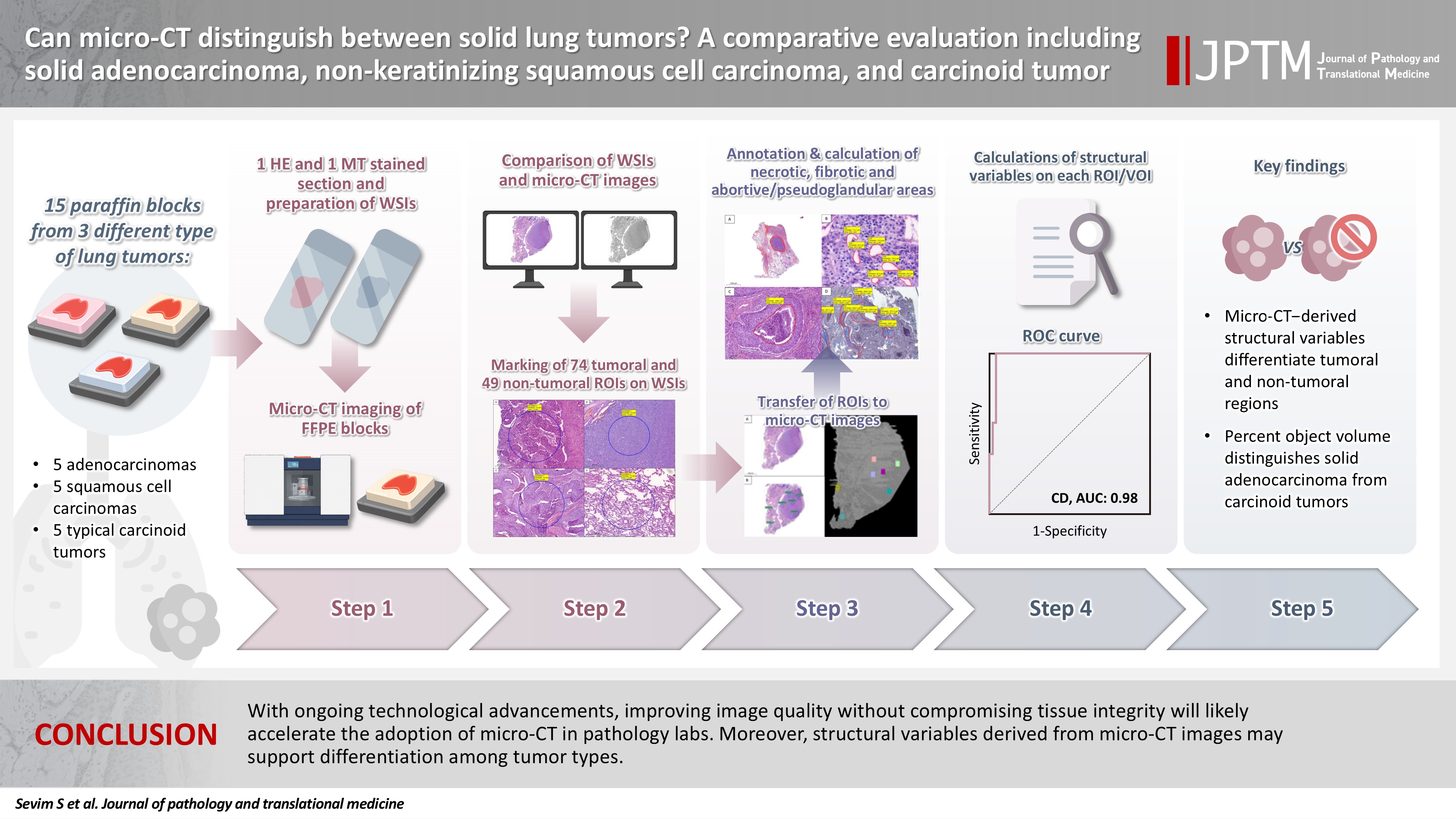
Some pulmonary carcinomas display a solid pattern, and immunohistochemistry is commonly used for tumor differentiation. Micro–computed tomography (micro-CT), with its ability to produce detailed three-dimensional images using small voxel sizes, may offer additional insights. This study investigates whether three solid tumor types, solid adenocarcinoma (sAC), non-keratinizing squamous cell carcinoma, and carcinoid tumor (CaT), can be differentiated using micro-CT. Methods: Fifteen paraffin blocks, five for each type, were scanned with micro-CT (Skyscan 1275, Bruker). These images were compared to whole slide images (WSIs) of the same tumors. Consequently, tumoral (n = 74) and non-tumoral (n = 49) regions of interest (tumor ROIs [tROIs] and non-tumor ROIs [ntROIs]) were selected on the micro-CT images and evaluated in terms of certain structural variables (percent object volume, structure model index, structure thickness, structure linear density, connectivity, connectivity density, open porosity, closed porosity) to investigate whether tumors can be differentiated from normal parenchyma and from each other. Results: Although detailed images comparable to WSIs could not be obtained, it was considered an important advantage to be able to examine the entire depth of the paraffin blocks. tROIs and ntROIs could be distinguished based on all variables (p < .001). Additionally, sAC showed a notable difference from CaT in “percent object volume” (p = .011). Conclusions: With ongoing technological advancements, improving image quality without compromising tissue integrity will likely accelerate the adoption of micro-CT in pathology labs. Moreover, structural variables derived from micro-CT images may support differentiation among tumor types.
Case Study
- Intrathyroidal metastasis of tonsillar squamous cell carcinoma masquerading as a primary thyroid tumor
- Jai-Hyang Go
- J Pathol Transl Med. 2023;57(4):242-245. Published online July 11, 2023
- DOI: https://doi.org/10.4132/jptm.2023.06.16
- 5,293 View
- 118 Download
- 2 Web of Science
- 2 Crossref
-
Abstract
PDF
- Intrathyroidal metastasis of tonsillar squamous cell carcinoma is rare. To date, only six cases have been reported in the literature. This case was unusual and presented with thyromegaly before the diagnosis of the primary tumor. A 55-year-old male patient was suspected to have a primary thyroid tumor with nodal metastasis. The thyroid gland was diffusely enlarged, with no discernible mass. Histologically, the thyroid parenchyma revealed extensive endolymphatic tumor emboli, which were positive for p40 and p16 in a background of chronic lymphocytic thyroiditis. Positron emission tomography–computed tomography revealed hypermetabolic activity in the right tonsillar region. Tonsillar biopsy revealed human papillomavirus–positive squamous cell carcinoma. The present case is the first reported case of intrathyroidal metastasis of tonsillar squamous cell carcinoma with an initial clinical presentation of thyroid enlargement before the primary tumor of tonsillar cancer was diagnosed.
-
Citations
Citations to this article as recorded by
- Metastasis to Thyroid from Recurrent Head and Neck Squamous Cell Carcinoma: A Case Series and Review of Literature
Avneet Kaur, Rohit Nayyar, Harit Kumar Chaturvedi, Akshat Malik
Indian Journal of Surgical Oncology.2025; 16(1): 122. CrossRef - Metastatic oropharyngeal squamous cell carcinoma to the thyroid: A case report and review of literature
Hannah Walker, Jed Speers, Milena Fabry, Sameep Kadakia
American Journal of Otolaryngology.2024; 45(4): 104306. CrossRef
- Metastasis to Thyroid from Recurrent Head and Neck Squamous Cell Carcinoma: A Case Series and Review of Literature
Original Article
- Age Estimation of Mummies by Dental Attrition: Application of Three-dimensional CT Images.
- Kwang Ho Jeong, Han Kyeom Kim, Chang Lyuk Yoon, Seong Jae Lee, Seung Yeon Ha
- Korean J Pathol. 2008;42(5):299-305.
- 2,707 View
- 51 Download
-
Abstract
PDF
- BACKGROUND
Because of the rarity of mummies in Korea and the difficulty in obtaining samples from mummies, studies to determine the ages of mummies are uncommon in Korea. This study was performed to determine the ages of mummies using the information obtained by nondestructive methods to minimize damages to the mummies. METHODS: Three mummies excavated between 2002 and 2004 were used. Three-dimensional reconstructed images of the total teeth were obtained by CT scanning. The age at death was determined according to the 'Age Estimation Table of Dental Attrition' as developed by Takei. Three teeth were extracted from each of three mummies and examined grossly and microscopically by serial sections using the Gustafson-Johanson method. RESULTS: The ages at death of the three mummies estimated by the Takei method were 23.57 years (Yoon mummy), 51.01 years (Bong mummy), and 64.45 years (Black mummy). These results were similar to the ages determined by the Gustafson method. CONCLUSION: Age determination method using a CT scan and three-dimensional reconstruction may be a valuable method because it minimizes the damages to valuable mummies and it gives reliable data similar to that obtained by other standard methods.
 First
First Prev
Prev


