
 E-submission

Search
- Page Path
- HOME > Search
Original Article
- Targeted next-generation sequencing–based mutational signature analysis in endometrial cancer: utility and limitations across molecular subtypes
- Uijin Kim, Ahwon Lee, Keun Ho Lee, Sung Jong Lee, Sook Hee Hong, Donghyuk Lee, Jun Kang, Miseon Lee
- J Pathol Transl Med. 2026;60(6):625-634. Published online November 10, 2026
- DOI: https://doi.org/10.4132/jptm.2026.09.07
- 92 View
- 1 Download
-
 Abstract
 PDF
 Supplementary Material
- Background
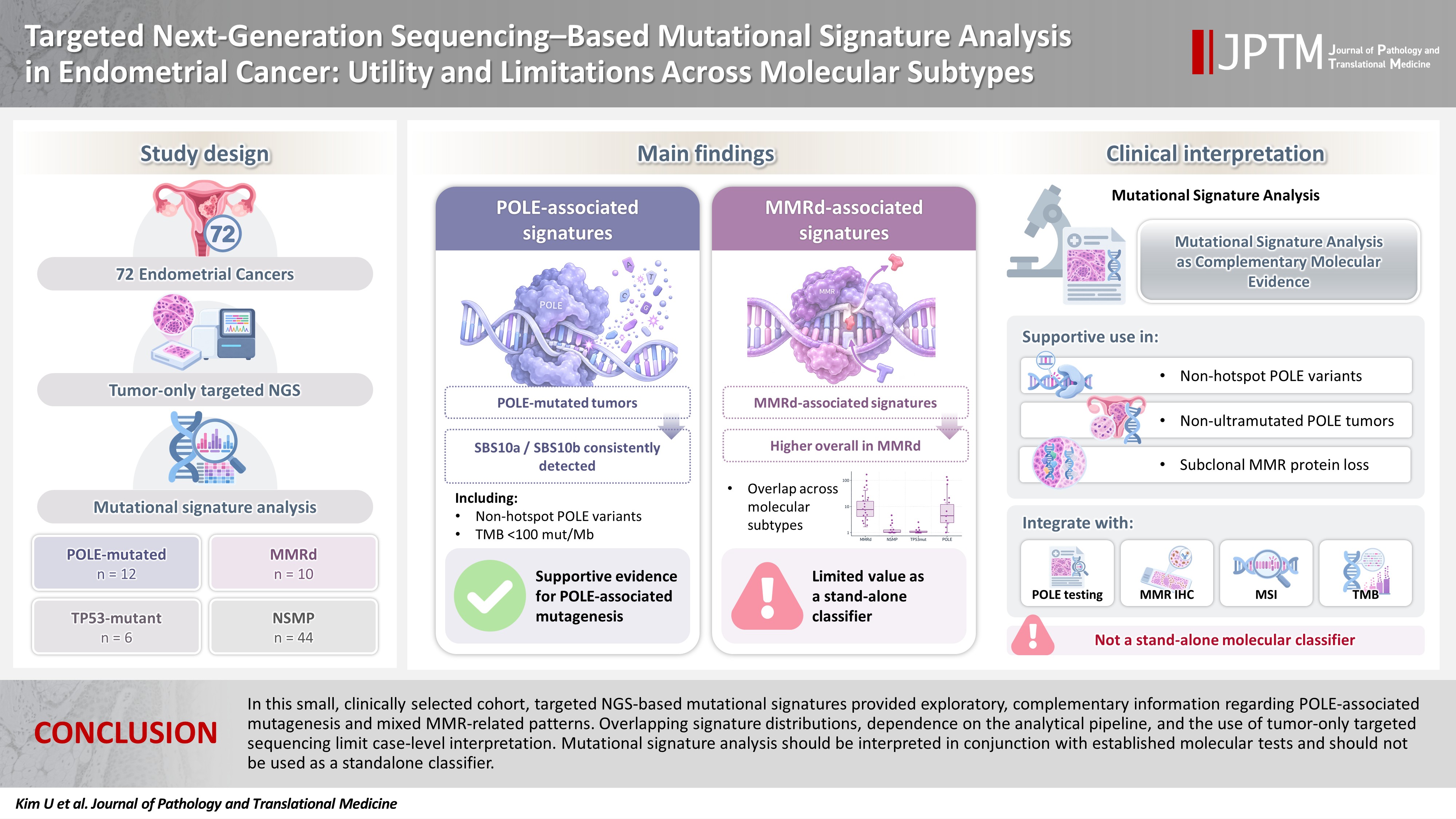
Molecular subtype classification is essential for staging endometrial cancer, planning adjuvant treatments, and stratifying prognosis. However, the correlation between mutational signature analysis using tumor-only targeted next-generation sequencing (NGS) data and molecular subtypes, as well as the interpretive value at the individual case level, remains inadequately characterized. Methods: We performed mutational signature analysis on tumor-only targeted NGS data from 72 endometrial cancer cases using the SATS R package. Molecular classification into four subtypes (POLE-mutated, mismatch repair deficient [MMRd], TP53-mutant, and no specific molecular profile [NSMP]) was established through a standard stepwise algorithm that integrated NGS and immunohistochemistry. Results: Out of the 72 endometrial cancers, 12 were classified as POLE-mutated, 10 as MMRd, six as TP53-mutant, and 44 as NSMP. All POLE-mutated tumors exhibited POLE-associated signatures (SBS10a/SBS10b), including cases with non-hotspot POLE exonuclease variants and tumor mutation burdens below the conventional ultramutation threshold. MMRd-associated signatures showed a broader distribution across molecular subtypes; while the summed MMRd-associated signatures were typically higher in MMRd tumors, low-level contributions were also noted in subsets of NSMP and TP53-mutant tumors. Within the POLE-mutated group, MMRd-associated signatures were more pronounced in cases displaying subclonal MMR protein loss as determined by immunohistochemistry. Conclusions: In this small, clinically selected cohort, targeted NGS-based mutational signatures provided exploratory, complementary information regarding POLE-associated mutagenesis and mixed MMR-related patterns. Overlapping signature distributions, dependence on the analytical pipeline, and the use of tumor-only targeted sequencing limit case-level interpretation. Mutational signature analysis should be interpreted in conjunction with established molecular tests and should not be used as a stand-alone classifier.
Case Report
- Primary Endometrial Squamous Cell Carcinoma: A Case Report and Review of Relevant Literature on Korean Women
- Sung Jong Lee, Hyun Joo Choi
- Korean J Pathol. 2012;46(4):395-398. Published online August 23, 2012
- DOI: https://doi.org/10.4132/KoreanJPathol.2012.46.4.395
- 12,223 View
- 93 Download
- 16 Crossref
-
Abstract
PDF
Primary endometrial squamous cell carcinoma (PESCC) is an extremely rare tumor with unclear pathogenesis. A 54-year-old postmenopausal woman presented with a 6-month history of vaginal bleeding. The patient was provisionally diagnosed with uterine submucosal leiomyoma. This was followed by total hysterectomy with a bilateral salpingo-oophorectomy under the laparoscopic guidance. Histopathologically, the tumor was PESCC which was accompanied by a lack of the tumor in the uterine cervix. The tumor showed positive immunoreactivity for p16INK4a. But there was no evidence of human papillomavirus (HPV) on
in situ hybridization and HPV DNA chip analysis. We also present a review of the relevant literature on Korean women.-
Citations
Citations to this article as recorded by
- Primary squamous cell carcinoma of the uterine wall associated with adenomyosis: A rare case report and literature review
Yang Li, Lanlan Xuan, Qiucheng Wang
International Journal of Gynecology & Obstetrics.2026; 172(1): 87. CrossRef - Primary endometrial squamous cell carcinoma with molecular Characterization: A case report
Yifei Zhan, Xiaojing Guo, Jinling Zhang
Gynecologic Oncology Reports.2026; 65: 102098. CrossRef - Primary endometrial squamous cell carcinoma (PESCC): Review of the literature and case report
Kuang-Han Liu, Chia-Chin Tsai, Krystal Baysan Lin, Pei-Shen Huang
Taiwanese Journal of Obstetrics and Gynecology.2025; 64(1): 159. CrossRef - Primary squamous cell carcinoma of the endometrium: A case report
Shuhui Cui, Zhe Dong
Medicine.2025; 104(46): e45920. CrossRef - p16 Block Type Overexpression, p53 Wild Type Reactivity, and Cervical Involvement do not Always Exclude the Diagnosis of Primary Endometrial Squamous Cell Carcinoma (PESCC)
Daniela Fanni, Clara Gerosa, Michele Peiretti, Valerio Mais, Elena Massa, Stefano Angioni, Gavino Faa
International Journal of Gynecological Pathology.2024; 43(2): 200. CrossRef - Pathogenetic characteristics of endometrioid adenocarcinoma of uterus at present stage
T. I. Moiseenko, S. V. Shatalova, E. M. Nepomnyashchaya, V. A. Bandovkina, M. L. Adamyan
Medical alphabet.2024; (36): 35. CrossRef - Case report: Clinicopathological characteristic of two cases of primary endometrial squamous cell carcinoma and review of the literature
Hui-Bin Zhang, Li-Hua Lin, Qiu-Ping Lin, Yuan-Qing Lin, Dan Luo, Shu-Xia Xu
Frontiers in Oncology.2024;[Epub] CrossRef - Treatment of primary squamous cell carcinoma of the endometrium and review of previous literature: A case report
Liyun Song, Qi Wu, Suning Bai, Ren Xu, Xiaona Wang, Yanyan Yang
Medicine.2023; 102(17): e33667. CrossRef -
Prevalence of
human papilloma virus
and
Chlamydia trachomatis
in endometrial and cervical carcinoma: a comparative study in North Indian women
Heena Gautam, Sumita Mehta, Nidhi Nayar, Neha Kumar, Syed Akhtar Husain, Mausumi Bharadwaj
Systems Biology in Reproductive Medicine.2023; 69(6): 399. CrossRef - PAX8 Positivity, Abnormal p53 Expression, and p16 Negativity in a Primary Endometrial Squamous Cell Carcinoma: A Case Report and Review of the Literature
Daniela Fanni, Michele Peiretti, Valerio Mais, Elena Massa, Clara Gerosa, Francesca Ledda, Maria Luisa Fais, Gavino Faa, Stefano Angioni
International Journal of Gynecological Pathology.2022; 41(4): 431. CrossRef - Molecular Analysis of HPV-independent Primary Endometrial Squamous Cell Carcinoma Reveals TP53 and CDKN2A Comutations
Mark R. Hopkins, Doreen N. Palsgrove, Brigitte M. Ronnett, Russell Vang, Jeffrey Lin, Tricia A. Murdock
American Journal of Surgical Pathology.2022; 46(12): 1611. CrossRef - Primary squamous cell carcinoma of the endometrium—Case report with cytological characteristics in direct and indirect endometrial samples
Sanda Rajhvajn, Ana Barišić, Lada Škopljanac‐Mačina, Danijela Jurič, Vesna Mahovlić
Cytopathology.2021; 32(6): 823. CrossRef - Primary squamous cell carcinoma of the endometrium associated with human papilloma virus in a young woman: a case report
Tchin Darré, Abdoul-Samadou Aboubakari, Lantam Sonhaye, Baguilane Douaguibe, Akila Bassowa, Gado Napo-Koura
Journal of Medical Case Reports.2019;[Epub] CrossRef - Primary squamous cell carcinoma of the endometrium in a woman of perimenopausal age
Chao Zhang, Hongyan Zhang, Linqing Yang, Yunfei Wang, Xiaoyu Li, Jinfeng Guo, Jing Xu
Medicine.2018; 97(48): e13418. CrossRef - S100P is Overexpressed in Squamous Cell and Adenosquamous Carcinoma Subtypes of Endometrial Cancer and Promotes Cancer Cell Proliferation and Invasion
Hongfei Jiang, Hang Hu, Fan Lin, Yoon Pin Lim, Yunfen Hua, Xiaomei Tong, Songying Zhang
Cancer Investigation.2016; 34(10): 477. CrossRef - Überraschung in der Abradatdiagnostik
U. Kellner, A. Kellner, U. Cirkel
Der Pathologe.2015; 36(3): 317. CrossRef
- Primary squamous cell carcinoma of the uterine wall associated with adenomyosis: A rare case report and literature review
Original Article
- Prognostic Significance of Amplification of the c-MYC Gene in Surgically Treated Stage IB-IIB Cervical Cancer.
- Tae Jung Kim, Ahwon Lee, Sung Jong Lee, Won Chul Lee, Yeong Jin Choi, Kyo Young Lee, Chang Suk Kang
- Korean J Pathol. 2011;45(6):596-603.
- DOI: https://doi.org/10.4132/KoreanJPathol.2011.45.6.596
- 5,991 View
- 42 Download
- 1 Crossref
-
Abstract
PDF
- BACKGROUND
Mutations of c-MYC have been described in cervical cancer. However, association between c-MYC gene status and its prognostic significance have not been clarified.
METHODS
Tissue microarray sections from 144 patients with stage IB-IIB cervical cancer treated by radical hysterectomy were analyzed by fluorescence in situ hybridization using a region-specific probe for c-MYC and a centromere-specific probe for chromosome 8.
RESULTS
Seventy five percent (108/144) of c-MYC gain and 6.9% (10/144) of c-MYC gene amplification were observed. c-MYC gene alteration was more frequently observed in squamous cell carcinoma than adenocarcinoma or adenosquamous carcinoma and were associated with low Ki67 labeling index (p=0.013). c-MYC amplification was not associated with clinicopathologic parameters except absence of bcl2 expression (p=0.048). Survival analysis revealed that patients with c-MYC amplification were significantly associated with higher risk of disease recurrence (p=0.007) and cancer related death (p=0.020). However, c-MYC gain was not associated with unfavorable outcome. Multivariate analysis proved c-MYC amplification as independent prognostic factors of shorter disease free survival and cancer-related death (p=0.028 and p=0.025, respectively).
CONCLUSIONS
c-MYC amplification, not gain, is an independent prognostic marker for shorter disease free and cancer specific survival in cervical cancer treated by radical hysterectomy. -
Citations
Citations to this article as recorded by- A Rare Case of Cutaneous Plasmacytosis in a Korean Male
Corey Georgesen, Meenal Kheterpal, Melissa Pulitzer
Case Reports in Pathology.2017; 2017: 1. CrossRef
- A Rare Case of Cutaneous Plasmacytosis in a Korean Male
 First
 Prev




