 E-submission
Search
- Page Path
- HOME > Search
Original Article
- Double cocktail immunostains with high molecular weight cytokeratin and GATA-3: useful stain to discriminate in situ involvement of prostatic ducts or acini from stromal invasion by urothelial carcinoma in the prostate
- Junghye Lee, Youngeun Yoo, Sanghui Park, Min-Sun Cho, Sun Hee Sung, Jae Y. Ro
- J Pathol Transl Med. 2020;54(2):146-153. Published online February 10, 2020
- DOI: https://doi.org/10.4132/jptm.2019.11.12
- 8,800 View
- 138 Download
- 2 Web of Science
- 2 Crossref
-
 Abstract
 PDF
PDF - Background
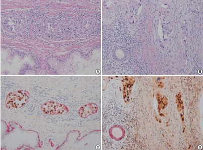
Distinguishing prostatic stromal invasion (PSI) by urothelial carcinoma (UC) from in situ UC involving prostatic ducts or acini with no stromal invasion (in situ involvement) may be challenging on hematoxylin and eosin stained sections. However, the distinction between them is important because cases with PSI show worse prognosis. This study was performed to assess the utility of double cocktail immunostains with high molecular weight cytokeratin (HMWCK) and GATA-3 to discriminate PSI by UC from in situ UC involvement of prostatic ducts or acini in the prostate.
Methods
Among 117 radical cystoprostatectomy specimens for bladder UCs, 25 cases showed secondary involvement of bladder UC in prostatic ducts/acini only or associated stromal invasion and of these 25 cases, seven cases revealed equivocal PSI. In these seven cases with equivocal PSI, HMWCK, and GATA-3 double immunohistochemical stains were performed to identify whether this cocktail stain is useful to identify the stromal invasion.
Results
In all cases, basal cells of prostate glands showed strong cytoplasmic staining for HMWCK and UC cells showed strong nuclear staining for GATA-3. In cases with stromal invasion of UC, GATA-3-positive tumor cells in the prostatic stroma without surrounding HMWCK-positive basal cells were highlighted and easily recognized. Among seven equivocal cases, two cases showed PSI and five in situ UC in the prostate. In two cases, the original diagnoses were revised.
Conclusions
Our study suggested that HMWCK and GATA-3 double stains could be utilized as an adjunct method in the distinction between PSI by UC from in situ UC involving prostatic ducts or acini. -
Citations
Citations to this article as recorded by
- Aberrant expression of GATA3 in metastatic adenocarcinoma of the prostate: an important pitfall
João Lobo, Nazario P Tenace, Sofia Cañete‐Portillo, Isa Carneiro, Rui Henrique, Roberta Lucianò, Lara R Harik, Cristina Magi‐Galluzzi
Histopathology.2024; 84(3): 507. CrossRef - Utility of D2-40, Cytokeratin 5/6, and High–Molecular-weight Cytokeratin (Clone 34βE12) in Distinguishing Intraductal Spread of Urothelial Carcinoma From Prostatic Stromal Invasion
Oleksii A. Iakymenko, Laurence M. Briski, Katiana S. Delma, Merce Jorda, Oleksandr N. Kryvenko
American Journal of Surgical Pathology.2022; 46(4): 454. CrossRef
- Aberrant expression of GATA3 in metastatic adenocarcinoma of the prostate: an important pitfall
Case Report
- Malignant Endometrioid Adenofibroma of the Ovary: A case report.
- Tae Jung Jang, Soon Hee Jung, Kyu Rae Kim, Hoguen Kim
- Korean J Pathol. 1990;24(4):497-501.
- 2,853 View
- 69 Download
-
Abstract
PDF
- Ovarian endometrioid adenofibroma is rare and characterized by prominent stroma. Its histologic classification is controversial but the malignant counterpart is distinguished from the borderline by the presence of confluent growth pattern of epithelium with invasion of the stroma by the endometrioid cells. A fifty-year-old woman was admitted with one month history of abdominal enlargement. Total abdominal hysterectomy with bilateral salpingo-oophorectomy was performed under the clinical diagnosis of ovarian malignancy. Grossly, the righy ovary had round, encapsulated, solid and whitish gray mass which measured 9 cm in the greatest dimension and showed peripheral small cysts. Microscopic examination revealed that the tumor consisted of endometria type glands set in fibrous stroma. The glands varied from tubules to cysts and the lining cells showed complicated architectural pattern with occasional papillary infoldings, atypical mitosis and malignant nuclear characteristics. Some cysts of glands showed intraluminal mucin products. Stromal invasions by individual epithelial cells showing malignant characteristics were often found. A brief summary of the histopathologic aspect of this tumor is presented together with review of literatures.
 First
First Prev
Prev


