 E-submission
Search
- Page Path
- HOME > Search
Case Study
- Intravascular schwannoma as an extremely unusual cause of vein obstruction: a case report
- Luis Miguel Chinchilla-Tábora, Beatriz Segovia Blázquez, José María Sayagués, Marta Rodríguez González, Joaquín González-Rivero, José Antonio Muñoz León, Andrea Beatriz Jiménez Pérez, Idalia González Morais, Diego Bueno-Sacristán, María Dolores Ludeña
- J Pathol Transl Med. 2024;58(5):249-254. Published online July 3, 2024
- DOI: https://doi.org/10.4132/jptm.2024.05.15
- 4,329 View
- 284 Download
-
 Abstract
 PDF
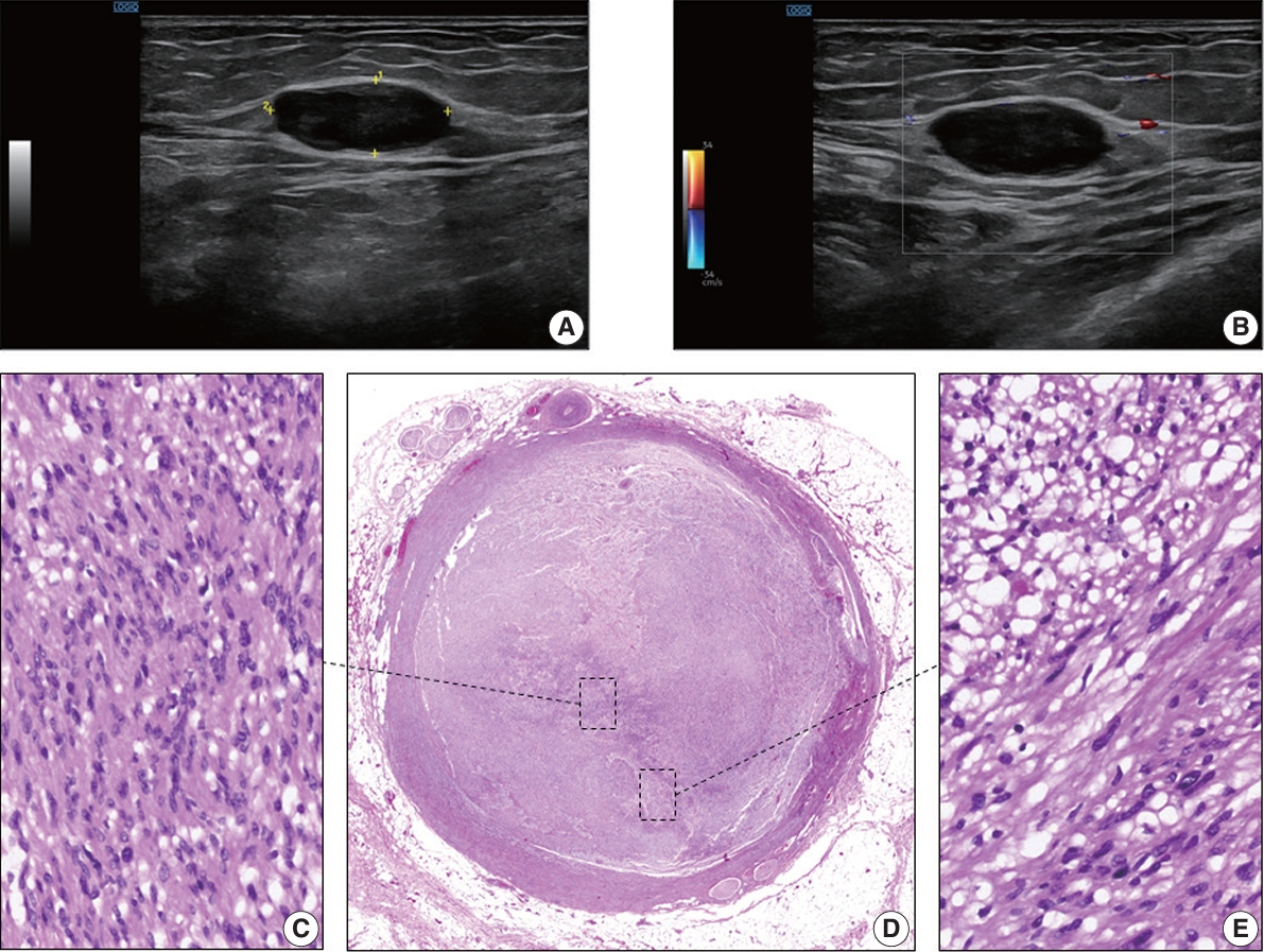
PDF - The blood vessel lumen is an extremely rare location for a benign peripheral nerve sheath tumor like schwannoma. Less than 10 cases have been previously reported. In this report, we present a case of a 68-year-old woman who had a soft tissue nodule at the posterior calf of her left leg during a physical examination. Pathological examination was performed after complete surgical excision. The patient underwent follow-up for 12 months after surgery without evidence of recurrence or any other complication. This is the first case of intravascular schwannoma reported as a cause of vein obstruction. Microscopically, the tumor was composed of Schwann spindle cells that were immunoreactive for S100 protein and SOX10. This tumor was surrounded by a well-defined vascular smooth muscle wall. Prospective series are required to improve the knowledge on the underlying mechanisms of intravascular schwannoma development.
Case Report
- Neuromuscular Choristoma of the Sciatic Nerve: A Case Report.
- Sun Young Kim, Hyuck Po Kwon, Kyoung Duck Kwak, Kee Baek Ahn
- Korean J Pathol. 2005;39(3):192-196.
- 2,452 View
- 20 Download
-
Abstract
PDF
- Neuromuscular choristoma is a rare benign tumor of the peripheral nerves. To the best of our knowledge, 21 cases have been reported to date. We describe here a 20-day-old female infant who presented with a buttock mass (4.5 x 4.1 x 3.2 cm on MRI) arising from the left sciatic nerve. Microscopically, it was characterized by an intimately disorganized mixture of nerve fibers and striated muscle fibers that were occasionally surrounded by the perineurium and separated by fibrous bands of varying thickness. In some areas, there appeared to be some cells in transitional forms between nerve fibers and muscle fibers, revealing variously positive expressions for S-100 protein in the muscular components. These findings are consistent with the hypothesis that neuroectodermal-derived Schwann cells can give rise to mature skeletal muscle. It appears that the fibrosis may be related to the degeneration of the neural components. The size of the mass on MRI has been unchanged during the 3-year follow-up period.
 First
First Prev
Prev


