 E-submission
Search
- Page Path
- HOME > Search
Original Articles
- Post-mortem assessment of vimentin expression as a biomarker for renal tubular regeneration following acute kidney injury
- Juan Carlos Alvarez Moreno, Hisham F. Bahmad, Christopher A. Febres-Aldana, Andrés Pirela, Andres Azuero, Ali Salami, Robert Poppiti
- J Pathol Transl Med. 2021;55(6):369-379. Published online October 14, 2021
- DOI: https://doi.org/10.4132/jptm.2021.08.03
- 8,439 View
- 151 Download
- 6 Web of Science
- 6 Crossref
-
 Abstract
 PDF
PDF - Background
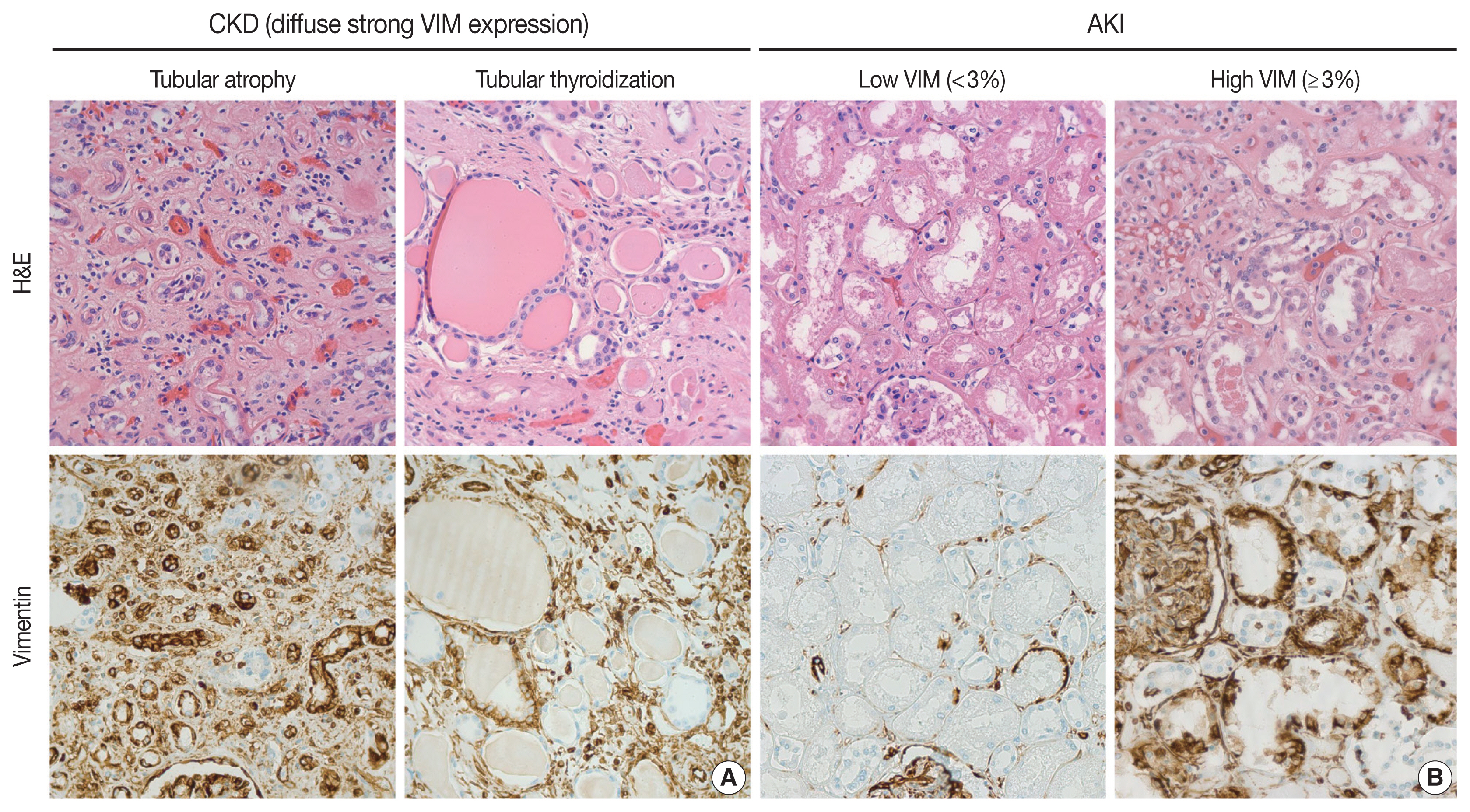
Acute kidney injury (AKI) is a common cause of morbidity and mortality. It mainly targets the renal tubular epithelium with pathological changes, referred to as acute tubular injury. The latter is followed by a regenerative response that is difficult to visualize on routine hematoxylin and eosin (H&E) stains. In this study, we examined the regenerative capacity of renal tubules by correlating vimentin (VIM) immunohistochemical (IHC) expression and pathological findings of AKI and renal tubular regeneration (RTR) on H&E.
Methods
We reviewed 23 autopsies performed in the clinical setting of AKI and RTR. VIM expression was scored in the renal cortical tubular epithelium using a statistical cutoff ≥ 3% for high expression and < 3% for low expression.
Results
Of the 23 kidney tissues examined, seven (30.4%) had low VIM expression, and 16 (69.6%) had high VIM expression. Kidney tissues with evidence of AKI and RTR had significantly higher VIM expression. Renal peritubular microenvironment features showing regenerative changes on H&E were associated with high VIM expression. In the univariate model, kidney tissues with RTR were 18-fold more likely to have high VIM expression.
Conclusions
In conclusion, our findings suggest that VIM could serve as an IHC marker for RTR following AKI. However, correlation with H&E findings remains critical to excluding chronic tubular damage. Collectively, our preliminary results pave the way for future studies including a larger sample size to validate the use of VIM as a reliable biomarker for RTR. -
Citations
Citations to this article as recorded by
- Influence of Vimentin and E-cadherin on the development of interstitial fibrosis in focal and segmental glomerulosclerosis – FSGS
Laura Penna Rocha, Monise Gini Urzêdo, Nayne Isabelli Zugolaro Donzelli, Bruna de Freitas Oliveira, Crislaine Aparecida Silva, Bruna Cunha Zaidan, Régia Caroline Peixoto Lira Fusco, Marlene Antônia dos Reis, Juliana Reis Machado
Tissue and Cell.2026; 102: 103555. CrossRef - Myocardial Infarction Injury Is Exacerbated by Nicotine in Vape Aerosol Exposure
Clarissa Savko, Carolina Esquer, Claudia Molinaro, Sophie Rokaw, Abraham G. Shain, Faid Jaafar, Morgan K. Wright, Joy A. Phillips, Tyler Hopkins, Sama Mikhail, Abigail Rieder, Ariana Mardani, Barbara Bailey, Mark A. Sussman
Journal of the American Heart Association.2025;[Epub] CrossRef - Spatio-temporal transcriptomic analysis reveals distinct nephrotoxicity, DNA damage, and regeneration response after cisplatin
Lukas S. Wijaya, Steven J. Kunnen, Panuwat Trairatphisan, Ciarán P. Fisher, Meredith E. Crosby, Kai Schaefer, Karen Bodié, Erin E. Vaughan, Laura Breidenbach, Thomas Reich, Diana Clausznitzer, Sylvestre Bonnet, Sipeng Zheng, Chantal Pont, James L. Stevens
Cell Biology and Toxicology.2025;[Epub] CrossRef - Characterization of macrophages in ischemia–reperfusion injury-induced acute kidney injury based on single-cell RNA-Seq and bulk RNA-Seq analysis
Qin Wang, Yuxing Liu, Yan Zhang, Siyuan Zhang, Meifang Zhao, Zhangzhe Peng, Hui Xu, Hao Huang
International Immunopharmacology.2024; 130: 111754. CrossRef - Renal tubular necrosis associated with anagrelide administration: a case report
Atsushi Sawase, Mineaki Kitamura, Misato Morimoto, Haruka Fukuda, Tadashi Uramatsu, Eisuke Katafuchi, Hiroshi Yamashita, Toshiyuki Nakayama, Hiroshi Mukae, Tomoya Nishino
CEN Case Reports.2024; 13(6): 510. CrossRef - Morin attenuates sepsis-induced acute kidney injury by regulating inflammatory responses, oxidative stress and tubular regeneration (morin and sepsis-induced acute kidney injury)
Aya M. Shehata, Nagui H. Fares, Basma H. Amin, Asmaa A. Mahmoud, Yomna I. Mahmoud
Environmental Toxicology and Pharmacology.2024; 111: 104543. CrossRef
- Influence of Vimentin and E-cadherin on the development of interstitial fibrosis in focal and segmental glomerulosclerosis – FSGS
- Ultrastructural Studies of Aortic Endothelial Injury and Regeneration.
- Gium Mi Jang, Dong Hoon Kim, Jyung Sik Kwak, Tae Joong Sohn
- Korean J Pathol. 1990;24(4):337-348.
- 2,110 View
- 16 Download
-
Abstract
PDF
- Author performed this experiment to define the most important factor preventing the intimal thickening. An endothelium of abdominal aorta in the rat was denuded by two different wires having same caliver. The degree of injury was limited to the endothelial cells in one, and extended to the internal elastic lamina in another. The results showed that at 72 hours, in the case of superficial injury, the entire injury site was covered by new regenerating cells, but in the case of disruption of the internal elastic lamina, the migrating smooth muscle cell completely reached into the intima and resulted in intemal thickening. Similar findings persisted to 1 week later. Above results suggest the most important factor preventing the intimal thickening in endothelial injury is the depth of the injury which limited within the endothelial cells without extending into the internal elastic lamina and medial smooth muscle cells.
- Factors Influencing Regeneration of Calvarial Defects in Rats.
- Sung Chul Lim, Young Sook Kim
- Korean J Pathol. 1999;33(11):999-1008.
- 1,936 View
- 14 Download
-
Abstract
PDF
- An experimental study was done to evaluate factors influencing guided regeneration of bone in standardized calvarial bony defect. An 8 mm circular transosseous calvarial bony defect was made. Various material such as demineralized freeze-dried bone (DFDB), BioMesh , Millipore filter and its combination was placed in the bony defect. A sequential histopathologic, histochemical, immunohistochemical, and histomorphometric studies were done on the guided bone regeneration in the calvarial bony defect. Bone formation was sigificantly enhanced when the DFDB was retained within the bony defect with a protective bioabsorbable membrane. Inframembranous DFDB-filling was required to prevent collapse of the membrane and preserve spaces for bone regeneration. The bioabsorbable membrane should presumably remain intact for longer than at least 5 weeks to facilitate bone regeneration. The new bone formation was dependent on the barrier-effect (preserving secluded spaces) and inflammation-inducing property of membrane, and guiding bone regeneration of the grafts. Macrophages recruited by grafts were partly involved in decrease of bone regeneration via the sequential events of release of fibronectin, chemotactic effect of the fibronectin to fibroblasts, and collagen lay-down.
- Apoptosis and Cell Proliferation in Experimental Acute Tubular Necrosis Induced by Intramuscular Glycerol Injection.
- Wan Seop Kim, Jung Woo Noh, Moon Hyang Park
- Korean J Pathol. 2003;37(1):41-49.
- 2,547 View
- 18 Download
-
Abstract
PDF
- BACKGROUND
Acute tubular necrosis (ATN) is the most common cause of acute renal failure. It is characterized by the destruction of tubular epithelial cells. To examine apoptosis and proliferative activity of tubular cells in the course of acute tubular necrosis, we induced acute renal failure by intramuscular hypertonic glycerol injection to New Zealand White rabbits.
METHODS
The immunohistochemistry was done for Ki-67 and tissue-transglutaminase (tTG), and the terminal deoxynucleotidyl transferase mediated nick end labeling (TUNEL) method was performed using a total of 77 renal specimens including 29 gun biopsies and 48 nephrectomiy specimens.
RESULTS
Widespread tubular injury with pigment casts and interstitial hemorrhage were noted. The tubular proliferation index was increased at 2 hours after glycerol injection, and the index peaked at 3 hours. The second cell proliferation peak was noted at 3 days. Apoptotic cells were identified by TUNEL and tTG staining. The apoptotic index was significantly increased, and it peaked at 24 hours after glycerol injection. There was a significant correlation between the proliferation index (MIB-1) the and the apototic index (TUNEL)(p= 0.001). A DNA ladder pattern was observed at 6 to 8 hours.
CONCLUSIONS
Tubular cell proliferation and apoptosis occur in the early phase after the induction of acute tubular necrosis, and the excess hyperplastic epithelial cells appear to be eliminated by apoptosis.
 First
First Prev
Prev


