 E-submission
Search
- Page Path
- HOME > Search
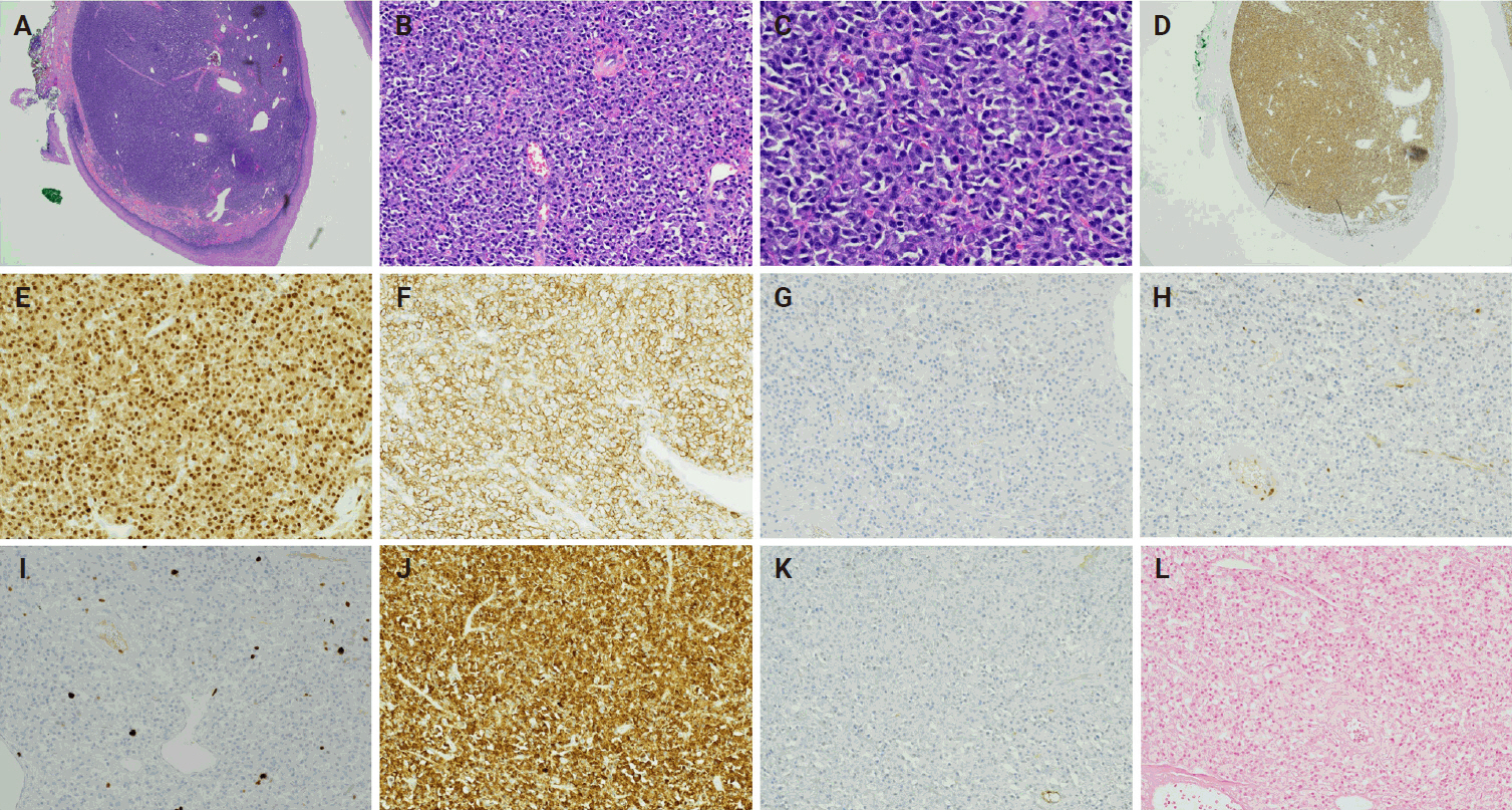
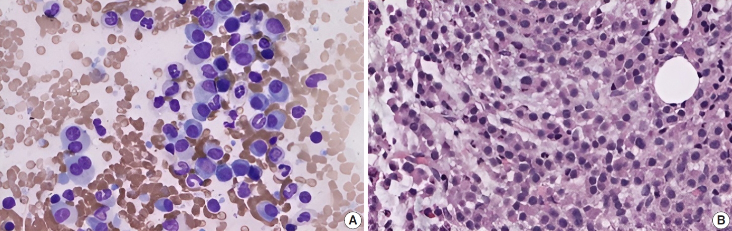
- Phenotypic plasticity in plasma cell myeloma: a CD138-negative case with a rare BRAF G469R mutation
- Sun-Ju Oh, So-Hak Chung
- Received November 21, 2025 Accepted February 2, 2026 Published online April 22, 2026
- DOI: https://doi.org/10.4132/jptm.2026.02.02 [Epub ahead of print]
- 277 View
- 26 Download
-
 Abstract
 PDF
PDF - CD138-negative plasma cell myeloma harboring a BRAF G469R mutation is described in a 76-year-old male presenting with multiple osteolytic lesions. Histologically, the lesion exhibited epithelioid to plasmacytoid morphology with prominent mitotic activity and vascular-like spaces. Immunophenotyping demonstrated strong vimentin and CD31 expression but absence of CD138 and other endothelial markers. Light-chain in situ hybridization confirmed a clonal κ-restricted plasma cell population. Bone marrow examination revealed near-complete replacement by atypical plasma cells, retaining CD138 negativity and demonstrating focal CD20 positivity, indicative of intratumoral heterogeneity. Next-generation sequencing identified a rare BRAF G469R variant. The patient exhibited poor response to bortezomib, lenalidomide, and dexamethasone therapy, necessitating a switch to carfilzomib-based treatment. This case underscores the diagnostic challenges of CD138-negative myeloma and highlights the importance of integrating morphology, immunophenotyping, and molecular profiling to inform accurate diagnosis and guide therapeutic strategies.
- Acquired aberrant partial CD3 expression in recurrent Epstein-Barr virus–negative solitary plasmacytoma of tonsil
- Chenchen Niu, Dong Ren, Truc Tran, Ashley Gamayo, Sherif Rezk, Xiaohui Zhao
- J Pathol Transl Med. 2025;59(4):262-268. Published online May 15, 2025
- DOI: https://doi.org/10.4132/jptm.2025.04.17
- 3,385 View
- 135 Download
-
Abstract
PDF
- The aberrant expression of specific T-cell maker CD3 in B-cell neoplasms can be a potential diagnostic pitfall leading to a misclassification of cell lineage. Here, we report a case of recurrent solitary plasmacytoma with new aberrant expression of CD3. The neoplastic plasma cells of the recurrent tumor were kappa restricted, positive for CD138, MUM1, negative for CD20, cyclin D1, and Epstein-Barr virus. CD79a was positive in majority of the tumor cells, except for a small focus which was strongly positive for CD3, but negative for other T-cell markers (CD2, CD5, CD7, CD4, and CD8) and CD56. The neoplastic plasma cells of the original tumor were negative for CD3. To the best of our knowledge, only one case of recurrent plasmacytoma with aberrant expression of CD3 has been published, which revealed disease progression in the recurrence. However, we did not observe morphologic evidence of disease progression in our case.
- Unusual biclonal IgA plasma cell myeloma with aberrant expression of high-risk immunophenotypes: first report of a new diagnostic and clinical challenge
- Carlos A. Monroig-Rivera, Clara N. Finch Cruz
- J Pathol Transl Med. 2023;57(2):132-137. Published online March 14, 2023
- DOI: https://doi.org/10.4132/jptm.2023.02.07
- 5,787 View
- 141 Download
-
Abstract
PDF
- IgA plasma cell myeloma (PCM) has been linked to molecular abnormalities that confer a higher risk for adverse patient outcomes. However, since IgA PCM only accounts for approximately 20% of all PCM, there are very few reports on high-risk IgA PCM. Moreover, no such reports are found on the more infrequent biclonal IgA PCM. Hence, we present a 65-year-old Puerto Rican female with acute abdominal pain, concomitant hypercalcemia, and acute renal failure. Protein electrophoresis with immunofixation found high IgA levels and detected a biclonal IgA gammopathy with kappa specificity. Histomorphologically, bone marrow showed numerous abnormal plasma cells (32%) replacing over 50% of the marrow stroma. Immunophenotyping analysis detected CD45-negative plasma cells aberrantly expressing CD33, CD43, OCT-2, and c-MYC. Chromosomal analysis revealed multiple abnormalities including the gain of chromosome 1q. Thus, we report on an unusual biclonal IgA PCM and the importance of timely diagnosing aggressive plasma cell neoplasms.
- IgG4-Related Disease Presented as a Mural Mass in the Stomach
- Chang Gok Woo, Jeong Hwan Yook, Ah Young Kim, Jihun Kim
- J Pathol Transl Med. 2016;50(1):67-70. Published online September 30, 2015
- DOI: https://doi.org/10.4132/jptm.2015.07.28
- 11,169 View
- 94 Download
- 21 Web of Science
- 22 Crossref
-
Abstract
PDF
- Isolated gastric IgG4-related disease (IgG4-RD) is a very rare tumefactive inflammatory condition, with only a few cases reported to date. A 48-year-old woman was incidentally found to have a subepithelial tumor in the stomach. Given a presumptive diagnosis of gastrointestinal stromal tumor or neuroendocrine tumor, she underwent wedge resection. The lesion was vaguely nodular and mainly involved the submucosa and proper muscle layer. Microscopically, all classical features of type I autoimmune pancreatitis including lymphoplasmacytic infiltration, storiform fibrosis, obliterative phlebitis, and numerous IgG4-positive plasma cells were seen. She had no evidence of IgG4-RD in other organs. Although very rare, IgG4-RD should be considered one of the differential diagnoses in the setting of gastric wall thickening or subepithelial mass-like lesion. Deep biopsy with awareness of this entity might avoid unnecessary surgical intervention.
-
Citations
Citations to this article as recorded by
- Compromiso gástrico por enfermedad relacionada con IgG4
Gilberto Jaramillo Trujillo, Oscar Fernando Ruiz, Melissa González Pabón, Maria Andrea Jaramillo Trujillo
Revista Repertorio de Medicina y Cirugía.2024; 33(3): 319. CrossRef - Value of High‐Frequency Ultrasonography in the Qualitative and Semi‐Quantitative Assessment of Immunoglobulin G4‐Related Submandibular Sialadenitis
Lei Chen, Lin Nong, Jumei Liu, Luzeng Chen, Yuhong Shao, Xiuming Sun
Journal of Ultrasound in Medicine.2023; 42(10): 2235. CrossRef - IgG4-related pseudotumours: a series of 12 cases and a review of the literature
Andrea Maccagno, Bianca Grosser, László Füzesi, Björn Konukiewitz, Dmytro Vlasenko, Dorothea Weckermann, Stephan Raab, Johannes Zenk, Abbas Agaimy, Bruno Märkl
Pathology.2022; 54(5): 563. CrossRef - IgG4-Related Disease With Gastrointestinal Involvement: Case Reports and Literature Review
Xinhe Zhang, Xing Jin, Lin Guan, Xuyong Lin, Xuedan Li, Yiling Li
Frontiers in Immunology.2022;[Epub] CrossRef - Clinicopathological characteristics of gastric IgG4‐related disease: Systematic scoping review
Haruki Sawada, Torrey Czech, Krixie Silangcruz, Landon Kozai, Adham Obeidat, Eric Andrew Wien, Midori Filiz Nishimura, Asami Nishikori, Yasuharu Sato, Yoshito Nishimura
Journal of Gastroenterology and Hepatology.2022; 37(10): 1865. CrossRef - Utility of gastric biopsy in diagnosing IgG4‐related gastrointestinal disease
Kaori Uchino, Kenji Notohara, Takeshi Uehara, Yasuhiro Kuraishi, Junya Itakura, Akihiro Matsukawa
Pathology International.2021; 71(2): 124. CrossRef - A reappraisal of sclerosing nodular and/or polypoid lesions of the gastrointestinal tract rich in IgG4‐positive plasma cells
Runjan Chetty
Histopathology.2020; 76(6): 832. CrossRef - Gastric IgG4-related disease presenting as a mass lesion and masquerading as a gastrointestinal stromal tumor
Banumathi Ramakrishna, Rohan Yewale, Kavita Vijayakumar, Patta Radhakrishna, Balakrishnan Siddartha Ramakrishna
Journal of Pathology and Translational Medicine.2020; 54(3): 258. CrossRef - IgG4-related Sclerosing Disease Forming a Gastric Submucosal Tumor Diagnosed after Laparoscopic Endoscopic Cooperative Surgery—Report of a Case—
Tatsuki ISHIKAWA, Katsunori NAKANO, Masafumi OSAKA, Yayoi KADOTANI, Kaori OKUGAWA, Kiyokazu AKIOKA, Kenta SHIGEMORI, Yohei HOSOKAWA
Nihon Rinsho Geka Gakkai Zasshi (Journal of Japan Surgical Association).2020; 81(2): 254. CrossRef - Calcifying fibrous tumor of the gastrointestinal tract: A clinicopathologic review and update
Donald Turbiville, Xu-Chen Zhang
World Journal of Gastroenterology.2020; 26(37): 5597. CrossRef - A Suspected Case of IgG4-Related Appendiceal Pseudotumor
Yudai Hojo, Yoshiharu Shirakata, Ai Izumi, Jun Matsui, Tokuyuki Yamashita, Hikaru Aoki, Makoto Kurimoto, Masaaki Hirata, Naoki Goda, Hiroaki Ito, Jun Tamura
The Japanese Journal of Gastroenterological Surgery.2020; 53(12): 976. CrossRef - Immunoglobulin G4-related gastric pseudotumor – An impostor
Manuel Santiago Mosquera, Andrea Suarez Gómez, Hugo Herrera, Karen Moreno-Medina, Alejandro González-Orozco, Carlos J-Perez Rivera
International Journal of Surgery Case Reports.2020; 75(C): 333. CrossRef - Imaging and pathological features of gastric lesion of immunoglobulin G4-related disease: A case report and review of the recent literature
Dai Inoue, Norihide Yoneda, Kotaro Yoshida, Hiromi Nuka, Jun Kinoshita, Sachio Fushida, Fumihito Toshima, Tetsuya Minami, Masayuki Takahira, Shoko Hamaoka, Hiroko Ikeda, Toshifumi Gabata, Mitsuhiro Kawano
Modern Rheumatology.2019; 29(2): 377. CrossRef - Immunoglobulin G4-Related Gastric Ulcer Mimicking Advanced Stomach Cancer in a Patient with Type I Autoimmune Pancreatitis
Joung Ha Park, Jin Hee Noh, Jang ho Lee, Goeun Lee, Seung-Mo Hong, Kwang Bum Cho, Myung-Hwan Kim
The Korean Journal of Medicine.2019; 94(3): 287. CrossRef - Review of IgG4-related disease
Raquel Sánchez-Oro, Elsa María Alonso-Muñoz, Lidia Martí Romero
Gastroenterología y Hepatología (English Edition).2019; 42(10): 638. CrossRef - Revisión de la enfermedad relacionada con la IgG4
Raquel Sánchez-Oro, Elsa María Alonso-Muñoz, Lidia Martí Romero
Gastroenterología y Hepatología.2019; 42(10): 638. CrossRef - Gastrointestinal manifestation of immunoglobulin G4-related disease: clarification through a multicenter survey
Kenji Notohara, Terumi Kamisawa, Kazushige Uchida, Yoh Zen, Mitsuhiro Kawano, Satomi Kasashima, Yasuharu Sato, Masahiro Shiokawa, Takeshi Uehara, Hajime Yoshifuji, Hiroko Hayashi, Koichi Inoue, Keisuke Iwasaki, Hiroo Kawano, Hiroyuki Matsubayashi, Yukitos
Journal of Gastroenterology.2018; 53(7): 845. CrossRef - IgG4-Related Disease Mimicking Crohn’s Disease: A Case Report and Review of Literature
Fabiana Ciccone, Antonio Ciccone, Mirko Di Ruscio, Filippo Vernia, Gianluca Cipolloni, Gino Coletti, Giuseppe Calvisi, Giuseppe Frieri, Giovanni Latella
Digestive Diseases and Sciences.2018; 63(4): 1072. CrossRef - IgG4-related Disease in the Stomach which Was Confused with Gastrointestinal Stromal Tumor (GIST): Two Case Reports and Review of the Literature
Ho Seok Seo, Yoon Ju Jung, Cho Hyun Park, Kyo Young Song, Eun Sun Jung
Journal of Gastric Cancer.2018; 18(1): 99. CrossRef - Multivisceral IgG4-related disease presenting as recurrent massive gastrointestinal bleeding: a case report and literature review
Xuexue Deng, Ronghua Fang, Jianshu Zhang, Rongqiong Li
BMC Gastroenterology.2018;[Epub] CrossRef - IgG4-Related Sclerosing Disease Presenting as a Gastric Submucosal Tumor
Takashi Masuda, Toshifumi Matsumoto, Yushi Kaishakuji, Hirotada Tajiri, Akinori Egashira, Hirofumi Kawanaka
The Japanese Journal of Gastroenterological Surgery.2018; 51(10): 599. CrossRef - A rare case of IgG4-related disease: a gastric mass, associated with regional lymphadenopathy
Dimitar Bulanov, Elena Arabadzhieva, Sasho Bonev, Atanas Yonkov, Diana Kyoseva, Tihomir Dikov, Violeta Dimitrova
BMC Surgery.2016;[Epub] CrossRef
- Compromiso gástrico por enfermedad relacionada con IgG4
- Castleman's Disease of the Renal Sinus Presenting as a Urothelial Malignancy: A Brief Case Report
- Se Min Jang, Hulin Han, Ki-Seok Jang, Young Jin Jun, Tchun Yong Lee, Seung Sam Paik
- Korean J Pathol. 2012;46(5):503-506. Published online October 25, 2012
- DOI: https://doi.org/10.4132/KoreanJPathol.2012.46.5.503
- 8,964 View
- 48 Download
- 5 Crossref
-
Abstract
PDF
Castleman's disease is a rare benign lymphoproliferative disorder that frequently affects lymph nodes of the mediastinal thorax and the neck. It very rarely affects the renal sinus. We report a case of Castleman's disease arising in the renal sinus in a 64-year-old man. The patient visited the hospital with the chief complaint of hematuria. Abdominal computed tomography revealed a homogeneous mass in the sinus of the left kidney, radiologically interpreted as a malignant urothelial tumor. Subsequently, nephroureterectomy was performed, after which microscopic examination of the specimen revealed a diffuse lymphoproliferative lesion with reactive lymphoid follicles of various sizes and prominent plasma cell infiltration of interfollicular spaces, highlighted by immunohistochemical staining for CD138. The lesion was diagnosed as Castleman's disease of the plasma cell type. Although preoperative diagnosis of Castleman's disease is difficult and the incidence is exceedingly rare, it should be considered in the differential diagnosis of renal sinus tumors.
-
Citations
Citations to this article as recorded by- Retrospective analysis of primary extranodal unicentric Castleman disease: a systematic review
Jianing Shen, Yongjun Zeng, Yuan Liu, Nie Xu
Frontiers in Medicine.2026;[Epub] CrossRef - Misdiagnosis of renal pelvic unicentric Castleman disease: a case report
Dian Fu, Bo Yang, Ming Yang, Zhenyu Xu, Wen Cheng, Zhijia Liu, Liming Zhang, Zhiguo Mao, Cheng Xue
Frontiers in Surgery.2023;[Epub] CrossRef - Case report: Castleman’s disease involving the renal sinus resembling renal cell carcinoma
Enlong Zhang, Yuan Li, Ning Lang
Frontiers in Surgery.2022;[Epub] CrossRef - Radiologic features of Castleman’s disease involving the renal sinus: A case report and review of the literature
Xiao-Wan Guo, Xu-Dong Jia, Shan-Shan Shen, Hong Ji, Ying-Min Chen, Qian Du, Shu-Qian Zhang
World Journal of Clinical Cases.2019; 7(8): 1001. CrossRef - Castleman’s Disease: a Suprarenal Surprise!
Praveen Sundar, Priyank Bijalwan, Ginil Kumar Pooleri
Indian Journal of Surgical Oncology.2018; 9(2): 254. CrossRef
- Retrospective analysis of primary extranodal unicentric Castleman disease: a systematic review
- The Increased Expression and Diagnostic Usefulness of CD56 Antigen in Paraffin Embedded Plasma Cell Neoplasm.
- Seok Hyung Kim, Chan Sik Park, Eun Young Choi, Hyun Wook Kang, Seong Hoe Park, Doo Hyun Chung
- Korean J Pathol. 2001;35(3):201-205.
- 2,519 View
- 29 Download
-
Abstract
PDF
- BACKGROUND
The natural killer cell antigen CD56 (NCAM) is a member of the immunoglobulin superfamily and is expressed on neurons, astrocytes, and Schwann cells. Recently, it has been reported that CD56 expression is detected on plasma cells of multiple myeloma by flow cytometry.
METHOD
In this study, to test the diagnostic usefulness of the anti-CD56 antibody for plasma cell neoplasm on paraffin-embedded materials, we performed immunohistochemical staining of samples from 19 patients with plasma cell neoplasms. These cases included 14 cases of multiple myeloma, 3 cases of solitary plasmacytoma of the bone, and two cases of extramedullary plasmacytoma.
RESULTS
The neoplastic plasma cells from 68 % of the patients with plasma cell neoplasms expressed CD56 highly. CD56 was expressed in all three cases of solitary plasmacytoma of the bone and one of two extramedullary plasmacytoma, and nine out of 14 multiple myeloma cases. In contrast, reactive plasma cells from the 18 patients with miscellaneous lesions were completely negative for CD56.
CONCLUSIONS
CD56 is aberrantly expressed on the neoplastic plasma cells, and it may be used as a useful marker for the diagnosis of plasma cell neoplasms in paraffin-embedded tissues.
- Giatn Lymph Node Hyperplasia : Analysis of 17 Cases with Special Reference to 5 Cases of Plasma Cell Type.
- Jeong Hee Cho, Seong Hoe Park, Yong Il Kim
- Korean J Pathol. 1990;24(3):204-214.
- 2,102 View
- 12 Download
-
Abstract
PDF
- This report describes the pathologic features of 17 cases of Castleman's disease, examined at the Department of Pathology, Seoul National University Hospital during a period from 1973 to 1989. The lesions in 12 cases were hyaline-vascular type and the remainders plasma cell type. The pathologic features favoring the plasma cell type over the hyaline vascular type included a sufficient number to large-sized follicles. However, a histologic overlapping between two types was present. In the hyaline vascular type the age of the patients ranged from 7 to 76 years and they appeared to be no particular sex predominence. The majority of the lesions occurred in the neck and within the chest. Almost all cases presented with a solitary mass except three cases. Neither conventional symptoms nor systemic manifestations were associated. The plasma cell type was characterized by presentation of constitutional symptoms, involvement of intra abdominal and inguinal lymphnodes, in association with unusual clinicopathologic features including IgA nephropathy, diabetes mellitus, systemic progressive sclerosis, peripheral neuropathy, and anemia. Immunohistochemical study was performed in three cases of the plasma cell type. Two cases revealed poly-clonal plasma cell infiltration. In a patient with IgA nephropathy, however, serum IgA was increase and a strong immunoreactivity to IgA heavy chain was found. Another case, associated with systemic progressive sclerosis and neuropathy, revealed monoclonal plasma cell infiltration (IgG and lambda light chain). The above results support a possibility that in some of the plasma cell type an altered immune mechanism is involved in its pathogenesis.
 First
First Prev
Prev


