 E-submission
Search
- Page Path
- HOME > Search
Case Report
- Solitary Peutz-Jeghers type harmartomatous polyp in duodenum with gastric foveolar epithelium: a case report
- Eugene Choi, Junghwan Lee, Youngsoo Park
- J Pathol Transl Med. 2023;57(2):128-131. Published online January 10, 2023
- DOI: https://doi.org/10.4132/jptm.2022.11.07
- 5,884 View
- 196 Download
- 1 Web of Science
- 1 Crossref
-
 Abstract
 PDF
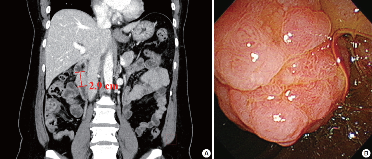
PDF - Peutz-Jeghers type hamartomatous polyp is known to be associated with Peutz-Jeghers syndrome, which shows characteristic multiple hamartomatous polyp involvement in the gastrointestinal tract, combined with mucocutaneous symptom, familial history of Peutz- Jeghers syndrome or STK11/LTB1 mutation. However, some cases showing histologic appearance of the polyps discovered in Peutz- Jeghers syndrome while lacking other diagnostic criteria of the syndrome have been reported, and these are called solitary Peutz- Jeghers type polyps. Herein, we report a case of solitary Peutz-Jeghers type polyp covered with heterotopic epithelium. The patient was 47-year-old female without any mucocutaneous symptoms nor familial history of Peutz-Jeghers syndrome. Microscopic examination revealed Peutz-Jeghers type hamartomatous polyp in duodenum covered with gastric type foveolar epithelium. Considering the definition of hamartomatous polyp, which is, the abnormal overgrowth of the indigenous epithelial component, the histological feature of current case is noteworthy in a point that it shows proliferation of heterotopic component, rather than the indigenous component.
-
Citations
Citations to this article as recorded by
- A Solitary Peutz-Jeghers Hamartomatous Polyp in the Gastric Body: A Case Report
Noelia Madera, Noemí Acevedo, Carmen González-Peralta, Rafael Castro, Vismelis Mezquita-Luna
Cureus.2024;[Epub] CrossRef
- A Solitary Peutz-Jeghers Hamartomatous Polyp in the Gastric Body: A Case Report
Case Study
- A Pyloric Gland-Phenotype Ovarian Mucinous Tumor Resembling Lobular Endocervical Glandular Hyperplasia in a Patient with Peutz-Jeghers Syndrome
- Eun Na Kim, Gu-Hwan Kim, Jiyoon Kim, In Ah Park, Jin Ho Shin, Yun Chai, Kyu-Rae Kim
- J Pathol Transl Med. 2017;51(2):159-164. Published online August 22, 2016
- DOI: https://doi.org/10.4132/jptm.2016.07.01
- 10,630 View
- 210 Download
- 10 Web of Science
- 9 Crossref
-
Abstract
PDF
- We describe an ovarian mucinous neoplasm that histologically resembles lobular endocervical glandular hyperplasia (LEGH) containing pyloric gland type mucin in a patient with Peutz-Jeghers syndrome (PJS). Although ovarian mucinous tumors rarely occur in PJS patients, their pyloric gland phenotype has not been clearly determined. The histopathologic features of the ovarian mucinous tumor were reminiscent of LEGH. The cytoplasmic mucin was stained with periodic acid-Schiff reaction after diastase treatment but was negative for Alcian blue pH 2.5, suggesting the presence of neutral mucin. Immunohistochemically, the epithelium expressed various gastric markers, including MUC6, HIK1083, and carbonic anhydrase-IX. Multiple ligation-dependent probe amplification detected a germline heterozygous deletion mutation at exons 1–7 of the STK11 gene (c.1-?_920+?del) in peripheral blood leukocytes and mosaic loss of heterozygosity in ovarian tumor tissue. Considering that LEGH and/or gastric-type cervical adenocarcinoma can be found in patients with PJS carrying germline and/or somatic STK11 mutations, our case indicates that STK11 mutations have an important role in the proliferation of pyloric-phenotype mucinous epithelium at various anatomical locations.
-
Citations
Citations to this article as recorded by- Molecular evidence of a clonal relationship of synchronous/multifocal gastric‐type lesions of the female genital tract
Min Shi, Hong Yang, Fang Zhang, Ting Hou, Huageng Huang, Yi Lu, Yehan Zhou, Ting Lan, Juan Ji, Jun Hou, Chengmin Zhou, Zhou Zhang, Sheng Qin, Zongyao Huang, Yang Liu
The Journal of Pathology.2026; 268(1): 27. CrossRef - Serine/threonine kinase 11 (STK11) associated adnexal tumors: from biology to therapeutic impact
Guanxiang Huang, Wenyu Lin, Tingting Jiang, Yuanjun Cai, Chengbin Lin, Pengming Sun
Human Genomics.2025;[Epub] CrossRef - Novel ultrasound features and diagnostic clues of gastric-type endocervical adenocarcinoma: a case series
Liwen Yang, Yangyang Wang, Jian Cai, Ying Xiong, Juan Li, Qi Zhou, Nan Ye, Hua Lai, Tianjiao Liu, Liuying Zhou
Frontiers in Oncology.2025;[Epub] CrossRef - Ovarian Mucinous Tumor Presenting Atypical Lobular Endocervical Glandular Hyperplasia-Like Appearance in a Patient With Germline STK11 p.F354L Variant: A Case Report
Hiroshi Yoshida, Kengo Hiranuma, Mariko Nakahara, Mayumi Kobayashi-Kato, Yasuhito Tanase, Masaya Uno, Kouya Shiraishi, Mitsuya Ishikawa, Tomoyasu Kato
International Journal of Surgical Pathology.2024; 32(2): 394. CrossRef - Preoperative multimodal ultrasonic imaging in a case of Peutz-Jeghers syndrome complicated by atypical lobular endocervical glandular hyperplasia: a case report and literature review
Liwen Yang, Duan Duan, Ying Xiong, Tianjiao Liu, Lijun Zhao, Fan Lai, Dingxian Gu, Liuying Zhou
Hereditary Cancer in Clinical Practice.2024;[Epub] CrossRef - Gastric‐type glandular lesions of the female genital tract excluding the cervix: emerging pathological entities
Richard W‐C Wong, Karen L Talia, W Glenn McCluggage
Histopathology.2024; 85(1): 20. CrossRef - Gastric-phenotype Mucinous Carcinoma of the Fallopian Tube with Secondary Ovarian Involvement in a Woman with Peutz-Jeghers Syndrome: A Case Report
Mónica Bronte Anaut, Javier Arredondo Montero, Maria Pilar Fernández Seara, Rosa Guarch Troyas
International Journal of Surgical Pathology.2023; 31(1): 92. CrossRef - Molecular characterization of gastric-type endocervical adenocarcinoma using next-generation sequencing
Swati Garg, Teddy S. Nagaria, Blaise Clarke, Orit Freedman, Zanobia Khan, Joerg Schwock, Marcus Q. Bernardini, Amit M. Oza, Kathy Han, Adam C. Smith, Tracy L. Stockley, Marjan Rouzbahman
Modern Pathology.2019; 32(12): 1823. CrossRef - The developing spectrum of gastric-type cervical glandular lesions
Karen L. Talia, W. Glenn McCluggage
Pathology.2018; 50(2): 122. CrossRef
- Molecular evidence of a clonal relationship of synchronous/multifocal gastric‐type lesions of the female genital tract
Case Report
- Peutz-Jeghers Syndrome with Extensive Epithelial Misplacements and Adenomatous and Carcinomatous Transformation: A case report.
- Jeong Ja Kwak, So Young Jin, Dong Wha Lee
- Korean J Pathol. 1993;27(6):630-637.
- 2,240 View
- 20 Download
-
Abstract
PDF
- Peutz-Jeghers syndrome is an autosomal dominant disease characterized by gastrointestinal ployposis and mucocutaneous melanin pigmentation involving the lip, oral mucosa, digits, palms and soles. The polyps are almost hamartomatous. The relationship of gastrointestinal carcinoma and the Peutz-Jeghers syndrome has been discussed for many years. The question is unsettled whether gastrointestinal carcinoma arise in hamartomatous polyps itself. Recently, there are a few reports that adenomatous and carcinomatous changes were superimposed upon the background of the hamartoma. Occasionally epithelial misplacement of the epithelium is found in the small intestinal polyps. Since the epithelial misplacement may involve submucosa, muscularis propria and serosa, a difficulty of histopathologic differential diagnosis between the epithelial misplacement and invasive adenocarcinoma cause overdiagnosis of cancer in the gastrointestinal polyps of Peutz-Jeghers syndrome. We present a case of Peutz-Jeghers syndrome of 39-year-old woman with multiple gastrointestinal polyps, two of which showed extensive epithelial misplacement even into the pancreas and another one at the colon showed carcinomatous change at the tip portion. Areas of hamartoma, adenoma and in situ carcinoma were noted in this colonic hamartomatous polyp. This case support that adenoma and carcinomatous changes may evolve directly within a hamartomaous polyp itself.
Original Article
- Gastrointestinal Polyposis in Koreans: A Nationwide Survey of Clinicopathologic Analysis of 112 Surgically Resected Cases.
- Mee Soo Chang, Hoguen Kim, Woo Ho Kim, Chan Il Park, Eun Kyung Hong, Han Kyeom Kim, In Soo Suh, Byung Kee Kim, Ja June Jang, Woon Sub Han, Hyung Sik Shin, So Young Jin, Dae Young Kang, Yong Il Kim
- Korean J Pathol. 1998;32(6):404-412.
- 2,460 View
- 11 Download
-
Abstract
- Gastrointestinal polyposis (GIP) is a rare disease characterized by formation of the numerous polyps in the gastrointestinal tract and presenting several extraintestinal manifestations. Most of the diseases are transmitted in an autosomal dominant pattern. In Korea, the epidemiological study as well as the pathological analysis of the GIP is not well established. We therefore analysed 38 items of GIP using surgically resected specimens. The materials in this study were collected from the 12 institutions and case reports in Korean literature between 1980 and 1991. The clinicopathologic findings were reevaluated by several members of the study group for gastrointestinal pathology. The results are as follows: (1) A total of 112 cases were included in this study: 83 cases were collected from 12 institutions and 29 cases were collected from Korean literature. The cases were classified as familial adenomatous polyposis (FAP), 59 cases; Gardner's syndrome, 3 cases; juvenile polyposis, 12 cases; Peutz-Jeghers syndrome, 35 cases; multiple colonic adenomas, 3 cases. (2) Among 59 cases of FAP, the range of age at operation was 14 to 61 years, and a family history was positive in 25 cases. The number of polyps in colorectum was 100~8,000. Of the 37 cases in which the examination of polyp density was available, 16 cases (43%) showed the highest density in the rectum and the sigmoid colon. The carcinomatous change within polyp(s) was present in 18 cases (31%), and associated advanced single or multiple colonic carcinomas existed in 37 cases (63%). Twenty-six (45%) tumors out of total 58 carcinomas were in the rectum. Twenty-five patients were evaluated for the upper gastrointestinal lesions, and 11 patients (44%) had pathologic lesions; multiple fundic gland polyps in 3 cases (12%), gastric and duodenal adenomas in 2 cases (8%), gastric adenomas in 2 cases (8%), duodenal adenomas in 2 cases (8%), gastric carcinoma and adenoma in 1 case (4%), gastric carcinoma in 1 case (4%). (3) Among 3 cases of Gardner's syndrome, the range of age at operation was 25 to 31 years, a family history was identified in 2 cases. One case was associated with an advanced colonic carcinoma and carcinomatous change within polyp. Extra gastrointestinal lesions were sebaceous cyst, epidermal cyst, osteoma and desmoid tumor. (4) Among 12 juvenile polyposis, the range of age at operation was 8 to 51 years and 5 patients had a family history. The carcinomatous change within polyp was found in 2 cases (17%) and associated advanced colonic carcinoma was in 4 cases (33%). The associated different type of polyps was tubular adenomas in 9 cases (75%), hyperplastic polyps in 4 cases (33%) and villous adenomas in 2 cases (17%). (5) Among 35 Peutz-Jeghers syndrome, the range of age at first operation was 6 to 42 years, family history was positive in 11 cases. The carcinomatous change within polyp was found in 1 case (3%), and associated advanced colonic carcinoma in 1 case (3%). The epithelial misplacement was observed in 4 cases (11%), and tubular or villous adenomatous feature in 4 cases (11%). In summary, the most frequent GIP for the surgical resection in Korea is FAP and the FAP is associated with high incidence of coexisting advanced and intramucosal carcinomas. Hamartomatous polyposis syndromes, such as juvenile polyposis and Peutz-Jeghers syndrome are another frequent disease for the surgical resection and are also associated with an increased risk of cancer.
 First
First Prev
Prev


