 E-submission
Search
- Page Path
- HOME > Search
Case Study
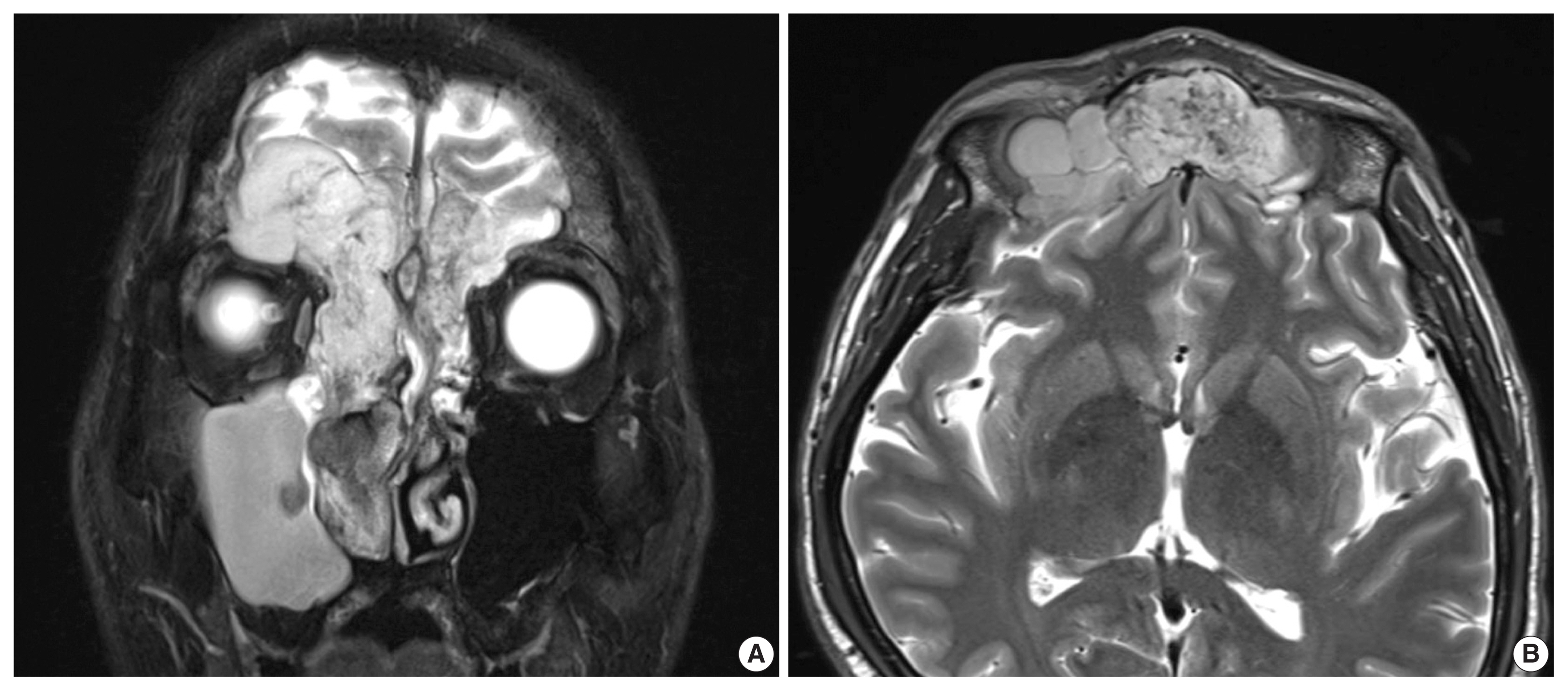
- A sinonasal yolk sac tumor in an adult
- Jaehoon Shin, Ji Heui Kim, Kyeong Cheon Jung, Kyung-Ja Cho
- J Pathol Transl Med. 2022;56(3):152-156. Published online January 26, 2022
- DOI: https://doi.org/10.4132/jptm.2021.12.09
- 7,896 View
- 225 Download
- 4 Web of Science
- 6 Crossref
-
 Abstract
 PDF
PDF - Yolk sac tumors (YSTs), which are also called endodermal sinus tumors, are malignant tumors of germ cell origin. These tumors usually occur in the gonads, but 20% of cases have been reported at extragonadal sites. The head and neck is a rarely affected region that accounts for just 1% of all malignant tumors of germ cell origin. In addition, YSTs arise mostly in childhood. We present a rare pathologically pure case of primary adult YST in the sinonasal area. A 45-year-old male patient presented with a rapidly growing mass in the nasal cavity, which caused nasal obstruction and bloody post-nasal drip. The histopathologic features indicated pure YST, and immunohistochemical analysis revealed positive reactivity for Sal-like protein 4 and alpha-fetoprotein. Herein, we discuss the clinical, radiologic, and histologic features of this YST and review other cases of sinonasal YST in adults.
-
Citations
Citations to this article as recorded by
- SMARCB1-deficient Sinonasal Carcinoma
Neha Mittal, Saurabh Nagar, Asawari Patil, Swapnil Ulhas Rane, Palgun Nisarga, Katha Rabade, Amit Janu, Deepa Nair, Shiva Thiagarajan, Sarbani Ghosh Laskar, Kumar Prabhash, Munita Bal
American Journal of Surgical Pathology.2025; 49(4): 381. CrossRef - International Consensus Statement on Allergy and Rhinology: Sinonasal Tumors
Edward C. Kuan, Eric W. Wang, Nithin D. Adappa, Daniel M. Beswick, Nyall R. London, Shirley Y. Su, Marilene B. Wang, Waleed M. Abuzeid, Borislav Alexiev, Jeremiah A. Alt, Paolo Antognoni, Michelle Alonso‐Basanta, Pete S. Batra, Mihir Bhayani, Diana Bell,
International Forum of Allergy & Rhinology.2024; 14(2): 149. CrossRef - Yolk sac tumor of postpubertal-type does not exhibit immunohistochemical loss of SMARCB1/INI1 and SMARCA4/BRG1…but choriocarcinoma?
Costantino Ricci, Francesca Ambrosi, Tania Franceschini, Francesca Giunchi, Eugenia Franchini, Francesco Massari, Veronica Mollica, Federico Mineo Bianchi, Maurizio Colecchia, Andres Martin Acosta, Michelangelo Fiorentino
Pathology - Research and Practice.2023; 241: 154269. CrossRef - Pure yolk sac tumor primarily in the nasal cavity: A case report
Zijun Liu, Baohong Wen, Yan Zhang
Asian Journal of Surgery.2023; 46(10): 4712. CrossRef - A case of Yolk sac tumor arising from paranasal sinus
Kaori Shinomura, Munehito Moriyama, Keigo Fujita, Takashi Hirano, Masashi Suzuki
JOURNAL OF JAPAN SOCIETY FOR HEAD AND NECK SURGERY.2023; 33(1): 41. CrossRef - A Novel Successful Case of Nasal and Sinus Yolk Sac Tumor With SMARCB1 (INI-1) Deficiency: A Case Report
Tianyu He, Zhiyu Wang, Hongbo Su, Sihan Li, Zheng He
Cureus.2022;[Epub] CrossRef
- SMARCB1-deficient Sinonasal Carcinoma
Case Report
- Glomus Tumor of the Sinonasal Tract: Two Case Reports and a Review of Literature.
- Young Wha Koh, Bong Jae Lee, Kyung Ja Cho
- Korean J Pathol. 2010;44(3):326-329.
- DOI: https://doi.org/10.4132/KoreanJPathol.2010.44.3.326
- 4,309 View
- 27 Download
- 8 Crossref
-
Abstract
PDF
- Herein we describe two cases of nasal glomus tumor. Histological findings were typical, save for one which was quite large (3.1 cm in its greatest dimension) with an invasive growth pattern and increased ki-67 labeling index (up to 10%). These features raised a red flag of similarity to a recently described "invasive glomus tumor of nasal cavity", suggesting a more aggressive form of glomus tumor. However, objective criteria for this possibility is lacking at present and more similar case studies are needed to establish a truly aggressive form of glomus tumor.
-
Citations
Citations to this article as recorded by- An Atypical Lesion in the Nasal Cavity: Glomus Tumor
Erbil Arık, Yasemin Gunduz, Gozde Cakirsoy Cakar, Halil Elden
Ear, Nose & Throat Journal.2025;[Epub] CrossRef - Nasal Septum Glomus Tumor: A Rare Cause of Unilateral Nasal Obstruction
Adamantios Kilmpasanis, Zoi Apazidi-Kesoglou, Alexandros Poutoglidis, Sotiria Sotiroudi, Konstantinos Vlachtsis, Nikolaos Tsetsos
Ear, Nose & Throat Journal.2023; 102(6): 402. CrossRef - A case of malignant glomus tumor (glomangiosarcoma) of the nasal cavity
Omar A Alhroub, Shimaa A Mahameed, Mohammad O Abdelhafez, Asil Alhroub, Hani Hour, Nabil Hasasna, Nazmi Kamal
Journal of Surgical Case Reports.2022;[Epub] CrossRef - Ethmoid glomangioma and oncogenic osteomalacia: a case report
Camila R. Muniz, Gabriela A. M. Bezerra, Viviane C. da Silva, Priscilla M. F. Aguiar, Gunter Gerson, Catarina B. D’Alva, André A. A. Nunes
Journal of Medical Case Reports.2021;[Epub] CrossRef - Large Glomus Tumor of The Lateral Nasal Wall : A Case Report
Bon Min Koo, Jong In Jeong, Dong Eun Kim
Journal of Clinical Otolaryngology Head and Neck Surgery.2019; 30(2): 243. CrossRef - Characteristics and prognosis of glomangiopericytomas: A systematic review
Eun Su Park, Jiyoung Kim, Sun-Young Jun
Head & Neck.2017; 39(9): 1897. CrossRef - Malignant Glomus Tumour (Glomangiosarcoma) with Additional Neuroendocrine Differentiation in a Horse
M. Peters, J. Grafen, C. Kuhnen, P. Wohlsein
Journal of Comparative Pathology.2016; 154(4): 309. CrossRef - A Case of Glomangioma of the Nasal Septum
Do Hun Kim
Korean Journal of Otorhinolaryngology-Head and Neck Surgery.2013; 56(9): 603. CrossRef
- An Atypical Lesion in the Nasal Cavity: Glomus Tumor
Original Article
- Metastatic Carcinomas to the Sinonasal Tract.
- Eun Ju Kim, Bong Jae Lee, Kyung Ja Cho
- Korean J Pathol. 2010;44(3):302-307.
- DOI: https://doi.org/10.4132/KoreanJPathol.2010.44.3.302
- 5,091 View
- 31 Download
- 1 Crossref
-
Abstract
PDF
- BACKGROUND
Metastases to the sinonasal tract are rare but occur for many malignancies. The demographics of sinonasal metastases in Korea aren't well known.
METHODS
Nine cases of metastases to the sinonasal tract identified at Asan Medical Center from January, 1995 to December, 2007 were reviewed.
RESULTS
Metastatic carcinomas accounted for 2.4% of sinonasal malignancies and 4.7% of carcinomas. Six kinds of cancer metastasized to the sinonasal tract. They included hepatocellular carcinomas (nasal cavity and maxillary sinus), colonic adenocarcinomas (sphenoid sinus and maxillary sinus), clear cell renal cell carcinoma (nasal cavity), pulmonary small cell carcinoma (nasal cavity), follicular carcinoma of thyroid (sphenoid sinus), and breast ductal carcinoma (maxillary sinus). Primary sites had been known in 7 cases, but follicular carcinoma and one adenocarcinoma were diagnosed after sinus metastases. Histologically, they had ill-defined borders and involved both mucosae and bones. Microscopic findings were not different from those for the primary tumors.
CONCLUSIONS
The pattern of sinonasal metastases in Korea are different from western data regarding incidence, site, and type, with hepatocellular carcinoma and the nasal cavity being the most common type and site, respectively. Awareness of the possibility of metastases and their pattern is encouraged when examining sinonasal tumors. -
Citations
Citations to this article as recorded by- Metastatic Carcinomas to the Oral Cavity and Oropharynx
Su-Jin Shin, Jong-Lyel Roh, Seung-Ho Choi, Soon Yuhl Nam, Sang Yoon Kim, Sung Bae Kim, Sang-wook Lee, Kyung-Ja Cho
Korean Journal of Pathology.2012; 46(3): 266. CrossRef
- Metastatic Carcinomas to the Oral Cavity and Oropharynx
Case Report
- Primary Meningioma of the Nasal Cavity and Paranasal Sinuses: A report of a case.
- Chang Ok Kim, Mi Kyung Jee, Ki Hwa Yang, Chang Suck Kang, Seok Jin Gang, Byoung Kee Kim, Sun Moo Kim
- Korean J Pathol. 1989;23(4):461-464.
- 2,301 View
- 16 Download
-
Abstract
PDF
- Primary extracranial and extraspinal meningiomas are rare.
Case
s involving the orbit, skin, nasal cavity, paranasal sinuses, oral cavity and parotid gland have been reported. The histogenesis of primary extracranial meningioma is still nucertain, but it has been thought that this tumor originates from arachnoid cell rests in displaced during embryonal development. The authors observed a case of primary meningioma of the nasal cavity and paranasal sinuses occurring in a thirty-eight year old male patient in Feb. 1989. He suffered from bulging in the medio-superior portion of left orbit for 15 years, and left nasal obstruction and headache for 5 years, A head CT scan revealed numberous polypoid masses filling the left frontal sinus left ethmoidal sinus, left maxillary sinus and left nasal cavity. During the operation, a connection to the dura was not found. Microscopically, there were discrete lobules or netst of meningothelial cells, beneath the nasal mucosa. They showed an occasional whorling pattern and psammoma bodies. Therefore, this case was diagnosed as primary meningioma, meningotheliomatous type involving the left nasal cavity and paranasal sinuses.
 First
First Prev
Prev


