 E-submission
Search
- Page Path
- HOME > Search
- Primary epithelioid inflammatory myofibroblastic sarcoma of the brain with EML4::ALK fusion mimicking intra-axial glioma: a case report and brief literature review
- Eric Eunshik Kim, Chul-Kee Park, Koung Mi Kang, Yoonjin Kwak, Sung-Hye Park, Jae-Kyung Won
- J Pathol Transl Med. 2024;58(3):141-145. Published online May 14, 2024
- DOI: https://doi.org/10.4132/jptm.2024.04.12
- 5,304 View
- 211 Download
- 2 Web of Science
- 2 Crossref
-
 Abstract
 PDF
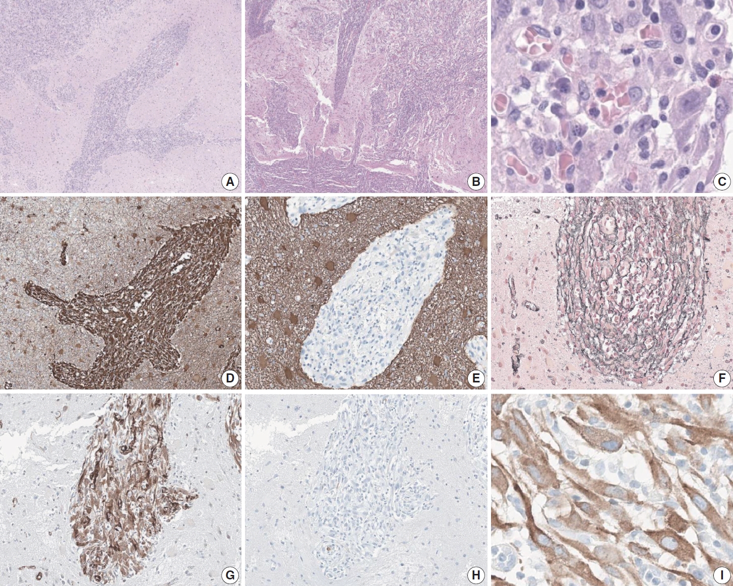
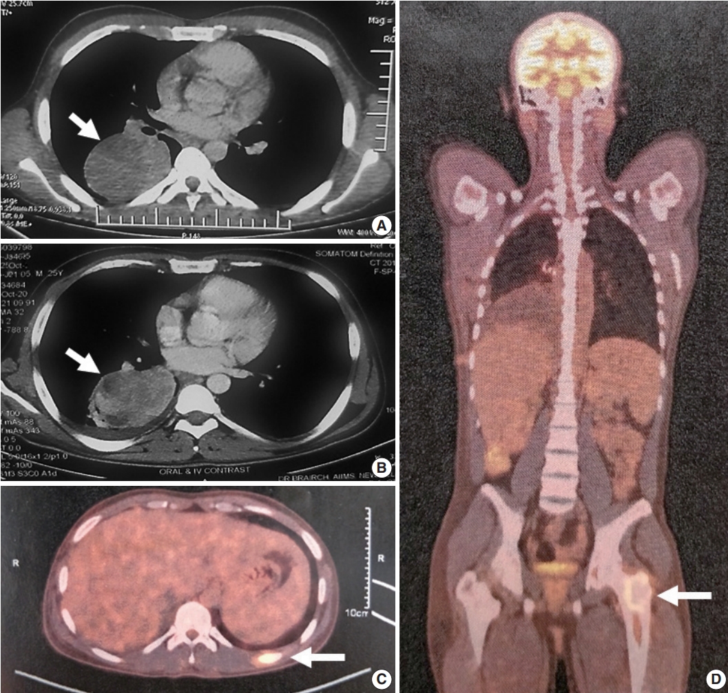
PDF - An aggressive subtype of inflammatory myofibroblastic tumor, epithelioid inflammatory myofibroblastic sarcoma occurs primarily inside the abdominal cavity, followed by a pulmonary localization. Most harbor anaplastic lymphoma kinase (ALK) gene rearrangements, with RANBP2 and RRBP1 among the well-documented fusion partners. We report the second case of primary epithelioid inflammatory myofibroblastic sarcoma of the brain, with a well-known EML4::ALK fusion. The case is notable for its intra-axial presentation that clinico-radiologically mimicked glioma.
-
Citations
Citations to this article as recorded by
- Primary pulmonary epithelioid inflammatory myofibroblastic sarcoma: a rare entity and a literature review
- Priyanka Singh, Aruna Nambirajan, Manish Kumar Gaur, Rahul Raj, Sunil Kumar, Prabhat Singh Malik, Deepali Jain
- J Pathol Transl Med. 2022;56(4):231-237. Published online July 7, 2022
- DOI: https://doi.org/10.4132/jptm.2022.05.08
- 7,447 View
- 133 Download
- 13 Web of Science
- 12 Crossref
-
Abstract
PDF
- Epithelioid inflammatory myofibroblastic sarcoma (EIMS) is an aggressive subtype of inflammatory myofibroblastic tumor (IMT) harboring anaplastic lymphoma kinase (ALK) gene fusions and is associated with high risk of local recurrence and poor prognosis. Herein, we present a young, non-smoking male who presented with complaints of cough and dyspnoea and was found to harbor a large right lower lobe lung mass. Biopsy showed a high-grade epithelioid to rhabdoid tumor with ALK and desmin protein expression. The patient initially received 5 cycles of crizotinib and remained stable for 1 year; however, he then developed multiple bony metastases, for which complete surgical resection was performed. Histopathology confirmed the diagnosis of EIMS, with ALK gene rearrangement demonstrated by fluorescence in situ hybridization. Postoperatively, the patient is asymptomatic with stable metastatic disease on crizotinib and has been started on palliative radiotherapy. EIMS is a very rare subtype of IMT that needs to be included in the differential diagnosis of ALKexpressing lung malignancies in young adults.
-
Citations
Citations to this article as recorded by- Inflammatory Myofibroblastic Tumor: An Updated Review
Joon Hyuk Choi
Cancers.2025; 17(8): 1327. CrossRef - Epithelioid Inflammatory Myofibroblastic Sarcoma: Case Series With a First Report of CLTC::ALK Fusion in an Aggressive Disease
Daisy Maharjan, Carina Dehner, Ali Alani, Robert Bell, Sheila Segura
Genes, Chromosomes and Cancer.2025;[Epub] CrossRef - ALK rearranged malignant mesenchymal neoplasms of thorax: therapeutically targetable ‘ALKomas’ beyond the spectrum of non-small cell lung carcinomas and thoracic inflammatory myofibroblastic tumors
Shreya Sadhu, Adarsh Barwad, Asit Ranjan Mridha, Prabhat Singh Malik, Aruna Nambirajan, Deepali Jain
Virchows Archiv.2025; 487(5): 1003. CrossRef - Mediastinal epithelioid inflammatory myofibroblastic sarcoma with the EML4‐ALK fusion: A case report and literature review
Tingyu Pan, Xinyu Sun, Xiao Wu, Futing Tang, Xianmei Zhou, Qian Wang, Shi Chen
Respirology Case Reports.2024;[Epub] CrossRef - Primary epithelioid inflammatory myofibroblastic sarcoma of the brain with EML4::ALK fusion mimicking intra-axial glioma: a case report and brief literature review
Eric Eunshik Kim, Chul-Kee Park, Koung Mi Kang, Yoonjin Kwak, Sung-Hye Park, Jae-Kyung Won
Journal of Pathology and Translational Medicine.2024; 58(3): 141. CrossRef - Epithelioid Inflammatory Myofibroblastic Sarcoma: A Report of a Rare Case
Varun Ronanki, Vaddatti Tejeswini, Inuganti Venkata Renuka, Shaik Raheema, Bakkamanthala S K Kanth
Cureus.2024;[Epub] CrossRef - Thoracic epithelioid inflammatory myofibroblastic sarcoma: a rare and aggressive disease with case report and literature review
Linke Yang, Pei Li, Runze Liu, Baomin Feng, Huiqing Mao, Xiaoyong Tang, Guangjian Yang
Discover Oncology.2024;[Epub] CrossRef - Epithelioid inflammatory myofibroblastic sarcoma with exceptionally long response to lorlatinib—a case report
Rafał Becht, Kajetan Kiełbowski, Justyna Żychowska, Wojciech Poncyljusz, Aleksandra Łanocha, Katarzyna Kozak, Ewa Gabrysz-Trybek, Paweł Domagała
Therapeutic Advances in Medical Oncology.2024;[Epub] CrossRef - Rare giant epithelioid inflammatory myofibroblastic sarcoma of the abdominal cavity in a child: a case report and review of the literature
Jinzhou Li, Haixing Su, Sheng Zhang, Xianyun Chen, Chongzhi Hou, Tao Cheng
Frontiers in Oncology.2024;[Epub] CrossRef - Case report: Epithelioid inflammatory myofibroblastic sarcoma treated with an ALK TKI ensartinib
Mengmeng Li, Ruyue Xing, Jiuyan Huang, Chao Shi, Chunhua Wei, Huijuan Wang
Frontiers in Oncology.2023;[Epub] CrossRef - Epithelioid Inflammatory Myofibroblastic Sarcoma With Poor Response to Crizotinib: A Case Report
Soheila Aminimoghaddam, Roghayeh Pourali
Clinical Medicine Insights: Case Reports.2023;[Epub] CrossRef - Epithelioid inflammatory myofibroblastic sarcoma: a case report and brief literature review
Weidong Dou, Yu Guan, Tao Liu, Hang Zheng, Shuo Feng, Yingchao Wu, Xin Wang, Zhanbing Liu
Frontiers in Oncology.2023;[Epub] CrossRef
- Inflammatory Myofibroblastic Tumor: An Updated Review
- Congenital Peribronchial Myofibroblastic Tumor: A Case Study and Literature Review
- Yuil Kim, Ha Young Park, Junhun Cho, Joungho Han, Eun Yoon Cho
- Korean J Pathol. 2013;47(2):172-176. Published online April 24, 2013
- DOI: https://doi.org/10.4132/KoreanJPathol.2013.47.2.172
- 10,016 View
- 63 Download
- 10 Crossref
-
Abstract
PDF
Congenital peribronchial myofibroblastic tumor (CPMT) is a benign pulmonary spindle cell neoplasm of intrauterine and perinatal period, which is thought to arise from primitive peribronchial mesenchyme. We present a case detected incidentally in a one-month-old infant. The solid and partially necrotic tumor involved the right middle and lower lobes of the lung with extension to the diaphragm. Histologically, the tumor was composed of fasciculated monotonous spindle cells, proliferating peribronchiolar cartilage and round cells with rich vasculature, and high mitotic activity was identified in the round cell area. Immunohistochemical and electron microscopic studies showed that the spindle cells were myofibroblastic in phenotype. Although the tumor showed several malignant pathological features, recurrence was not observed in the two-year follow-up period, consistent with the benign clinical behavior of CPMT.
-
Citations
Citations to this article as recorded by- Congenital Peribronchial Myofibroblastic Tumor: Clinical Features, Pathology, and Surgical Considerations
Kavya Rajesh, Drew Bolster, Mariam Naqvi, Sonya Fabricant, Raghavendra Pillappa, Andrew Brownlee, Carlos Pelayo, Eugene Kim, David Bliss, Eveline Shue
Pediatric Blood & Cancer.2026;[Epub] CrossRef - Congenital Peribronchial Myofibroblastic Tumors Harbor a Recurrent EGFR Kinase Domain Duplication
Sheren Younes, Carlos J. Suarez, Jennifer Pogoriler, Tricia Bhatti, Archana Shenoy, Raya Saab, Lea F. Surrey, Serena Y. Tan
Modern Pathology.2025; 38(2): 100661. CrossRef - EGFR‐KDD Myofibroblastic Neoplasm or Congenital Peribronchial Myofibroblastic Tumor (CPMT)? Report of a Congenital Myofibroblastic Neoplasm With Unusual Histologic Features
Emma Rullo, Sabina Barresi, Sabrina Rossi, Sara Patrizi, Evelina Miele, Marta Barisella, Michela Casanova, Andrea Ferrari, Stefano Chiaravalli, Gloria Pelizzo, Rita Alaggio
Genes, Chromosomes and Cancer.2025;[Epub] CrossRef - Congenital peribronchial myofibroblastic tumor (CPMT): a case report with long term follow-up and next-generation sequencing (NGS)
Ping Zhou, Shuang Li, Weiya Wang, Yuan Tang, Lili Jiang
BMC Pediatrics.2023;[Epub] CrossRef - Neonatal congenital lung tumors — the importance of mid-second-trimester ultrasound as a diagnostic clue
Stephan L. Waelti, Laurent Garel, Dorothée Dal Soglio, Françoise Rypens, Michael Messerli, Josée Dubois
Pediatric Radiology.2017; 47(13): 1766. CrossRef - Congenital peribronchial myofibroblastic tumor: Case report and review of literature
Jolanta Jedrzkiewicz, Eric Scaife, Bo Hong, Sarah South, Mouied Alashari
Journal of Pediatric Surgery Case Reports.2015; 3(4): 154. CrossRef - Perinatal Thoracic Mass Lesions: Pre- and Postnatal Imaging
Evan J. Zucker, Monica Epelman, Beverley Newman
Seminars in Ultrasound, CT and MRI.2015; 36(6): 501. CrossRef - Prenatal imaging and immunohistochemical analysis of congenital peribronchial myofibroblastic tumor
Y.‐A. Tu, W.‐C. Lin, H.‐J. Chen, J.‐C. Shih
Ultrasound in Obstetrics & Gynecology.2015; 46(2): 247. CrossRef - A Congenital Peribronchial Myofibroblastic Tumor Detected in a Premature Infant at 28 Weeks but That Resolved in the Late Stage of Pregnancy
Bo Xia, Gang Yu, Chun Hong, Lei Zhang, Jing Tang, Cuifen Liu
Medicine.2015; 94(42): e1842. CrossRef - Congenital peribronchial myofibroblastic tumor
Yuka Hotokebuchi, Kenichi Kohashi, Satoshi Toyoshima, Naoko Matsumoto, Toshinori Nakashima, Yoshinao Oda
Pathology International.2014; 64(4): 189. CrossRef
- Congenital Peribronchial Myofibroblastic Tumor: Clinical Features, Pathology, and Surgical Considerations
- Fine Needle Aspiration Cytology of Inflammatory Myofibroblastic Tumor in Mesentery: A Case Report.
- Hyun Jin Son, Joo Heon Kim, Woo Sung Moon, Myoung Jae Kang, Ho Yeul Choi
- J Pathol Transl Med. 2000;11(1):35-40.
- 2,221 View
- 17 Download
-
Abstract
PDF
- Since inflammatory myofibroblastic tumor was initially recognized in the lung, this tumor has been described in other extrapulmonary sites. In spite of relatively uniform histologic findings in various organs, a rarity in extrapulmonary sites and highly vascular characteristics frequently lead to a misdiagnosis in preoperative radiology and fine needle aspiration cytology. We present a case of inflammatory myofibro blastic tumor occurring in the mesentery of a 4-month-old girl. Fine needle aspira tion cytology smear disclosed characteristic spindle cells intermixed with prominent mature plasma cells and lymphocytes. According to the immunohistochemical staining, we recognized that the intervening spindle cells are myofibroblasts which have reactivity for the both actin and vimentin.
- Fine Needle Aspiration Cytologic Findings of Gastric Inflammatory Myofibroblastic Tumor: A case report.
- Ji Hye Lee, Bong Kyung Shin, Chung Yeul Kim, Seong Jin Cho, Han Kyeom Kim, In Sun Kim
- J Pathol Transl Med. 2001;12(2):117-120.
- 2,025 View
- 12 Download
-
Abstract
PDF
- Inflammatory myofibroblastic tumor, histologically characterized by the presence of bland-looking spindle cells and infiltration of chronic inflammatory cells, is extremely rare in the gastric wall. We report a case of gastric inflammatory myofibroblastic tumor in a 27-month-old boy. The fine needle aspiration biopsy from the mass showed loose clusters or scattered spindle cells and inflammatory cells, predominantly of lymphocytes and plasma cells. The spindle cells resembled fibroblasts or myofibroblasts. Differential diagnosis from benign and malignant diseases involving abdominal cavity was discussed.
- Inflammatory Myofibroblastic Tumor of the Breast: A Case Report.
- Myoung Ja Chung, So Yeong Oh, Kyu Yun Jang, Woo Sung Moon, Myoung Jae Kang, Dong Geun Lee
- Korean J Pathol. 2005;39(1):54-58.
- 2,389 View
- 23 Download
-
Abstract
PDF
- Inflammatory myofibroblastic tumor (IMT) is characterized by a clonal proliferation of myofibroblasic spindle cells, and this is accompanied by a lymphoplasmacytic infiltration. In the majority of cases, this disease has occurred in the lungs and only 9 cases of IMT in the breast have been previously reported. We report here on an IMT in a 25-year-old-female who presented with a palpable mass in the right breast. Histologically, it was characterized by plump spindle cells admixed with prominent inflammation, that was composed of lymphocytes and plasma cells. Immunohistochemically, the spindle cells were positive for vimentin and -smooth muscle actin.
- Immunohistochemical Findings in 10 Cases of Inflammatory Myofibroblastic Tumor.
- Soo Jin Jung, Mi Seon Kang, Chang Hoon Lee, Sook Hee Hong, Hye Kyoung Yoon
- Korean J Pathol. 1999;33(9):717-722.
- 2,477 View
- 22 Download
-
Abstract
PDF
- A wide range of denomination has been used for inflammatory myofibroblastic tumor (IMT). IMT is not entirely homogeneous, even though it shows some overlapping histologic features such as haphazard proliferation of spindle cell and polymorphic chronic inflammatory cell infiltraion. The spindle cell is considered to be of myofibroblastic origin but follicular dendritic cell origin was reported recently. IMT is known as nonneoplastic, aberrant inflammatory response. However, IMT could show local invasion, recurrence, vascular invasion, and malignant transformation, and clonal characteristics and aneuploidy of IMT support the hypothesis that IMT may be a neoplastic process. In order to define the nature of spindle cell of IMT, immunohistochemical stains for smooth muscle actin (SMA), vimentin (VMT), lysozyme, S-100 protein, cytokeratin, CD21 were done. Additional immunohistochemical stains for MIB-1 for proliferating activity and LMP (latent membrane protein) for Epstein-Barr virus (EBV) were done. IMTs were composed of each 2 cases from lung, liver and lymph node and one case from common bile duct, maxillary sinus, bladder and thigh, and were histologically subclassified according to Coffin et al. Nine cases (90%) were positive for SMA and VMT, but no correlation between SMA and VMT immunoreactivity and histologic types was identified. Five cases (50%) were positive for lysozyme and S-100 protein, and histologic type III was negative for lysozyme and S-100 protein, and immunoreactivity for S-100 protein was different according to the histologic subtypes. All 11 cases were negative for CD21 and EBV LMP. MIB-1 labelling index was less than 1% in all cases. In summary, the spindle cell is regarded as myofibroblastic origin rather than follicular dendritic cell origin. Relationship with EBV is not clear, and negligible MIB-1 reaction suggests that IMT might have a good prognosis.
- Inflammatory Myofibroblastic Tumor of the Mesentery: A case report.
- Sung Jig Lim, Gou Young Kim, Jae Hoon Park, Youn Wha Kim, Yong Koo Park, Ju Hie Lee, Moon Ho Yang
- Korean J Pathol. 1999;33(9):729-732.
- 2,203 View
- 32 Download
-
Abstract
PDF
- Inflammatory myofibroblastic tumor or inflammatory pseudotumor is characterized by spindle cell proliferation with inflammatory cell infiltration, predominantly plasma cells and lymphocytes. We have experienced a case of inflammatory myofibroblastic tumor of the mesentery in a 57-year-old male patient with intermittent abdominal pain. On computer tomography, a well demarcated mass was seen in the mesenteric side of the ascending colon. Right hemicolectomy was performed under the impression of the metastatic tumor of lymph nodes. Grossly, a rather well-circumscribed gray white mass was noted in the mesentery of the ascending colon. Microscopically, the lesion consisted of plump spindle cells and accompanying inflammatory cellular infiltrates. The spindle cells were positive for vimentin.
- Inflammatory Pseudotumor of the Urinary Bladder: An Immunohistochemical and Ultrastructural Study.
- Seung Sam Paik, Joo Seob Keum, Moon Hyang Park, Jung Dal Park
- Korean J Pathol. 1996;30(5):447-452.
- 2,155 View
- 23 Download
-
Abstract
PDF
- Inflammatory pseudotumor of the urinary bladder is an unusual, benign mesenchymal proliferative lesion of the submucosal stroma easily mistaken for a malignant neoplasm clinically and histologically. We present a case and describe the clinical presentation and radiologic, histologic, histochemical, immunohistochemical, and ultrastructural findings. A 23-year old patient presented with sudden onset of gross painless hematuria for 3 months. There was no previous instrumentation or surgery involving the genitourinary tract. Cystoscopy revealed a large polypoid and ulcerated bladder mass. The lesion consisted of plump spindle shaped, fibroblast-like cells embedded in a myxoid stroma. Mitotic figures were negligible and the lesion showed encroachment on the superficial muscle bundles. The spindle cells were immunoreactive for vimentin and muscle specific actin. Immunohistochemical and ultrastructural findings revealed the fibroblastic-myofibroblastic nature of this lesion. Complete surgical excision by partial cystectomy was successful in eradicating the lesion. The findings are described with a discussion of the pathogenesis and review of the literature.
- Inflammatory Myofibroblastic Tumor of the Stomach: A Case Report.
- Joon Mee Kim, In Suh Park, Lucia Kim, Suk Jin Choi, Jee young Han, Young Chae Chu, Kyoung Rae Kim
- Korean J Pathol. 2006;40(2):148-150.
- 2,293 View
- 25 Download
-
Abstract
PDF
- Inflammatory myofibroblastic tumor of the stomach is a rare tumor-like, benign disease with an uncertain pathogenesis. A 15-year-old male presented with epigastric pain. Endoscopic ultrasonography revealed a 2.3 cm sized ovoid intramuscular mass in the lower body of the stomach at the lesser curvature. Histologically, the tumor was composed of smooth muscle actin positive- and vimentin positive spindle cells and there were a large number of lymphocytes, plasma cells and histiocytes in the fibrotic background. The spindle cells were also positive for ALK1, but negative for EBV in situ hybridization.
- Follicular Lymphoma with Monoclonal Plasma Cell Differentiation: A Case Report.
- Hyun chul Kim, Young Seok Lee, Jung woo Choi, Ae ree Kim, Bom Woo Yeom, Han kyeom Kim, In sun Kim
- Korean J Pathol. 2006;40(2):151-155.
- 2,406 View
- 27 Download
-
Abstract
PDF
- We present a case of recurrent follicular lymphoma with an extensive plasma cell component involving infra-auricular lymph nodes in a 64 year-old woman. Immunohistochemical staining showed a strongly positive reaction of the follicles with CD20, bcl-2, bcl-6, CD10 and CD21 on the first biopsy specimen. The intrafollicular and interfollicular plasma cells showed monoclonality for IgG heavy chain and lambda light chain. The histological and immunohistochemical findings in the recurrent tumor were identical with those of the original. Discussion is focused on the importance of the differential diagnosis between reactive lymphoid hyperplasia and other lymphomas having plasmacytic differentiation.
- Inflammatory Myofibroblastic Tumor (InflammatoryFibrosarcoma) of the Lung: A Case Report.
- Minseob Eom, Tae Heon Kim, Jin Kyu Park, Kwang Hwa Park, Soon Hee Jung, Kwang Gil Lee
- Korean J Pathol. 2003;37(4):291-295.
- 2,221 View
- 21 Download
-
Abstract
PDF
- Inflammatory myofibroblastic tumor, also known as inflammatory fibrosarcoma, has been frequently diagnosed as inflammatory pseudotumor. Although there are six cases reported as inflammatory pseudotumors or inflammatory myofibroblastic tumors in the lung, no cases of pulmonary inflammatory myofibroblastic tumor with features of inflammatory fibrosarcoma have been reported in Korea. We experienced a case of inflammatory myofibroblastic tumor (inflammatory fibrosarcoma)characterized by high cellularity, severe nuclear pleomorphism, necrosis, or increased mitoticcounts. A 31-year-old male patient with a solitary pulmonary nodule on the routine chest x-rayreceived a right lower lobectomy. The tumor was an ovoid solid mass with multifocal necrosis, showing diffuse irregular proliferation of spindle cells with high cellularity and focal nuclear pleomorphism, admixed with dense lymphoplasmacytic cells. Although spindle cells are focally immunoreactivefor smooth muscle actin, the ultrastructural examination failed to demonstrate smooth muscledifferentiation. In cases of inflammatory myofibroblastic tumor (inflammatory fibrosarcoma), a completeexcision and close follow-up without radical surgery, radiation, or chemotherapy are needed.
- Inflammatory Myofibroblastic Tumor of the Maxillary Sinus: A case report.
- Hyun Jin Son, Seung O Ko, Myoung Ja Chung, Ho Yeul Choi
- Korean J Pathol. 2000;34(8):601-604.
- 2,007 View
- 20 Download
-
Abstract
PDF
- Inflammatory myofibroblastic tumor (IMT) is a space occupying lesion which is composed of myofibroblasts, plasma cells, and lymphocytes. IMT of the maxillary sinus is rare and its etiology is unknown. We present a case of inflammatory myofibroblastic tumor occurring in the right maxillary sinus of a 57-year-old woman. Radiologically, this tumor was interpreted as malignant neoplasm. On histologic examination, bundles of spindle cells were admixed with inflammatory cells including mature plasma cells and lymphocytes. On the basis of the immunohistochemical findings and ultrastructural features, we recognized that the intervening spindle cells were myofibroblasts. We discussed etiology and prognostic factors of this tumor.
- Inflammatory Myofibroblastic Tumor in Posterior Mediastinum.
- Seung Sam Paik, Seok Hoon Jeon, Se Jin Jang, Moon Hyang Park, Jung Dal Lee
- Korean J Pathol. 1997;31(1):63-67.
- 2,232 View
- 31 Download
-
Abstract
PDF
- Inflammatory myofibroblastic tumor(IMT) or inflammatory pseudotumor is a rare, solid tumor that most often affects children. This tumor is characterized by a spindle cell proliferation admixed with a variety of inflammatory cells. Although it has disputed nosology, a distinctive fibroinflammatory and even pseudosarcomatous appearance have been well appreciated. Herein, we report a case of IMT in the posterior mediastinum in a 19-year-old girl with clinical findings. The immunohistochemical and ultrastructural studies on the tumor cells are reported, and their distinctive characteristics are discussed in details.
 First
First Prev
Prev


