 E-submission
Search
- Page Path
- HOME > Search
Case Study
- Mucocele of the rectal stump: mucinous cystic neoplasm with low-grade dysplasia simulating low-grade appendiceal mucinous neoplasm
- Hasan Basri Aydin, Maria Faraz, A. David Chismark, Haiyan Qiu, Hwajeong Lee
- J Pathol Transl Med. 2025;59(2):139-146. Published online February 26, 2025
- DOI: https://doi.org/10.4132/jptm.2024.12.27
- 3,654 View
- 174 Download
-
 Abstract
 PDF
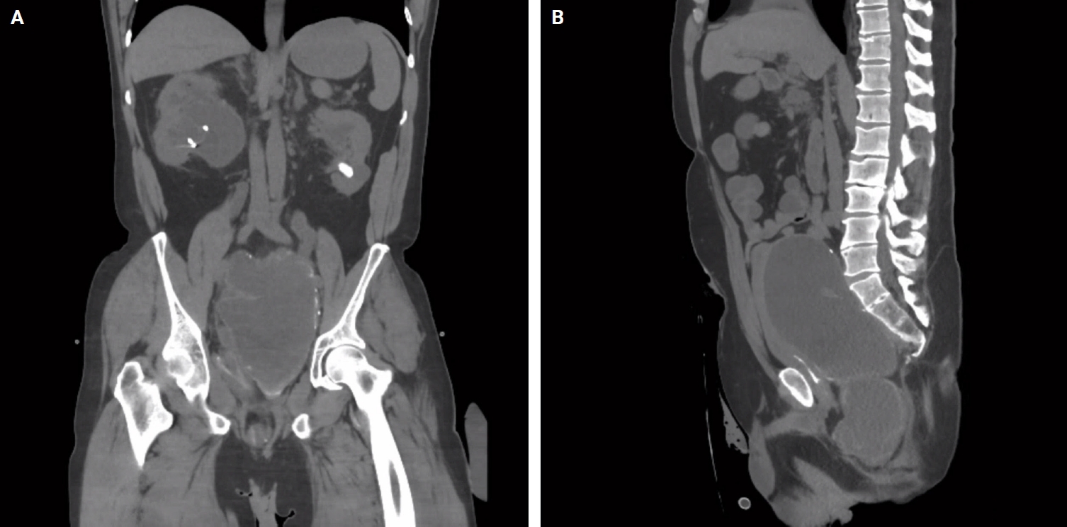
PDF - Mucoceles, commonly observed in the appendix, are mucin-filled, dilated structures arising from a range of etiologies. Cases associated with dysplastic or neoplastic epithelium can rupture and disseminate within the abdominopelvic cavity. Similar lesions in other parts of the colon are exceedingly rare, with only 16 colonic mucoceles having been reported. The first case of a colonic mucinous neoplasm with dysplasia resembling a low-grade appendiceal mucinous neoplasm involving rectal stump was described in 2016. Here, we present the second such case arising in the rectal stump, identified in a 44-year-old male with extensive surgical history. Microscopic examination revealed low-grade dysplastic epithelium lining the cyst and mucin dissecting into the stroma, without evidence of rupture or extramural mucin. The patient was followed for 16 months without recurrence or peritoneal disease. The exact etiology and outcome of these rare lesions remain unknown, requiring close follow-up.
Case Reports
- Myxoglobulosis of the Appendix: A Case Report.
- Jong Yup Bae, Jong Woo Kim
- Korean J Pathol. 2001;35(3):256-258.
- 2,148 View
- 37 Download
-
Abstract
PDF
- Myxoglobulosis of the appendix is a rare and peculiar form of appendiceal mucocele. It is characterized by pearl or fish egg-like mucin globules in the lumen. We report a case of myxoglobulosis of the appendix in a 31-year-old man. Myxoglobules are composed of eosinophilic necrotic cell debris and mucin in the central nidus and lamellar structures alternating with necrotic cell debris and mucin in the peripheral zone. A stirring effect produced by vigorous appendiceal peristalsis may contribute to its lamellar growth.
- Obstructive Mucocele of the Appendix Secondary to Endometriosis: A Case Report.
- Chang Hun Lee, Dong Hoon Shin, Jun Woo Lee
- Korean J Pathol. 2004;38(6):419-422.
- 2,247 View
- 18 Download
-
Abstract
PDF
- Appendiceal mucoceles are usually associated with hyperplastic or neoplastic mucosal proliferation and obstructive lesions such as postinflammatory scarring, fecalith, carcinoid tumor, and endometriosis. Among these, an association with endometriosis is known to be very exceptional. We herein report on a rare case of obstructive mucocele of the appendix that was secondary to endometriosis in a 42-year-old patient with pelvic endometriosis. A computed tomography scan demonstrated a periappendiceal abscess-like lesion with a left adnexal mass that was suggestive of endometriosis. On gross examination, the periappendiceal lesion consisted of a mucin-filled cavity (the so-called mucocele) that was 1.8 cm in diameter, and it protruded into the cecal lumen. Microscopically, the lining epithelium of the cavity was almost totally denuded. A small amount of mucus spilled over outside the mucocele, but pseudomyxoma peritonei was not present. The wall of the mucocele showed the characteristic multiple foci of endometriosis involving predominantly the muscularis propria and the serosa of the appendix and adjacent cecal walls.
- Vestibular Adenoma of the Vulva: A case report.
- Kyu Rae Kim, Sei Yul Han
- Korean J Pathol. 1992;26(2):197-200.
- 2,115 View
- 23 Download
-
Abstract
PDF
- A vestibular adenoma arising in mucin-secreting glands of the vulva is described. Grossly, the adenoma was a firm, well-demarcated solid mass with mucoid cut surface, measuring 3.5x3x3 cm, which was associated with an adjacent Bartholin's duct cyst. Microscopically, the solid mass was composed of proliferated mucous acini separted by fibromuscular septa and ducts lined by mucin-secreting columnar epithelium, transitional epithelium or metaplastic squamous epithelium. To our knowledge, less than 20 cases of such cases have been reported in the English literatures. However, whether the nature of proliferation is neoplastic or non-tumorous is still unclear.
 First
First Prev
Prev


