 E-submission
Search
- Page Path
- HOME > Search
Case Study
- Diagnostic challenge in Burkitt lymphoma of the mandible initially misdiagnosed as osteomyelitis: a case report
- Jiwon Do, Jin-Young Choi
- J Pathol Transl Med. 2025;59(6):460-466. Published online November 14, 2025
- DOI: https://doi.org/10.4132/jptm.2025.09.18
- 3,640 View
- 115 Download
-
 Abstract
 PDF
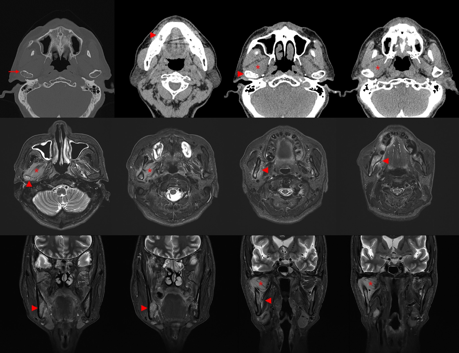
PDF - Burkitt lymphoma (BL) is a highly aggressive B-cell neoplasm that rarely involves the mandible in elderly without apparent immunodeficiency. We report a case of a 72-year-old male who presented with persistent mandibular pain following extraction of tooth #46. Initial imaging findings were consistent with incipient osteomyelitis, and the patient was treated with antibiotics. Despite treatment, pain persisted, and follow-up imaging revealed swelling and diffusion restriction in the lateral pterygoid muscle without evidence of a distinct mass. Biopsy revealed BL confirmed by immunohistochemistry: CD10+, BCL6+, c-MYC+, Ki-67 >95%, and negative for BCL2, MUM-1, and Epstein-Barr virus. Although c-MYC immunopositivity was demonstrated, fluorescence in situ hybridization for MYC rearrangement could not be performed due to limited tissue, representing a diagnostic limitation. Notably, the patient had no trismus despite deep muscle involvement, but complained of facial paresthesia and showed remote swelling in the scapular area during hospitalization. Systemic staging with imaging, cerebrospinal fluid cytology, and imaging revealed disseminated nodal and extranodal involvement including the central nervous system, corresponding to stage IV disease by Lugano classification. This case highlights the diagnostic challenge of distinguishing lymphoma from osteomyelitis and underscores the importance of considering malignancy in cases of refractory mandibular inflammation with atypical features.
Case Reports
- Fine Needle Aspiration Cytology of Langerhans Cell Histiocytosis of Mandible: A Case Report.
- Sang Ryung Lee, Jae Hee Suh, Hee Jung Cha, Young Min Kim, Hye Jeong Choi
- Korean J Pathol. 2010;44(1):106-109.
- DOI: https://doi.org/10.4132/KoreanJPathol.2010.44.1.106
- 4,504 View
- 25 Download
- 1 Crossref
-
Abstract
PDF
- We present a case of mandibular involvement with Langerhans cell histiocytosis (LCH), diagnosed by ultrasound-guided aspiration and subsequently confirmed by incisional biopsy and immunohistochemistry in an eight-year-old boy. The cytologic findings included the presence of characteristic Langerhans cells of both mononucleate and multinucleate form. Diagnostic confirmation was obtained by immunopositivity for S-100 protein and CD1a of Langerhans histiocytes on paraffin-embedded sections obtained during incisional biopsy of the right mandibular area. By reporting a case of childhood LCH, we correlate the cytologic findings with histologic features and discuss the role of aspiration cytologic diagnosis in such a rare and cytomorphologically characteristic case.
-
Citations
Citations to this article as recorded by
- Bronchial Washing Cytology of Pulmonary Langerhans Cell Histiocytosis: A Case Report
Taeyeong Kim, Hyeong Ju Kwon, Minseob Eom, Sang Wook Kim, Min Hi Sin, Soon-Hee Jung
Journal of Pathology and Translational Medicine.2017; 51(4): 444. CrossRef
- Bronchial Washing Cytology of Pulmonary Langerhans Cell Histiocytosis: A Case Report
- A Case of Desmoplastic Fibroma of the Mandible.
- Dong Won Kim, Tae Jung Kwon, Dong Wha Lee
- Korean J Pathol. 1988;22(3):340-347.
- 2,024 View
- 15 Download
-
Abstract
PDF
- A case of desmoplastic fibroma of mandible in a 18 years old woman is presented. She had complained progressive swelling of right mandible for 4 years. Radiographically, a multilocular radiolucent of right hemimandibulectomy showed multinodular external surface without cortical destruction. Cut surfaces revealed grayish white, fibrous homogeneous appearance with firm consistency, sparing head portion. The maximum diameter of the tumor was 13 cm. Microscopically, the tumor was composed of interlacing bundles of monomorphic spindle-shaped cells with abundant intercellular collagen. Ultrastructurally, most tumor cells were fibroblastic-like cells with abundant RER and cytoplasmic fibrils, but a few disclosed transition to myofibroblasts. However, no fully developed myofibroblasts were seen.
 First
First Prev
Prev


