 E-submission
Search
- Page Path
- HOME > Search
Case Study
- Primary renal BCOR::CCNB3 sarcoma in a female patient: case report
- Somang Lee, Binnari Kim
- J Pathol Transl Med. 2025;59(1):84-90. Published online January 15, 2025
- DOI: https://doi.org/10.4132/jptm.2024.09.30
- 5,933 View
- 180 Download
- 1 Web of Science
- 1 Crossref
-
 Abstract
 PDF
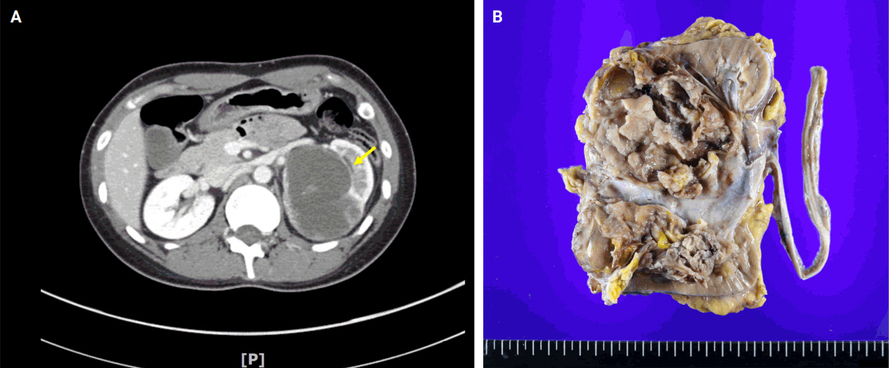
PDF - BCOR-rearranged sarcoma was classified by the World Health Organization in 2020 as a new subgroup of undifferentiated small round-cell sarcoma. It is known to occur very rarely in the kidney. This report presents the first case of a primary renal BCOR::CCNB3 sarcoma in a 22-year-old woman. An 8-cm cystic mass was identified in the left kidney by abdominal pelvic computed tomography. Histopathologic examination revealed the mass to be composed of small round to oval or spindle cells with fibrous septa and a delicate vascular network. A BCOR::CCNB3 fusion was detected by next-generation sequencing–based molecular testing. BCOR::CCNB3 sarcoma presents diagnostic difficulties, highlighting the importance of recognizing its histological features. Immunohistochemical markers are helpful for diagnosis, but genetic molecular testing is necessary for accurate diagnosis. These tumors have a very poor and aggressive prognosis, and an optimal therapeutic regimen has not yet been defined. Therefore, further studies are needed.
-
Citations
Citations to this article as recorded by
- Update on the management of BCOR::CCNB3 sarcoma
Jungo Imanishi, Kenji Sato, Yoshinao Kikuchi, Asako Yamamoto, Shiori Watabe, Taisuke Matsuyama, Chiaki Sato, Hiroshi Kobayashi, Hirotaka Kawano
Japanese Journal of Clinical Oncology.2025; 55(10): 1097. CrossRef
- Update on the management of BCOR::CCNB3 sarcoma
Review
- Neuroendocrine Tumors of the Female Reproductive Tract: A Literature Review
- Yi Kyeong Chun
- J Pathol Transl Med. 2015;49(6):450-461. Published online October 13, 2015
- DOI: https://doi.org/10.4132/jptm.2015.09.20
- 20,605 View
- 284 Download
- 28 Web of Science
- 29 Crossref
-
Abstract
PDF
- Neuroendocrine tumors of the female reproductive tract are a heterogeneous group of neoplasms that display various histologic findings and biologic behaviors. In this review, the classification and clinicopathologic characteristics of neuroendocrine tumors of the female reproductive tract are described. Differential diagnoses are discussed, especially for non-neuroendocrine tumors showing high-grade nuclei with neuroendocrine differentiation. This review also discusses recent advances in our pathogenetic understanding of these disorders.
-
Citations
Citations to this article as recorded by- Mixed neuroendocrine-non-neuroendocrine neoplasm (MiNEN) of the cervix in a 38-year-old female: a case report and review of literature
Josh Matthew B. Chen, Denise B. Andal, Benedict Jose P. Canora, Claire Anne Therese M. Hemedez
Human Pathology Reports.2026; 43: 300815. CrossRef - Neuroendocrine Neoplasms of the Gastrointestinal Tract: Morphology, WHO 2022 Grading, and Prognostic Perspectives
Hussein Qasim, Shaima' Dibian, Mohammad Abu Shugaer, Karis Khattab, Mudhaffer Touqan, Matteo Luigi Giuseppe Leoni , Giustino Varrassi
Cureus.2026;[Epub] CrossRef - A rare case report of primary ovarian carcinoid presenting with constipation
Xiaofeng Deng, Qian Huang, Bangfang Xie, Hailong Huang, Jianguo Chen
Frontiers in Oncology.2025;[Epub] CrossRef - Clinical, pathological characteristics, and therapeutic outcomes of primary ovarian carcinoid tumors: a case series of 15 cases
Xinyue Dai, Suidan Chen, Simeng Yang
World Journal of Surgical Oncology.2025;[Epub] CrossRef - Smart Red Blood Cell Carriers: A Nanotechnological Approach to Cancer Drug Delivery
Ioannis Tsamesidis, Georgios Dryllis, Sotirios P. Fortis, Andreas Sphicas, Vasiliki Konstantinidou, Maria Chatzidimitriou, Stella Mitka, Maria Trapali, Petros Skepastianos, Anastasios G. Kriebardis, Ilias Pessach
Current Issues in Molecular Biology.2025; 47(9): 711. CrossRef - Imaging of Gynecologic Neuroendocrine Tumors: A Case-Based Pictorial Essay
Ana Paula Bavaresco, Ulysses S. Torres, Mayara S. Cruz, Vitor V.C. Machado, Cynthia L.P. Borborema, Giovanna S. Torre, Jhonata Soares Da Silva, Tulio A. Kawai, Gustavo R.A. Focchi, Eduardo O. Pacheco, Aley Talans, Daniel Bekhor, Ana Paula C. Moura, Lucas
Seminars in Ultrasound, CT and MRI.2025;[Epub] CrossRef - Challenges in Diagnosis and Management of Ovarian Neuroendocrine Carcinoma: A Case of Aggressive Disease With Multimodal Treatment Approach
Javeria Haider, Humera Mahmood, Muhammad Faheem, Shaista Khurshid, Abdullah, Biruk Demisse Ayalew, Humza Saeed
Clinical Case Reports.2025;[Epub] CrossRef - Neuroendocrine Marker Expression in Primary Non-neuroendocrine Epithelial Tumors of the Ovary: A Study of 551 Cases
Michaela Kendall Bártů, Kristýna Němejcová, Romana Michálková, Quang Hiep Bui, Jana Drozenová, Pavel Fabian, Oluwole Fadare, Jitka Hausnerová, Jan Laco, Radoslav Matěj, Gábor Méhes, Adam Šafanda, Naveena Singh, Petr Škapa, Zuzana Špůrková, Simona Stolnicu
International Journal of Gynecological Pathology.2024; 43(2): 123. CrossRef - Diagnostic and therapeutic challenge of neuroendocrine endometrial carcinoma: a case report
Hariyono Winarto, David Calvin, Fitriyadi Kusuma, Kartiwa Hadi Nuryanto, Yuri Feharsal, Dewita Nilasari, Hartono Tjahjadi
The Pan African Medical Journal.2024;[Epub] CrossRef - Neuroendocrine carcinoma of ovary: Hitherto rare entity in primary ovarian tumors
Md A. Osama, Seema Rao, Punita Bhardwaj, Geeta Mediratta, Sunita Bhalla, Sonia Badwal
Indian Journal of Pathology and Microbiology.2023; 66(4): 855. CrossRef - Mixed neuroendocrine–non-neuroendocrine neoplasm with mucinous adenocarcinoma and amphicrine carcinoma components in the bile duct: an autopsy case
Toji Murabayashi, Yoshihide Kanno, Takashi Odaira, Shinsuke Koshita, Takahisa Ogawa, Hiroaki Kusunose, Toshitaka Sakai, Keisuke Yonamine, Kazuaki Miyamoto, Fumisato Kozakai, Kazuki Endo, Yutaka Noda, Takashi Sawai, Kei Ito
Clinical Journal of Gastroenterology.2023; 16(2): 310. CrossRef - Coexistence of Papillary Thyroid Carcinoma and Strumal Carcinoid Arising from Struma Ovarii in Pregnant Women: a Case Report and Review
Myungsoo Im, Doohwa Kim, Soree Ryang, Bo Hyun Kim
International Journal of Thyroidology.2023; 16(1): 134. CrossRef - Role of radiotherapy in the management of rare gynaecological cancers
R. Morcet-Delattre, S. Espenel, P. Tas, C. Chargari, A. Escande
Cancer/Radiothérapie.2023; 27(8): 778. CrossRef - Small cell carcinoma of the ovary, pulmonary type: A role for adjuvant radiotherapy after carboplatin and etoposide?
Anase S. Asom, Ricardo R. Lastra, Yasmin Hasan, Lori Weinberg, Gini F. Fleming, Katherine C. Kurnit
Gynecologic Oncology Reports.2022; 39: 100925. CrossRef - MicroRNA and Metabolic Profiling of a Primary Ovarian Neuroendocrine Carcinoma Pulmonary-Type Reveals a High Degree of Similarity with Small Cell Lung Cancer
Stefano Miglietta, Giulia Girolimetti, Lorena Marchio, Manuela Sollazzo, Noemi Laprovitera, Sara Coluccelli, Dario De Biase, Antonio De Leo, Donatella Santini, Ivana Kurelac, Luisa Iommarini, Anna Ghelli, Davide Campana, Manuela Ferracin, Anna Myriam Perr
Non-Coding RNA.2022; 8(5): 64. CrossRef - Neuroendocrine Carcinomas of the Uterine Cervix, Endometrium, and Ovary Show Higher Tendencies for Bone, Brain, and Liver Organotrophic Metastases
Hyung Kyu Park
Current Oncology.2022; 29(10): 7461. CrossRef - Uterine carcinoma admixed with neuroendocrine carcinoma
Maria Victoria Olinca, Anca Potecă, Elvira Brătilă, Mihai Mitran
Ginecologia.ro.2022; 4(38): 32. CrossRef - The puzzle of gynecologic neuroendocrine carcinomas: State of the art and future directions
Giuseppe Caruso, Carolina Maria Sassu, Federica Tomao, Violante Di Donato, Giorgia Perniola, Margherita Fischetti, Pierluigi Benedetti Panici, Innocenza Palaia
Critical Reviews in Oncology/Hematology.2021; 162: 103344. CrossRef - Pitfalls and challenges in managing neuroendocrine carcinoma of gynecological origin: A case series and brief review
Lauren E. Farmer, Rutmi U. Goradia, Nisha A. Lakhi
Clinical Case Reports.2021;[Epub] CrossRef - Primary mixed large cell neuroendocrine and high grade serous carcinoma of the endometrium
Liesel Elisabeth Hardy, Zia Chaudry, King Wan, Chloe Ayres
BMJ Case Reports.2020; 13(9): e234977. CrossRef - Neuroendocrine carcinoma of the endometrium: Disease course, treatment, and outcomes
Kathryn Schlechtweg, Ling Chen, Caryn M. St. Clair, Ana I. Tergas, Fady Khoury-Collado, June Y. Hou, Alexander Melamed, Alfred I. Neugut, Dawn L. Hershman, Jason D. Wright
Gynecologic Oncology.2019; 155(2): 254. CrossRef - Peritoneal Fluid Cytology of Disseminated Large Cell Neuroendocrine Carcinoma Combined with Endometrioid Adenocarcinoma of the Endometrium
Yong-Moon Lee, Min-Kyung Yeo, Song-Yi Choi, Kyung-Hee Kim, Kwang-Sun Suh
Journal of Pathology and Translational Medicine.2019; 53(6): 407. CrossRef - Pro-Gastrin Releasing Peptide: A New Serum Marker for Endometrioid Adenocarcinoma
Mine Kiseli, Gamze Sinem Caglar, Asli Yarci Gursoy, Tolga Tasci, Tuba Candar, Egemen Akincioglu, Emre Goksan Pabuccu, Nurettin Boran, Gokhan Tulunay, Haldun Umudum
Gynecologic and Obstetric Investigation.2018; 83(6): 540. CrossRef - Tumeur neuroendocrine à petite cellule de l’endomètre : prise en charge originale
E. Galmiche, N. Hudry, P. Sagot, P. Ginod, S. Douvier
Gynécologie Obstétrique Fertilité & Sénologie .2017; 45(6): 381. CrossRef - Twist on a classic: vitamin D and hypercalcaemia of malignancy
Juan C Osorio, Masha G Jones, Nina Schatz-Siemers, Stephanie J Tang
BMJ Case Reports.2017; 2017: bcr-2017-220819. CrossRef - Mixed Neuroendocrine-Nonneuroendocrine Neoplasms (MiNENs): Unifying the Concept of a Heterogeneous Group of Neoplasms
Stefano La Rosa, Fausto Sessa, Silvia Uccella
Endocrine Pathology.2016; 27(4): 284. CrossRef - Neuroendocrine tumours in rare sites: differences in nomenclature and diagnostics—a rare and ubiquitous histotype
Elia Guadagno, Gaetano De Rosa, Marialaura Del Basso De Caro
Journal of Clinical Pathology.2016; 69(7): 563. CrossRef - Primary ovarian neuroendocrine tumor arising in association with a mature cystic teratoma: A case report
Nicolas M. Orsi, Mini Menon
Gynecologic Oncology Reports.2016; 17: 83. CrossRef - Benign Endometrial Polyp and Primary Endometrial Small Cell Neuroendocrine Carcinoma Confined to the Polyp: A Rare Association
Pembe Oltulu, Ceyhan Uğurluoğlu, Ayşenur Uğur, Sıdıka Fındık, Lema Tavlı
Journal of Clinical and Experimental Investigations.2016;[Epub] CrossRef
- Mixed neuroendocrine-non-neuroendocrine neoplasm (MiNEN) of the cervix in a 38-year-old female: a case report and review of literature
Case Report
- Primary Malignant Melanoma of the Male Urethra.
- Seung Wook Lee, Eun Kyung Kim, Won Mi Lee, Jung Man Jo, Tag Keun Yoo, Jeong Yoon Kang
- Korean J Pathol. 2010;44(6):662-665.
- DOI: https://doi.org/10.4132/KoreanJPathol.2010.44.6.662
- 3,862 View
- 30 Download
-
Abstract
PDF
- Primary malignant melanoma occurring within the male urethra is very rare. Here we report a case of malignant melanoma of the urethra in a 74-year-old man. He presented with asymptomatic gross hematuria for 5 months. His glans penis and adjacent penile skin had become discolored black 10 years ago. Cystourethroscopy showed a smooth oval-shaped elevated mass in the fossa navicularis. There were no abnormal findings in the proximal urethra and urinary bladder. Computed tomography did not detect any inguinal lymph node enlargement or distant metastases. The patient underwent partial penectomy and ilioinguinal lymph node dissection. Grossly, the distal urethra revealed an ovoid pigmented nodule, that measured 1 x 0.5 cm. Microscopic findings showed a nodular malignant melanoma arising in the urethral mucosa with pagetoid spread to the epidermis of the glans penis. There were no recurrences over a period of 12 months after surgery without chemotherapy. This is the second case of a primary malignant melanoma of the male urethra in Korea.
Original Article
- Signet Ring Cell Variant of Invasive Lobular Carcinoma of Male Breast.
- Seung Sam Paik, Seok Hoon Jeon, Moon Hyang Park, Pa Jong Jung, Jung Dal Lee
- Korean J Pathol. 1997;31(2):179-181.
- 2,233 View
- 20 Download
-
Abstract
PDF
- Lobular carcinoma of the male breast is very rare, because of the absence of lobules in the normal male breast. Herein, a case of lobular carcinoma of the male breast with cellular features of signet ring cells is described. A 57-year-old man presented with a left breast mass. Histologic examination showed classic invasive lobular carcinoma with in situ component. Most infiltrating tumor cells had a prominent signet ring cell appearance. The patient was phenotypically male and had fathered children. There was no history of predisposing factors to breast lesion, such as hormone use or gynecomastia.
Case Reports
- B-cell Prolymphocytic Leukemia Involving Entire Female Genital Tract: A case report.
- Hee Jung Lee, Young Shin Kim, Yong Gu Kim, Kyung Ja Han, Kyo Young Lee, Chang Suk Kang, Sang In Shim, Jong Wook Lee, Woong Shick Ahn, Soo Pyung Aim, Seung Il Kim
- Korean J Pathol. 1999;33(2):145-148.
- 2,230 View
- 10 Download
-
Abstract
- Prolymphocytic leukemia is a chronic lymphoproliferative disorder, characterized by prominent splenomegaly, prolymphocytes accounting for more than 55% of circulating lymphocytes, no significant peripheral lymphadenopathy and short term survival with terminal fatal multi-organ failure. We report a case of B-cell prolymphocytic leukemia in a 57-year-old woman who presented with easy bruising and arthritis for 1 year and low abdominal pain for 2 months. Physical examination revealed gingival hypertrophy and mild splenomegaly. On peripheral blood smears the leukocytes were markedly increased in number due to leukemic cells that count about 62% of leukocytes. The bone marrow aspiration smear and biopsy revealed diffuse infiltration of medium to large prolymphocytes having moderate amount of basophilic cytoplasm, round to oval nuclei with coarse chromatin, and prominent nucleoli. Abdominal pain aggravated despite chemotherapy, and pelvic computed tomography (CT) revealed a huge lobular pelvic mass which had increased in size on the follow-up CT. Total hysterectomy with bilateral adnexectomy was performed. Microscopic findings included massive infiltration of prolymphocytic cells in the uterus, upper vaginal wall, bilateral ovaries, and bilateral mesosalpinges. On immunohistochemistry, the leukemic cells showed B cell gamma light chain phenotype.
- Cellular Angiofibroma of the Vulva: A Report of Three Cases.
- Hye Jeong Choi, Sung Nam Kim, Kyu Rae Kim
- Korean J Pathol. 2001;35(3):259-262.
- 2,267 View
- 28 Download
-
Abstract
PDF
- Cellular angiofibroma is a recently described, distinctive soft tissue tumor of the vulvovaginal region which is characterized by small, well-circumscribed tumors with fibroblastic differentiation. We report three cases of cellular angiofibroma of the vulva in middle-aged women. All three patients presented with painless swelling in the labium majora. The age of the three patients ranged from 43 to 56 years old (mean: 48 years old) and the size of the tumor ranged from 2 to 5 cm. The microscopic appearance was characterized by a cellular, well-circumscribed mass composed of uniform, bland, spindle stromal cells, numerous thick-walled, hyalinized vessels, and a scarce component of mature adipocytes. Immunohistochemical stains of the tumor cells show positivity for vimentin but negativity for smooth muscle actin, S-100 protein, desmin, factor VIII-related antigen and epithelial membrane antigen. The tumor should be differentiated from aggressive angiomyxoma and angiomyofibroblastoma because of its different clinicopathologic features, cells of origin and immunohistochemical findings.
- Adenoid Cystic Carcinoma of the Male Breast: A case report.
- Mi Kyung Lee, In Chul Hong, Woo Ick Yang, Sang Ho Cho
- Korean J Pathol. 1999;33(5):389-392.
- 2,198 View
- 21 Download
-
Abstract
PDF
- A 65 year-old male patient presented with a large palpable mass beneath the areola of the right breast for 7 years. The resected breast tissue was almost totally replaced by a round large solid mass (9 6 cm) with a pink-gray to yellow firm, partly nodular cut surface. Microscopically, the tumor revealed the diagnostic biphasic cellular pattern of adenoid cystic carcinoma, which consisted of both cribriform pattern of myoepithelial cells and tubular pattern of epithelial cells. On immunohistochemistry, the tumor revealed immunoreactivities for alpha-smooth muscle actin and S-100 protein in the myoepithelial cells and for AE1/AE3 in the epithelial cells. Mitoses were scarce. Multifocal lymphatic permeation and foci of perineural invasion were also found. Underlying resection margins and overlying skin were invaded by the tumor. We diagnosed this tumor as grade II adenoid cystic carcinoma according to the system utilized for the salivary gland tumors.
- Fine Needle Aspiration Cytology of Gynecomastia: Review of 14 Cases.
- Hye Kyoung Yoon, Seol Mi Park, Jong Eun Joo
- J Pathol Transl Med. 1994;5(2):143-147.
- 2,694 View
- 81 Download
-
Abstract
PDF
- Fine needle aspiration cytologic findings in 14 cases of gynecomastia are described. General cytomorphologic features resemble those of fibrocystic disease in women than those of fibroadenoma. Among the cytologic parameters, three-dimensional structure of epithelial cell clusters, presence of micronucleoli and irregularities of nuclear size and shape are suggestive of epithelial proliferative activity. In addition, 4 cases are proliferative breast disease without atypia and 10 cases are nonproliferative breast disease depending on cytologic criteria grading system.
- Myofibroblastoma of the Male Breast: Report of a case.
- Ji Eun Kim, Yeon Lim Suh, Howe Jung Ree
- Korean J Pathol. 1996;30(7):623-629.
- 2,172 View
- 10 Download
-
Abstract
- A case of myofibroblastoma of the breast in a 55-year-old man is described. Myofibroblastoma is a relatively recently recognized benign stromal tumor, and predominantly occurs in middle aged men. The pateint presented with a nontender firm mass in his right breast. Fine needle aspiration biopsy revealed bland looking stromal cell clusters without epithelial cells. Simple excision was done and the patient discharged uneventfully. The mass was well demarcated, lobulated and sligtly myxoid. Microscopically bipolar elongated spindle cell fascicles with interspersing broad collagen bands are so characteristic. Ultrastructurally the tumor cell show features of fibroblast as well as smooth muscle cell.
Original Article
- Carcinosarcoma (Malignant M llerian Mixed Tumor) of the Female Genital Tract: A clinical and pathologic study of ten carcinosarcomas.
- Sung Ran Hong, Yee Jeong Kim, Hy Sook Kim, Jae Uk Shim, Chong Taik Park
- Korean J Pathol. 1998;32(5):362-369.
- 2,258 View
- 10 Download
-
Abstract
- Carcinosarcomas of the female genital tract have generally been regarded as a type of sarcoma. Recent studies, however, suggest the tumor may be more closely related to carcinoma and may represent metaplastic carcinoma in histogenesis. We analyzed clinicopathologic and immunohistochemical features of 10 carcinosarcomas to evaluate the relative importance of the carcinomatous and sarcomatous components in metastasis and recurrence. The primary tumor originated in the uterine body in seven cases, the uterine cervix in two and the ovary in one. Patient,s ages ranged from 54 to 71 years (mean, 64). The most common symptom of the uterine mass was vaginal bleeding. The median survival time was 21 months following diagnosis in five cases. Surgico-pathologic FIGO stages of five patients who received an operation were stage III and IV, but clinical FIGO stage of three patients (60%) among them were I. Lymphovascular invasions were identified in seven areas; five vascular invasion lesions showed the carcinomatous component alone, one the sarcomatous component alone, and remained one admixture of both components. Metastatic and recurrent lesions to the paraaortic lymph node, ovary, pelvic wall, or vaginal vault showed characteristically carcinomatous component only. Immunohistochemically, positive reactions for cytokeratin and epithelial membrane antigen were noted in the sarcomatous component of five cases. Vimentin positivity was detected in carcinomatous component of three cases. We conclude that the dominant element in carcinosarcomas of the female genital tract is the carcinomatous component. The survival rate of carcinosarcoma is extremely poor. The surgico-pathologic stage is better indicator of survival than the clinical stage. Immunohistochemical findings suggest that carcinosarcoma may represent a metaplastic carcinoma in histogenesis.
Case Reports
- Female Adnexal Tumor of probable Wolffian origin: A case report.
- Yee Jeong Kim, Sung Ran Hong, Hy Sook Kim, Hyon U Lee
- Korean J Pathol. 1994;28(4):427-429.
- 2,131 View
- 15 Download
-
Abstract
PDF
- We report a case of right broad ligament tumor with features of female adnexal tumor of probable wolffian origin. A 40-year-old woman presented with dysfunctional uterine bleeding. Ultrasonography revealed 1 10cm sized right parovarian solid mass. On microscopic examination, the tumor showed mixed pattern of tightly packed tubular structures and diffuse spindle cell proliferation. Immunohistochemical study demonstrated cytokeratin-and vimentin-positivity and carcino-embryonic antigen-negativity. The ultrastructural study showed prominent tubular structures, continuous basal lamina, definite junctional complex but no secretory granules or glycogen particles, favoring wolffian origin.
- Liposarcoma of the Breast in Male: Report of a case.
- Jong Boum Choi, Sung Churl Lim, Keun Hong Kee, Ho Jong Jeon, Chae Hong Suh
- Korean J Pathol. 1992;26(3):293-297.
- 2,260 View
- 34 Download
-
Abstract
PDF
- Liposarcoma of the male breast is rare. In English literature, nearly 90 tumors of this type can be found, but the majority of citation is female without detail descriptions of gross or microscopic features. Recently, we experienced a case with pleomorphic liposarcoma of the breast. This 62-year-old male patient presented with tumorous mass of right breast for 3 months ago. He underwent radical mastectomy under the impression of breast cancer. Received specimen was a breast and attached nodular bulging mass. The serial section reveal a relatively defined nodular mass consists of yellowish and glistened bulging tumorous lesion, measuring 5x4.5x5cm. Microscopically, there were multiple pleomorphic giant cells composed of plump esoinophilic or microvesiculated cytoplasm and bizarre nuclei with prominent nucleoli. These cells were positive staining for oil-red O. Ultrastructurally, variable sized numerous fatty vacuoles in the cytoplasm were seen.
- Primary Osteosarcoma of the Breast: A case report.
- Dong Chool Kim, Yun Kyung Lee, Ho Jong Jeon, Sung Chul Lim
- Korean J Pathol. 2000;34(9):677-679.
- 2,160 View
- 15 Download
-
Abstract
PDF
- We report a case of primary osteosarcoma of the breast which is rare and exhibits poor prognosis. A 52 years-old-woman was admitted with rapidly growing right mammary mass. A huge lobulated dense mass with speckled calcifications, suggesting malignancy, was observed on mammography. She underwent a radical mastectomy. Grossly, the mass measured 16 14 6 cm and showed grayish white hard lobulated tissue with focal hemorrhage and necrosis. Light microscopically, the tumor was confirmed as an osteosarcoma devoid of any epithelial components. In postoperative whole body bone scan, there was no evidence of the other malignancy. To the best of our knowledge, the present case is the first report of primary osteosarcoma of the breast in Korea.
 First
First Prev
Prev


