 E-submission
Search
- Page Path
- HOME > Search
Original Article
- Blocking Toll-like receptor 9 attenuates bleomycin-induced pulmonary injury
- Badr Alzahrani, Mohamed M. S. Gaballa, Ahmed A. Tantawy, Maha A. Moussa, Salma A. Shoulah, Said M. Elshafae
- J Pathol Transl Med. 2022;56(2):81-91. Published online March 2, 2022
- DOI: https://doi.org/10.4132/jptm.2021.12.27

- 8,635 View
- 150 Download
- 12 Web of Science
- 12 Crossref
-
 Abstract
 PDF
PDF - Background
Acute respiratory distress syndrome (ARDS) is one of the most common complications in coronavirus disease 2019 patients suffering from acute lung injury (ALI). In ARDS, marked distortion of pulmonary architecture has been reported. The pulmonary lesions in ARDS include hemodynamic derangements (such as alveolar edema and hemorrhage), vascular and bronchiolar damage, interstitial inflammatory cellular aggregations, and eventually fibrosis. Bleomycin induces ARDS-representative pulmonary damage in mice and rats; therefore, we used bleomycin model mice in our study. Recently, Toll-like receptor 9 (TLR9) was implicated in the development of ARDS and ALI.
Methods
In this study, we evaluated the efficiency of a TLR9 blocker (ODN2088) on bleomycin-induced pulmonary damage. We measured the apoptosis rate, inflammatory reaction, and fibroplasia in bleomycin- and bleomycin + ODN2088-treated mice.
Results
Our results showed a significant amelioration in bleomycin-induced damage to pulmonary architecture following ODN2088 treatment. A marked decrease in pulmonary epithelial and endothelial apoptosis rate as measured by cleaved caspase-3 expression, inflammatory reaction as indicated by tumor necrosis factor α expression, and pulmonary fibrosis as demonstrated by Van Gieson staining and α-smooth muscle actin immunohistochemistry were observed following ODN2088 treatment.
Conclusions
All these findings indicate that blocking downstream TLR9 signaling could be beneficial in prevention or mitigation of ARDS through hemodynamic derangements, inflammation, apoptosis, and fibrosis. -
Citations
Citations to this article as recorded by
- Nano-sized DNase scavenges cell-free DNA for acute lung injury treatment
Ruijie Chen, Yitianhe Xu, Zhanzheng Ye, Yixuan Zhu, Mengxue Zhang, Fangfang Lv, Yunzhi Wang, Xinyu Di, Yinhao Lin, Shengnan Song, Zihao Huang, Shize Li, Zhinan He, Hailin Zhang, Longfa Kou
Journal of Controlled Release.2026; 394: 114846. CrossRef - A novel mouse model of myositis-associated interstitial lung disease was established by using TLR9 agonist combined with muscle homogenate
Ling Bai, Jiarui Zhu, Wenlan Ma, Peipei Zhao, Feifei Li, Cen Zhang, Sigong Zhang
Clinical and Experimental Immunology.2025;[Epub] CrossRef - Toll-like Receptor 9 Inhibition Mitigates Fibroproliferative Responses in Translational Models of Pulmonary Fibrosis
Glenda Trujillo, Alicia Regueiro-Ren, Chunjian Liu, Buqu Hu, Ying Sun, Farida Ahangari, Vitoria Fiorini, Genta Ishikawa, Karam Al Jumaily, Johad Khoury, John McGovern, Chris J. Lee, Xue Yan Peng, Taylor Pivarnik, Huanxing Sun, Anjali Walia, Samuel Woo, Sh
American Journal of Respiratory and Critical Care Medicine.2025; 211(1): 91. CrossRef - CD103+ dendritic cell–fibroblast crosstalk via TLR9, TDO2, and AHR signaling drives lung fibrogenesis
Hannah Carter, Rita Medina Costa, Taylor S. Adams, Talon M. Gilchrist, Claire E. Emch, Monica Bame, Justin M. Oldham, Steven K. Huang, Angela L. Linderholm, Imre Noth, Naftali Kaminski, Bethany B. Moore, Stephen J. Gurczynski
JCI Insight.2025;[Epub] CrossRef - The Mitigated Effect of the Combination of Metformin and Stearic Acid to Ameliorate Bleomycin‐Induced Pulmonary Fibrosis in Rats via Inhibiting Gal‐3/Smad3/α‐SMA and TNF‐α/NF‐κβ Signaling Pathways
Maha M. Salem, Nermin S. Youssef, Mai El Keiy, Abeer A. Khamis
Journal of Biochemical and Molecular Toxicology.2025;[Epub] CrossRef - Unraveling the Interactive Role of Mitochondrial Dysfunction in Promoting Macrophage Polarization in Pulmonary Fibrosis
Jiajia Zou, Junling Jian, Dehua Ge, Bin Zhou, Jiaxiang Zhang
Journal of Biochemical and Molecular Toxicology.2025;[Epub] CrossRef - Mechanisms underlying dose-limiting toxicities of conventional chemotherapeutic agents
Mohammad Amin Manavi, Mohammad Hosein Fathian Nasab, Razieh Mohammad Jafari, Ahmad Reza Dehpour
Journal of Chemotherapy.2024; 36(8): 623. CrossRef - Innate Immune Response-Mediated Inflammation in Viral Pneumonia
Weiwei Ni, Xin Wei, Rui Wu
Journal of Pediatric Infectious Diseases.2024; 19(03): 140. CrossRef - Combination of losartan with pirfenidone: a protective anti-fibrotic against pulmonary fibrosis induced by bleomycin in rats
Arian Amirkhosravi, Maryamossadat Mirtajaddini Goki, Mahmoud Reza Heidari, Somayyeh Karami-Mohajeri, Maryam Iranpour, Maryam Torshabi, Mitra Mehrabani, Ali Mandegary, Mehrnaz Mehrabani
Scientific Reports.2024;[Epub] CrossRef - Suppression of miR-17 Alleviates Acute Respiratory Distress-associated Lung Fibrosis by Regulating Mfn2
Mei-xia Xu, Tao Xu, Ning An
Current Medical Science.2024; 44(5): 964. CrossRef - Study of Recombinant Interleukin-1 Receptor Antagonist Compositions Biological Activity After Injection and Inhalation in Mouse Model of Pulmonary Inflammation
Alexander M. Ischenko, Ksenia A. Nekrasova, Denis S. Laptev, Dmitry V. Bobkov, Alexander A. Kolobov, Andrey S. Simbirtsev
Cytokines and inflammation.2024; 21(3): 153. CrossRef - TLR9: A friend or a foe
Mona M. Saber, Nada Monir, Azza S. Awad, Marwa E. Elsherbiny, Hala F. Zaki
Life Sciences.2022; 307: 120874. CrossRef
- Nano-sized DNase scavenges cell-free DNA for acute lung injury treatment
Case Study
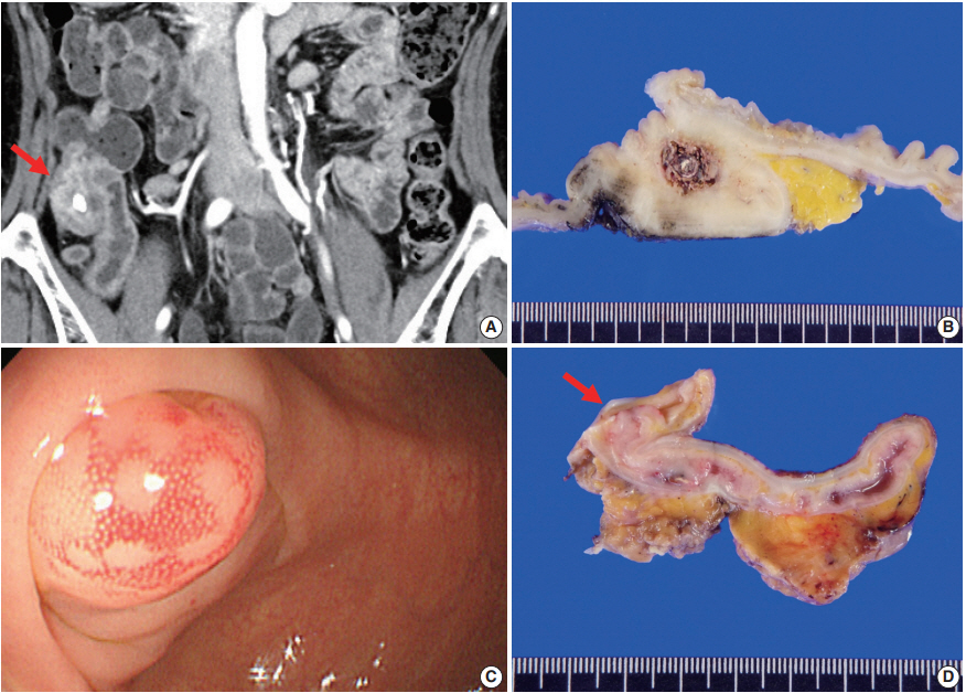
- Appendiceal actinomycosis mimicking appendiceal tumor, appendicitis or inflammatory bowel disease
- You-Na Sung, Jihun Kim
- J Pathol Transl Med. 2021;55(5):349-354. Published online June 26, 2020
- DOI: https://doi.org/10.4132/jptm.2020.05.17
- 9,276 View
- 171 Download
- 5 Web of Science
- 4 Crossref
-
Abstract
PDF
- Appendiceal actinomycosis is very rare and its diagnosis is often difficult even in surgically resected specimens. Here we report two cases of appendiceal actinomycosis confirmed by pathologic examination of surgically resected specimens. Characteristic histologic features included transmural chronic inflammation with Crohn-like lymphoid aggregates and polypoid mucosal protrusion into cecal lumen through fibrous expansion of the submucosa. Chronic active inflammation involved the mucosa of the appendix and cecum around the appendiceal orifice. Crohn’s disease with predominant cecal involvement and inflammatory pseudotumor were considered as differential diagnoses. Careful examination revealed a few actinomycotic colonies in the mucosa, confirming the diagnosis. A high index of suspicion with awareness of the characteristic histologic features might prompt careful inspection for the actinomycotic colonies, leading to the appropriate diagnosis of this rare disease.
-
Citations
Citations to this article as recorded by- Persistent intra-abdominal abscess with intestinal obstruction following seven failed drainage procedures over 3.5 years: a case report
Ayman Shemes, Salma Samra, Ahmed Mohamed, Amr A. Elgharib
BMC Surgery.2025;[Epub] CrossRef - Appendicular actinomycosis: The first reported case of an uncommon finding of a common ailment from Nepal
Sujan Bohara, Manoj Khadka, Pawan Singh Bhat, Prajwal Syangtang, Badal Karki, Bhagawan Shrestha, Shoshan Arja Acharya, Khusbhu Khetan, Jyoti Rayamajhi, Sushil Bahadur Rawal
Clinical Case Reports.2023;[Epub] CrossRef - Abdominopelvic actinomycosis: An unexpected diagnosis in an elderly female with a destructive-appearing soft tissue mass
Elise Hyser, Drashti Antala, Harvey Friedman, Jonathan Stake
IDCases.2022; 28: e01479. CrossRef - Diagnosing granulomatous disease during appendectomy
Atilla Şenaylı
Clinical Case Reports.2021;[Epub] CrossRef
- Persistent intra-abdominal abscess with intestinal obstruction following seven failed drainage procedures over 3.5 years: a case report
Original Article
- Does Polymerase Chain Reaction of Tissue Specimens Aid in the Diagnosis of Tuberculosis?
- Yoo Jin Lee, Seojin Kim, Youngjin Kang, Jiyoon Jung, Eunjung Lee, Joo-Young Kim, Jeong Hyeon Lee, Youngseok Lee, Yang-seok Chae, Chul Hwan Kim
- J Pathol Transl Med. 2016;50(6):451-458. Published online October 10, 2016
- DOI: https://doi.org/10.4132/jptm.2016.08.04
- 14,962 View
- 260 Download
- 7 Web of Science
- 9 Crossref
-
Abstract
PDF
- Background
Mycobacterial culture is the gold standard test for diagnosing tuberculosis (TB), but it is time-consuming. Polymerase chain reaction (PCR) is a highly sensitive and specific method that can reduce the time required for diagnosis. The diagnostic efficacy of PCR differs, so this study determined the actual sensitivity of TB-PCR in tissue specimens.
Methods
We retrospectively reviewed 574 cases. The results of the nested PCR of the IS6110 gene, mycobacterial culture, TB-specific antigen-induced interferon-γ release assay (IGRA), acid-fast bacilli (AFB) staining, and histological findings were evaluated.
Results
The positivity rates were 17.6% for PCR, 3.3% for the AFB stain, 22.2% for mycobacterial culture, and 55.4% for IGRA. PCR had a low sensitivity (51.1%) and a high specificity (86.3%) based on the culture results of other studies. The sensitivity was higher (65.5%) in cases with necrotizing granuloma but showed the highest sensitivity (66.7%) in those with necrosis only. The concordance rate between the methods indicated that PCR was the best method compared to mycobacterial culture, and the concordance rate increased for the methods using positive result for PCR or histologic features.
Conclusions
PCR of tissue specimens is a good alternative to detect tuberculosis, but it may not be as sensitive as previously suggested. Its reliability may also be influenced by some histological features. Our data showed a higher sensitivity when specimens contained necrosis, which indicated that only specimens with necrosis should be used for PCR to detect tuberculosis. -
Citations
Citations to this article as recorded by-
The Need for Persistence in the Diagnosis of Mycobacterium Tuberculosis Mono-arthritis: A Unique Case Presentation
T. Bekoulis, P. Christodoulou, K. Dogramatzis, E. Markopoulou, Emmanouel Antonogiannakis, E. Kokkinakis, Alexandros P. Apostolopoulos, A. Manimanaki
Journal of Long-Term Effects of Medical Implants.2024; 34(1): 35. CrossRef - A Case Report on Scrofuloderma: A Cutaneous Manifestation of Tuberculosis
Soham R Meghe, Adarshlata Singh, Drishti M Bhatt, Shreya N Gupta, Varun Hanumanthaiah, Shree Ramya Talasila
Cureus.2024;[Epub] CrossRef - An overview of infectious disease laboratory methods: an update for the histopathologist
Daniel R. Stevenson
Diagnostic Histopathology.2024; 30(10): 534. CrossRef - Diagnostic Utility of Biplex/Multiplex Polymerase Chain Reaction in Infectious Granulomatous Dermatitis in North Indian Population
Mayur Parkhi, Mukin Kumar S, Dipankar De, Rakesh Yadav, Sunil Sethi, Bishan Dass Radotra, Uma Nahar Saikia
The American Journal of Dermatopathology.2021; 43(8): 567. CrossRef - Reduction of turnaround time for non-tuberculous mycobacteria detection in heater–cooler units by propidium monoazide–real-time polymerase chain reaction
S. Ditommaso, M. Giacomuzzi, G. Memoli, R. Cavallo, A. Curtoni, M. Avolio, C. Silvestre, C.M. Zotti
Journal of Hospital Infection.2020; 104(3): 365. CrossRef - Ergonomic Diagnostic Tool based on Chip Mini RT-PCR for Diagnosis of Pulmonary and Extra Pulmonary Tuberculosis
V Mangayarkarasi, Sneka P, Sujith R, Jayaprakash Jayaprakash
Journal of Pure and Applied Microbiology.2019; 13(2): 1185. CrossRef - Cutaneous Tuberculosis: Clinicopathologic Arrays and Diagnostic Challenges
Priyatam Khadka, Soniya Koirala, Januka Thapaliya
Dermatology Research and Practice.2018; 2018: 1. CrossRef - Utility of Real-Time Quantitative Polymerase Chain Reaction in DetectingMycobacterium tuberculosis
Zhongquan Lv, Mingxin Zhang, Hui Zhang, Xinxin Lu
BioMed Research International.2017; 2017: 1. CrossRef - Primary Appendicular Tuberculosis
Vipul D Yagnik
Gastroenterology & Hepatology: Open Access.2017;[Epub] CrossRef
-
The Need for Persistence in the Diagnosis of Mycobacterium Tuberculosis Mono-arthritis: A Unique Case Presentation
Case Study
- Oncocytic Renal Cell Carcinoma with Tubulopapillary Growth Having a Fat Component
- Na Rae Kim, Hyun Yee Cho
- J Pathol Transl Med. 2015;49(5):413-417. Published online July 30, 2015
- DOI: https://doi.org/10.4132/jptm.2015.07.01
- 12,092 View
- 83 Download
- 1 Web of Science
- 1 Crossref
-
Abstract
PDF
- We report a rare case of oncocytic renal cell carcinoma (RCC) with tubulopapillary growth in the background of tuberculous end-stage kidney disease. Histology of the renal mass consisted of oncocytic cells forming solid, thin tubules and rare papillae. The tumor had abundant eosinophilic oncocytic cells containing occasional cytoplasmic Mallory body–like hyaline globules and a tiny focus of clear cells with intervening mature fat. Both the oncocytic cells and clear cells were immunoreactive for a-methylacyl-CoA racemase, vimentin, pancytokeratin, and CD10, and negative for transcription factor E3, CD15, human melanoma black 45, and c-kit. Mallory body–like hyaline globules were positive for CAM 5.2 and periodic acid–Schiff with or without diastase. Ultrastructurally, the tumor cells had abundant cytoplasmic mitochondria. The present case is a rare case of oncocytic RCC with tubulopapillary growth pattern. The case is unique in that the tumor was mixed with fat component, which is not common in RCC and thus can lead to misdiagnosis.
-
Citations
Citations to this article as recorded by- Oncocytic papillary renal cell carcinoma (OPRCC): 2 case report and literature review
Yanchen Wang, Lihui Guan, Yaming Liu, Yuxuan Liu, Xiaoyan Guo, Yaofei Sun
Frontiers in Oncology.2025;[Epub] CrossRef
- Oncocytic papillary renal cell carcinoma (OPRCC): 2 case report and literature review
Original Article
- Prognostic Significance of Amplification of the c-MYC Gene in Surgically Treated Stage IB-IIB Cervical Cancer.
- Tae Jung Kim, Ahwon Lee, Sung Jong Lee, Won Chul Lee, Yeong Jin Choi, Kyo Young Lee, Chang Suk Kang
- Korean J Pathol. 2011;45(6):596-603.
- DOI: https://doi.org/10.4132/KoreanJPathol.2011.45.6.596
- 5,271 View
- 42 Download
- 1 Crossref
-
Abstract
PDF
- BACKGROUND
Mutations of c-MYC have been described in cervical cancer. However, association between c-MYC gene status and its prognostic significance have not been clarified.
METHODS
Tissue microarray sections from 144 patients with stage IB-IIB cervical cancer treated by radical hysterectomy were analyzed by fluorescence in situ hybridization using a region-specific probe for c-MYC and a centromere-specific probe for chromosome 8.
RESULTS
Seventy five percent (108/144) of c-MYC gain and 6.9% (10/144) of c-MYC gene amplification were observed. c-MYC gene alteration was more frequently observed in squamous cell carcinoma than adenocarcinoma or adenosquamous carcinoma and were associated with low Ki67 labeling index (p=0.013). c-MYC amplification was not associated with clinicopathologic parameters except absence of bcl2 expression (p=0.048). Survival analysis revealed that patients with c-MYC amplification were significantly associated with higher risk of disease recurrence (p=0.007) and cancer related death (p=0.020). However, c-MYC gain was not associated with unfavorable outcome. Multivariate analysis proved c-MYC amplification as independent prognostic factors of shorter disease free survival and cancer-related death (p=0.028 and p=0.025, respectively).
CONCLUSIONS
c-MYC amplification, not gain, is an independent prognostic marker for shorter disease free and cancer specific survival in cervical cancer treated by radical hysterectomy. -
Citations
Citations to this article as recorded by- A Rare Case of Cutaneous Plasmacytosis in a Korean Male
Corey Georgesen, Meenal Kheterpal, Melissa Pulitzer
Case Reports in Pathology.2017; 2017: 1. CrossRef
- A Rare Case of Cutaneous Plasmacytosis in a Korean Male
Case Report
- Mycophenolate Mofetil-Related Colitis: A Case Report.
- Kyungeun Kim, Jerad M Gardner, Mary Schwartz, Matthew L Tompson, Jae Y Ro
- Korean J Pathol. 2010;44(3):333-337.
- DOI: https://doi.org/10.4132/KoreanJPathol.2010.44.3.333
- 6,278 View
- 70 Download
- 4 Crossref
-
Abstract
PDF
- Mycophenolate mofetil (MMF)-related colitis is one of the common causes of afebrile diarrhea in transplant patients. Pathologic diagnosis of MMF-related colitis is difficult because microscopic findings of MMF effects resemble those of graft-versus-host disease, inflammatory bowel disease and ischemic colitis. However, if diagnosed, MMF-induced colitis can be markedly improved by discontinuing the drug. A 70-year-old man having a history of transplantation presented with a one month history of afebrile diarrhea. Colonoscopy revealed patchy erosions. The colonoscopic biopsy specimen showed not only crypt disarray with degenerated crypts and scattered epithelial cell apoptosis, but also stromal inflammatory cell infiltration. A review of his medical records showed that he had been taking immunosuppressive drugs including MMF since his heart transplantation 6 years prior. The histologic findings of colonic mucosa were consistent with MMF-related colitis. After discontinuing MMF, the diarrhea quickly resolved and has not recurred for 10 months.
-
Citations
Citations to this article as recorded by- Colitis eosinofílica inducida por micofenolato: reporte de caso
Mario Alberto Caviedes-Cleves, Ariel Antonio Arteta-Cueto
Iatreia.2023;[Epub] CrossRef - Mycophenolate Mofetil-Induced Colonic Injury Manifesting Endoscopically As Ischemic Colitis
Clive J Miranda, Murad H Ali, Muddasir Ayaz, Raheel M Khan, Mayada Ismail
Cureus.2023;[Epub] CrossRef - Transformation of the Amino Acid Pool in the Rat Brain under Conditions of Experimental Immunodeficiency
N. I. Filina, M. N. Kurbat
Neurochemical Journal.2022; 16(2): 174. CrossRef - A Case Series of De Novo Inflammatory Bowel Disease After Kidney Transplantation
P. Azevedo, C. Freitas, P. Aguiar, H. Silva, T. Santos, P. Farrajota, M. Almeida, S. Pedroso, L.S. Martins, L. Dias, R. Vizcaíno, A. Castro Henriques, A. Cabrita
Transplantation Proceedings.2013; 45(3): 1084. CrossRef
- Colitis eosinofílica inducida por micofenolato: reporte de caso
Original Articles
- Comparison of Various Detection Methods of Mycobacterium Species in Formalin-Fixed Paraffin-Embedded Tissue with Chronic Granulomatous Inflammation.
- Hyun Seung Lee, Hyoungnam Lee, Soyoung Im, Yun Su Lee, Kyo Young Lee, Yeong Jin Choi
- Korean J Pathol. 2010;44(3):259-266.
- DOI: https://doi.org/10.4132/KoreanJPathol.2010.44.3.259
- 5,026 View
- 52 Download
- 2 Crossref
-
Abstract
PDF
- BACKGROUND
To determine the most effective method for detecting mycobacteria in formalin- fixed paraffin-embedded (FFPE) tissue, we compared the results of Ziehl-Neelsen stain (ZNS) and mycobacterial culture with those of polymerase chain reaction (PCR) and real-time quantitative PCR (RQ-PCR).
METHODS
We analyzed 54 cases diagnosed as chronic granulomatous inflammation. In all cases, ZNS and nested PCR using three different primers, IS6110, Mpb64 and IS6110/Rpobeta were done. RQ-PCR with the IS6110/Rpobeta primer was done in 51 cases.
RESULTS
Mycobacteria were identified by ZNS in 15/54 (27.8%) cases. RQ-PCR had the highest sensitivity (80.0%) compared to PCR with IS6110 (73.3%), Mpb64 (60.0%) and IS6110/Rpobeta (73.3%). Specificity was higher in all PCR experiments (79.5-82.1%) than in RQ-PCR (69.4%) experiments. The false negative rate was lowest for RQ-PCR (20.0%) than for PCR with IS6110 (26.7%), Mpb64 (40.0%) and IS6110/Rpobeta (26.7%). The false positive rate was highest for RQ-PCR (30.6%) compared to PCR with IS6110 (20.5%), Mpb64 (17.9%) and IS6110/Rpobeta (20.5%).
CONCLUSIONS
RQ-PCR had the highest sensitivity, and the lowest false negative rate, but it also had a higher false positive rate than PCR for detection of mycobacteria in FFPE tissues. -
Citations
Citations to this article as recorded by- Clinical Usefulness of PCR for Differential Diagnosis of Tuberculosis and Nontuberculous Mycobacterial Infection in Paraffin-Embedded Lung Tissues
Yo Na Kim, Kyoung Min Kim, Ha Na Choi, Ju Hyung Lee, Ho Sung Park, Kyu Yun Jang, Woo Sung Moon, Myoung Jae Kang, Dong Geun Lee, Myoung Ja Chung
The Journal of Molecular Diagnostics.2015; 17(5): 597. CrossRef - Usefulness of PCR to Mycobacterium Tuberculous and Nontuberculous Mycobacteria from Paraffin-embedded Tissues
Yeon-Il Choi, Hye-Young Kim
Korean Journal of Clinical Laboratory Science.2014; 46(2): 47. CrossRef
- Clinical Usefulness of PCR for Differential Diagnosis of Tuberculosis and Nontuberculous Mycobacterial Infection in Paraffin-Embedded Lung Tissues
- Pathologic Findings of Surgically Resected Nontuberculous Mycobacterial Pulmonary Infection.
- Hye Jong Song, Jung Suk An, Joungho Han, Won Jung Koh, Hong Kwang Kim, Yong Soo Choi
- Korean J Pathol. 2010;44(1):56-62.
- DOI: https://doi.org/10.4132/KoreanJPathol.2010.44.1.56
- 3,601 View
- 40 Download
- 1 Crossref
-
Abstract
PDF
- BACKGROUND
Surgical resection of the involved lung for nontuberculous mycobacteria (NTM) has become an important curative therapy. However, there is limited information on the histopathological features of NTM pulmonary disease in Korean patients with NTM infection.
METHODS
We evaluated 51 specimens from 49 patients who were treated at our referral center from 2002 to 2009.
RESULTS
Almost all the cases showed bronchiectasis with lymphocytic infiltration. Variable features of granulomatous inflammation were found; well-defined granulomas in the parenchyma (68.6%), pneumonia-like granulomatous lesions (49.0%) and granulomatous inflammation in the bronchial wall (41.2%) were identified. The microscopic findings of cavitary lesions (37.3%) showed that these lesions were composed of granulomas and necrosis.
CONCLUSIONS
The differentiation of tuberculosis from NTM could not be accurately made based solely on the histological features. However, the airway centered tendency of NTM reflected an airborn etiology, and this could be correlated with the classification according to the radiological findings. In addition, coexisting constitutional lung diseases, and especially bronchiectasis, were suspected to be predisposing conditions for NTM organisms to colonize and progress to true NTM pulmonary disease. -
Citations
Citations to this article as recorded by- Post-bronchoscopy sputum culture improves detection of nontuberculous mycobacterial pulmonary disease: A retrospective Study
Kohei Yamamoto, Tatsuya Imabayashi, Toshiyuki Tanaka, Kazuki Jinno, Shunya Tanaka, Sayaka Uda, Tatsuya Yuba, Chieko Takumi
Respiratory Investigation.2026; 64(2): 101379. CrossRef
- Post-bronchoscopy sputum culture improves detection of nontuberculous mycobacterial pulmonary disease: A retrospective Study
- Experimental Study of the Progressive Glomerulosclerosis Induced by Long-term Administration of Puromycin Aminonucleoside in Rats.
- Mi Kyung Kim, Hyun Soon Lee
- Korean J Pathol. 1993;27(1):1-10.
- 2,068 View
- 17 Download
-
Abstract
PDF
- Pathogenetic mechanisms of progressive glomerulosclerosis are not clear. We studied the long-term(10 weeks) effects of puromycin aminonucleoside(PAN) in Sprague-Dawley rats with or without uninephrectomy(UN). Compared to rats with PAN injections only, rats with uninephrectomy and PAN injections showed significantly higher serum levels of urea nitrogen(153 +/- 155 mg/dl vs. 16 +/- 4 mg/dl, p<0.01), ceatinine(2.96 +/- 1.21 mg/dl vs. 0.92 +/- 0.36 mg/dl, p<0.01), cholesterol(466 +/- 125 mg/dl vs. 94 +/- 27 mg/dl, p<0.01), and triglyceride(337 +/- 237 mg/dl vs. 111 +/- 36 mg/dl, p<0.05) as well as increased amounts of proteinuria(428 +/- 90 mg/day vs. 136 +/- 130 mg/day, p<0.01). Lesions of focal segmental glomerulosclerosis(FSGS) were more frequently observed in rats with UN and PAN injections than rats with PAN infections only(39.5 +/- 17.2% vs. 4.3 +/- 4.7%, p<0.01). Ultrastructural examination of the glomeruli from rats with UN and PAN injections revealed severe epithelial cell changes including foot process effacement, vaculoar change or pseudocyst formation and focal detachment of epithelial cells from the underlying basement membrane. The results suggest that chronic nephrosis induced by PAN showed functional and morphologic features similar to those of human FSGS. Cytotoxic effect of PAN on the glomerular epithelial cells may be an initiating factor for the development of FSGS. which may be aggravated by some hemodynamic changes induced by uninephrectomy.
- Expression of Cancer-Related Genes in Epstein Barr Virus-Infected Burkitt's Lymphoma Cell Line Treated with Mitomycin C.
- Woo Bom Yeom, Seol Hee Park, Min Kyung Kim, Chul Hwan Kim, In Sun Kim, Dale Lee
- Korean J Pathol. 2001;35(4):271-277.
- 2,282 View
- 19 Download
-
Abstract
PDF
- BACKGROUND
Infection of Epstein Barr virus (EBV) into B cells drives the infected cells into the cell cycle and frequently results in lymphoblastoid cells. Mitomycin C inhibits DNA synthesis of epithelial cells as well as lymphoid cells by cross-linking with DNA. Many of the cancer cells have various pathways for escaping the responsiveness to the negative growth-regulatory effects of mitomycin C and gaining the immortalized property. The auther performed a cell culture of an EBV infected Jijoye lymphoma cell line, and compared the cell cycle and cancer related genes between the mitomycin treated- and non-treated group.
METHODS
DNA and RNA were extracted from the Jijoye cells; and EBV nuclear antigen (EBNA)-1, 2 and latent membrane protein (LMP) of EBV and p53 and p21 mRNA analyse was performed.
RESULTS
Mitomycin C blocked G2/M phase, however, mitomycin did not affect the expression of EBNA-1, 2 and LMP. Mitomycin C also increased the p21 mRNA expression without p53 mRNA increase.
CONCLUSIONS
Mitomycin C induces B cell apoptosis by blocking the G2/M phase and by increasing p21 mRNA independent to p53, which reveals the presence of an alternative pathway of p21 induction by mitomycin C in EBV positive lymphoma cells
- Comparative Assessment of Immunohistochemical and Zieh1-Neelsen Stains for Demonstration of Mycobacterium Tuberculosis.
- Mee Yon Cho, Soon Hee Jung, Woo Ick Yang
- Korean J Pathol. 1993;27(3):243-248.
- 2,204 View
- 20 Download
-
Abstract
PDF
- To obtain a useful method for the identification of mycobacteria in tissue section, we evaluated 118 cases of tuberculosis: 48 pulm onary, 14 lymph nodal and 56 synovial tuberculosis. Seventy nine of these cases underwent the culture study. Sections stained with anti-Mycobacterium bovis were compared with the results of the Zieh1-Neelsen stain and culture. The immunohistochemical stain for Mycobacterium bovis in al examined cases was not any more sensitive than the Zieh1-Neelsen stain(p>0.05). Neverthless, the immunohistochemical stain was a useful method for the localization of mycobacteria because of the striking contrast between its background and the wider dimension of a positive area. Immunoreactive areas demonstrated a few intact mycobacteria showing a positive reaction in the Zieh1-Neelsen stain. In conclusion, double staining method using the immunohischemical stain for Mycobacterium bovis and the Zieh1-Neelsen stain is an efficient technique in oder to confirm the diagnosis of tuberculosis.
Case Reports
- Actinomycosis of the Penile Shaft Coexisting with Fibrous Pseudotumor of the Testis.
- Eun Jung Cha, Kyu Yun Jang, Ho Sung Park, Jong Kwan Park, Chang Seop Lee, Myoung Ja Chung, Woo Sung Moon, Dong Geun Lee, Myoung Jae Kang
- Korean J Pathol. 2008;42(1):50-53.
- 2,481 View
- 19 Download
-
Abstract
PDF
- Here, we present an uncommon case of the penile shaft actinomycosis with coexisting fibrous pseudotumors of the testis. A 37-year-old, circumcised man presented with one penile and eight scrotal masses. The penile mass having a healed surface ulceration was located at the right side of the penile shaft. It was relatively circumscribed without a fibrous capsule. The cut surface showed a yellow-brown color with central focal necrosis. The scrotal tumors were circumscribed, whorled, white masses 0.3-2.0 cm in diameters, and were attached to the tunica vaginalis and tunica albuginea. Microscopically, the penile mass showed active inflammatory changes containing actinomyces displaying characteristic sulfur granules. Testicular masses were fibrous pseudotumors composed of bland spindle and stellate cells lying in dense collagenous stroma. Actinomycosis of the penis has been reported to occur at the corona of the uncircumcised penis associated with pilonidal sinus. The present case was not associated with pilonidal sinus and, unusually, displayed co-existence with fibrous pseudotumors of the testis.
- Granulomatous Mycosis Fungoides: A case report.
- Kyung Sin Lee, Young Oak Kim, Kee Suck Suh, Sang Tae Kim, Man Ha Huh
- Korean J Pathol. 1995;29(5):694-697.
- 2,034 View
- 10 Download
-
Abstract
- Granulomatous mycosis fungoides is an extremely rare and unusual histologic variant of mycosis fungoides. This form is clinically characterized by spontaneous resolution of ulcerated nodular lesions into poikiloderma. Histologically, a strong granulomatous component can obscure the underlying cutaneous lymphoma, which is frequently mistaken for non-neoplastic dermatitides or cutaneous sarcoidosis. We report a case of granulomatous mycosis fungoides occurring on the left cheek of 34-year-old man confirmed histologically with an aid of immunohistochemistry and clinical course (immediate response to PROMACE-CYTOBAM chemotherapy), with emphasis on differential diagnosis, along with a review of literature. This is the first documented report in the Korean literature.
Original Articles
- Microsatellite Instability and the Expression of Tumor-associated Genes in Multiple Cancer.
- Kyung Soo Kim, Chan Choi, Chang Soo Park, Sang Woo Juhng
- Korean J Pathol. 1997;31(7):617-627.
- 2,085 View
- 17 Download
-
Abstract
PDF
- Genetic changes associated with oncogenes or tumor suppressor genes are frequently observed in human cancers. These changes may be more frequent in multiple primary cancers than sporadic cancers. These experiments were designed in order to know the genetic changes using microsatellite PCR technique and the expression of tumor-associated genes by immunohistochemistry for c-myc and p53 in 17 cases of multiple primary carcinomas. The niicrosatellite instability (MSI) were found in 8 of 17 cases (47.1 %); six cases showed MSI in more than two microsatellite loci and two cases revealed MSI in one locus. MSI was found in 2 out of 7.patients (28.6%) of multiple primary carcinomas arising from the unrelated organs, and 6 out of 10 patients (60.0%) arising from the same or related organs. When each case of multiple primary carcinomas was examined, immunohistochemistry for c-myc was positive in 25 cases (71.4%) and p53 was positive in 21 cases (60.0%) out of 35 cases. But there was no correlation between MSI and expression of tumor-associated genes. From the above the results, MSI is more important in carcinogenesis of multiple primary carcinomas arising from the same or related organs than those from unrelated organs.
- Detection of the c-m c Oncogene Amplification in Ovarian Carcinomas by Differential Polymerase Chain Reaction.
- Geun Shin Lyu, Chan Kum Park, Chun Geun Lee, Youl Hee Cho, Youn Yeoung Hwang, Jung Dal Lee
- Korean J Pathol. 1997;31(7):644-654.
- 2,171 View
- 13 Download
-
Abstract
PDF
- The amplification of c-myc oncogene was evaluated in 42 cases of ovarian carcinomas to correlate with clinical parameters. Using oligonucleotide primers, sequences from the c-myc exon-3 gene and from a control gene, tissue plasminogen activator (tPA), were amplified simultaneously by polymerase chain reaction (PCR). After the products of differential PCR (d-PCR) were electrophoresed, slot blot hybridization was performed, and hybridized with P32 dATP-labeled myc and tPA oligonucleotide probes and then autoradiographed. The signal intensities of the two products were quantitated by densitometry and the ratios of two products (c-myc/tPA) were measured. The ovarian carcinomas showed significantly increased amplification of c-myc oncogene Oligonucleoti compared to normal control group (p<0.05). 15 of 42 cases (35.7%) showed various degrees of the MYC gene amplification up to 27 folds in various histologic types of ovarian carcinomas. No significant differences of the MYC gene amplification according to histologic subtypes, tumor action) grades and clinical stages of ovarian carcinomas were present.
- Apoptosis Induced by Adriamycin in HeLa Cells.
- Sun Young Kim, Sang Sook Lee
- Korean J Pathol. 1993;27(5):433-442.
- 2,287 View
- 19 Download
-
Abstract
PDF
- This study was carried out to demonstrate the mode of ADR-induced cell death(apoptosis) on the light and electron microscopic features, to measure the apoptotic index dependent on various doses of ADR, to investigate the possible mechanism of apoptosis induced by ADR, and to evaluate ISNT method for the detection of DNA strand break. HeLa cells were treated with various doses of ADR 0.1~100.0 microgram/ml and observed under the light and transmission electron microscopes at 6 hours, 1 day and 3 days after ADR treatment. In addition, DNA strand breaks induced by ADR were detected in HeLa cells using the in situ nick translation(ISNT) method. The results were as follows: 1) The cell viability of HeLa cells decreased and the apoptotic index increased following exposure to ADR in a dose-dependent manner, resulting in about 44% of apoptotic index at 100.0 microgram/ml of ADR treatment. 2) Light microscopically, HeLa cells treated with ADR showed shrinkage or condensation of nucleus and cytoplasm. There were various unclear changes showing irregular, large, delineated masses of condensed chromatin abutting on the nuclear envelopes. Later stage of apoptosis revealed contracted and condensed cytoplasm with irregular cell membrane. Electron microscopically, margination of condensed chromatin, dilatation of endoplasmic reticulum under the plasma membrane, aggregation of cytoplasmic organelles with morphologically intact mitochondria, and irregular cell surface with blebbing were observed. 3) ISNT using biotinylated dUTP exhibited strong positive nuclear staining in HeLa cells treated with ADR. There was a marked response at 10.0~20.0 microgram/ml of ADR treatment. It is concluded from the above results that the death of HeLa cells induced by ADR was apoptotic in type based on light and electron microscopic appearance. The apoptotic index correlated with the increasing dose of ADR. ISNT with biotinylated dUTP led to visible evidence of DNA strand breaks following ADR treatment of HeLa cells. ISNT can be used for detection of DNA degradation, caused by activation of endogenous endonuclease, which is an early and specific characteristic of apoptosis.
- Deep-seated Fungal Infections in Biopsy Specimens.
- Kyung Ae Yoon, Je G Chi
- Korean J Pathol. 1990;24(4):349-357.
- 2,308 View
- 20 Download
-
Abstract
PDF
- A total of 197 cases of fungal infections in biopsy specimens obtained from the pathology file of the Department of Pathology, Seoul National University Hospital, for a period of 28 years from 1960 to 1987 were studied by histopathologic investigations with analysis of clinical records. The following results are obtained; 1) While most fungal infections increased in number, annual relative frequency of them were between 0.1% and 0.2%. 2) Among 197 cases of fungal infections, deep-seated mycosis was 175 cases (88.8%). Aspergillosis was the most common mycosis, accounting for 29.4 percent. 3) The age and sex distribution of fungal infection was even throughout the ages, but it was more common in age group over fifty, and the male was more frequently affected than the female. 4) Aspergillosis was encountered in 58 cases (29.4%) and the preferred localization was the lung. The candidiasis was seen in 48 cases (24.4%) and the preferred localization of cryptococcosis was lymph node and skin.
- Expression of p53, c-myc, Transforming Growth Factor-alpha and -beta in Human Epithelial Ovarian Tumors.
- Jae Hwa Lee, Young Ok Lee, Man Ha Huh
- Korean J Pathol. 1996;30(1):23-31.
- 2,027 View
- 18 Download
-
Abstract
PDF
- The author examined expression of tumor-related antigens, such as p53 tumor supressor protein, c-myc, TGF-alpha, and TGF-beta proteins in 75 cases of surgically resected epithelial ovarian tumors. Peroxidase immunohistochemistry was used to determine the frequency of expression, the relationship among expression of these antigens and histopathological spectrums, and clinical stage, and their potential prognostic significance. The results are summarized as follows. A positive correlation was found between expression of p53(P=0.02), c-myc(P=0.03), and TGF-alpha(P=0.001) and histological degrees of malignancy(benign, borderline, or malignant) in epithelial ovarian tumors. A significant correlation was found between expression of p53 and histological degrees of malignancy in serous ovarian tumors(P=0.003) and mucinous tumors (P=0.049). A significant correlation was also found between expression of c-myc and the histological grade of serous carcinomas(P=0.02). A correlation between expression of these antigenic proteins and clinical stage of epithelial ovarian tumors was not demonstrated. Expression of p53 and c-myc was closely correlated with expression of TGF-alpha irrespective of the histological degrees of malignancy and type of epithelial ovarian tumors(0.4 < or = K < or = 0.7). The results of this study support the ideas that expression of c-myc and TGF-alpha might be a useful prognostic indicator in human ovarian carcinomas, and expression of p53 could be another indicator of prognosis, as the expression of p53 is characteristic in that the expression is mostly seen in invasive ovarian carcinomas.
Case Reports
- Chromomycosis of the Skin: A case report.
- Tae Sook Kim, Kye Yong Song, Je G Chi
- Korean J Pathol. 1993;27(5):531-534.
- 2,152 View
- 23 Download
-
Abstract
PDF
- Cutaneous chromomycosis is a slow growing verrucous skin infection caused by five species, identified as Philadophara verruscosa, Fonsecaea dermatitidis, Fonsecaea pedrosoi, Fonsecaea compactum, Cladosporium, carionii. We report a case of chromomycosis of the skin causing by Cladosporium genus, in view of its rarity and unique light microscopic and ultrastructural features. This 65-year-old female had suffered from eczematous and lichenified skin lesion for the last two years since she burned her dorsum of the left hand by boiling oil. The lesion was slowly progressed, and ended up to the brownish hardening of the skin covered with crusts. Skin punch biopsy was done. Histologically, the sections revealed typical features of chronic granulomatous inflammation along with the heavy infiltration of the lymphocytes, plasma cells and histiocytes. Pseudocarcinomatous downgrowth of the epidermis and microabscesses in the upper dermis were also found. Numerous thick walled fungal organisms were noted within the giant cells and freely in the dermis, which were positive ant PAS stain, and diastase resistant. Thick mucous capsule is not observed. The organisms showed very faint yellowish walls with a few buddings and a few short elongated hyphae were also noted. Ultrastructurally, the fungus wall was multilayered and inner organelles were sparse. There is no evidence of systemic involvement, especially brain.
- A Case Report of Cutaneous Coccidioidomycosis.
- Sunhee Chang, Sang Hwa Shim, Ji Eun Kwak, Mee Joo, Hanseong Kim, Hai Jin Park, Yee Gyung Kwak, Je G Chi
- Korean J Pathol. 2008;42(4):223-225.
- 2,443 View
- 36 Download
-
Abstract
PDF
- Coccidioidomycosis, which results from inhaling the spores of Coccidioides species, is endemic in the southwestern United States. The primary infection site is the lung, and dissemination of the disease can occur. We report a case of cutaneous coccidioidomycosis in a 79-year-old Korean woman who presented with purpura on both lower extremities, but no pulmonary symptoms and no history of visiting an endemic area. Microscopically, skin biopsy showed multiple aggregates of granulomas in the dermis. Numerous multinucleated giant cells were associated with the granulomas. Mature and immature fungal spherules, which were 20 to 30 micrometer in diameter, were present inside and outside the multinucleated giant cells.
- Actinomycosis of the Intrahepatic Bile Duct, Superimposed on Hepatolithiasis: A Case Report.
- Ji Han Jung, Hyun Joo Choi, Jinyoung Yoo, Seok Jin Kang, Chang Suk Kang
- Korean J Pathol. 2005;39(2):140-144.
- 2,297 View
- 32 Download
-
Abstract
PDF
- Actinomycosis is a chronic suppurative infection caused by Actinomyces, a filamentous, grampositive, anaerobic bacterium that is a normal inhabitant of the oral cavity and gastrointestinal tract. Actinomycosis of the biliary duct is very rare and the pathogenesis of this infection is poorly understood. We report here on a case of actinomycosis in the intrahepatic bile duct that was superimposed on hepatolithiasis. A 55-year-old woman presented with epigastric discomfort and episodic upper abdominal pain for 1 year. The radiologic findings revealed multiple hepatolithiasis and cholelithiasis. On performing left lateral segmentectomy of the liver, the markedly dilated intrahepatic bile duct contained several brown pigmented stones, and periductal chronic inflammation with fibrosis and proliferation of the bile ductules were observed. In addition to the intrahepatic stones, there were sulfur granules with neutrophilic infiltration and necrotic debris. The gram stain and methenamine silver stain revealed tangled filamentous and branching bacteria, which were consistent with Actinomyces. Acid-fast staining result was negative and no malignancy was detected.
- Chromomycosis of the Nasolacrimal Duct: Report of a case.
- So Dug Lim, Ji Eun Kim, Kyu Rae Kim
- Korean J Pathol. 1996;30(4):358-360.
- 2,075 View
- 17 Download
-
Abstract
PDF
- Chromomycosis is a chronic cutaneous fungal infection characterized by the presence of dark brown parasitic forms of fungi in biopsied tissues. It is usually resulted from the entry of the causative organism through a minor trauma, and therefore, most lesions are found on the exposed part of the body, such as limbs or face, and occurrence on the nasolacrimal duct is extremely rare. We report a case of chromomycosis of the right nasolacrimal duct, which was presented with clinical symptoms of nasolacrimal duct obstruction. The patient was a 40 year-old male who had had minor injuries on his left eye twice by paper and branches of the tree two months prior to epiphora in right eye. On dacryocystorhinostomy, right nasolacrimal duct was obstructed by pinkish brown mucoid ball. Microscopically, the mucoid ball revealed eosinophilic, amorphous, necrotic materials admixed with brown pigmented fungi. The organism showed characteristic dark brown, round, thick-walled, often septated, sclerotic bodies and brown pigmented long bead-like hyphae. The adjacent mucosa shows marked chronic nonspecific inflammation with fibrosis.
Original Article
- Detection of bcl-2/IgH Gene Rearrangement and Expression of c-myc and p53 Oncoprotein in B-cell Lymphoma.
- Ghee Young Kwon, Chul Woo Kim
- Korean J Pathol. 1996;30(5):437-446.
- 2,203 View
- 25 Download
-
Abstract
PDF
- Many kinds of genetic changes have been known to be associated with malignant lymphoma and bcl-2, p53 and c-myc are some examples. We investigated the expression of p53 and c-myc protein in follicular and diffuse B cell lymphoma by immunohistochemistry to study the possible role of these proteins in the lymphomagenesis and transformation of the tumor. The rearrangement of bcl-2 oncogene and the immunoglobulin heavy chain gene was searched for in those cases by polymerase chain reaction(PCR). Paraffin-embedded tissues of fifteen follicular lymphomas and 14 diffuse lymphoma cases were used. The results of immunohistochemical staining are summarized as follows: 1) p53 positivity is significantly higher in diffuse lymphoma than in follicular lymphoma(P=0.001); 2) c-myc expression is not increased in diffuse lymphoma compared with follicular lymphoma; 3) PCNA index is significantly higher in diffuse lymphoma than in follicular lymphoma(P=0.03) but there was no statistically significant correlation between PCNA index and p53 positivity(P=0.44); 4) Eight out of 14 cases of follicular lymphoma and 12 of 14 cases of diffuse lymphoma showed rearrangement of the immunogloblulin heavy chain gene; 5) bcl-2 oncogene rearrangement was identified in only one case of follicular lymphoma and all the diffuse type lymphomas were negative in bcl-2/IgH rearrangement. In conclusion, assuming that the follicular pattern of B-cell lymphoma often transforms to diffuse type in later stages, c-myc over-expression might be an earlier event than p53 mutation in the process of tumor progression in B-cell lymphoma. bcl-2/IgH gene rearrangement in follicular lymphoma is a rare finding in Korea compared to that of Western countries.
Case Report
- Submandibular Soft Tissue Actinomycosis Diagnosed by Fine Needle Aspiration Cytology: A Case Report.
- Ho Jung Lee, Dong Hoon Kim, Won Mi Lee, Eun Kyung Kim, Jong Eun Joo
- J Pathol Transl Med. 2005;16(1):57-60.
- 2,214 View
- 31 Download
-
Abstract
PDF
- A patient with actinomyces infection of the submandibular soft tissue was diagnosed by fine needle aspiration cytology (FNAC). A 38-year-old woman presented with a right submandibular mass which slowly grew in size over one month. Clinically and radiologically, the lesion was considered as tuberculous lymphadenitis or cellulitis. The polymerase chain reaction for tuberculosis was done by aspirated specimen but the result was negative. The smears of aspiration cytology showed characteristic colonies(sulfur granules) of actinomyces in inflammatory background. After antibiotic therapy for eight months, the patient has been well, showing no detectable mass. This patient was simply and rapidly diagnosed by FNAC and can avoid unnecessary surgical biopsy.
Original Articles
- Ultrastructural Changes in Glomerular Anionic Sites in Puromycin Aminonucleoside Nephropathy.
- Hyun Chul Kim, Chan Oh Choi, Young Ho Kim, Kwan Kyu Park
- Korean J Pathol. 2000;34(1):56-67.
- 1,983 View
- 12 Download
-
Abstract
PDF
- An ultrastructural study was done on puromycin aminonucleoside (PAN) nephropathy which was induced in a group of Sprague-Dawley rats by a single intraperitoneally injected dose. To study the ultrastructural alteration of glomerular anionic sites renal tissue was stained with polyethyleneimine (PEI) as a cationic probe. The PEI method seemed to selectively stain heparan sulfate proteoglycan in the basement membrane and has been widely used to evaluate the changes of the basement membrane in human diseases as well as in experimental work. The experimental rats developed proteinuria three days after the PAN injection. Electron microscopic studies of glomeruli showed the loss of epithelial foot processes, formation of cytoplasmic vacuoles, microvillous formation, and increased numbers of lysosomes in the cytoplasm of podocytes. The anionic sites on the basement membrane with foot process fusion were mostly indistinguishable from those seen in control rats, but focal areas of loss or disarray of anionic sites were noted. The anionic sites were not seen on the basement membrane where the overlying epithelium was detached. The results suggest that proteinuria in PAN nephrosis may be primarily due to a glomerular epithelial lesion, leading to focal disarray of anionic sites or focal defects in the epithelial covering of the basement membrane. The loss of anionic sites in the basement membrane may result partially from the foot process fusion, but mostly from the epithelial detachment.
- The Expression of ras and myc Oncogene in Transitional Cell Carcinoma of the Urinary Bladder.
- Chang Soo Park, Byoung Dong Juhng, Sang Woo Juhng, Kyu Hyuk Cho
- Korean J Pathol. 1987;21(4):233-239.
- 1,951 View
- 11 Download
-
Abstract
PDF
- The oncogenes, which have been detected in various human solid tumors, transform culture cells, and the level of m-RNA specific for an oncogene increases in the cellular extract of the tumor cells. These findings suggested that oncogene expression was closely related with carcinogenesis. Recently, oncogen products were considered as tumor markers, but it was not confirmed that the relationship between quantitative change of oncogene product and malignant potential of a neoplasm. To evaluate the relationship between the quantitative change of oncogene product and malignant potential, immunohistochemical staining for the ras and myc oncogene products was performed in the sections of papilloma and transitional cell carcinoma of urinary bladder. 1) Positive reaction of c-ras oncogene product was noted along the cell membrane and in the cytoplasm, and c-myc oncogene product in the nucleus, and along the unclear membrane and cell membrane. 2) Tissue expression of c-ras oncogene was homogeneous and strong in the transitional cell carcinoma rather than in papilloma. 3) The ratio of the positive cells with c-ras oncogene product was 35.1% in the papilloma, 79.4% in the grade I, 81.9% in the grade II, 87.6%, in the grade III of transitional cell carcinoma of the urinary bladder. There was a tendency for the ratio to increase with the degree of histological grading. 4) By the immunoperoxidase staining of c-myc oncogene product, the number of the cells showing positive nuclear staining incrased with the tumor grading.
- An Ultrastructural Study of Bleomycin-Induced Interstitial Pulmonary Fibrosis in the Rat.
- Seung Che Cho, Kwan Kyu Park, Kun Young Kwon, Eun Sook Chang
- Korean J Pathol. 1991;25(6):539-550.
- 2,354 View
- 21 Download
-
Abstract
PDF
- This study was carried out to investigate the mechanisms of interstitial pulmonary fibrosis of rats after the intratracheal administration of bleomycin. Both lungs after bleomycin injection were examined by light and electron microscopy. The results are as follows: Light microscopically, 1 or 2 weeks after bleomycin injection acute and chronic inflammatory infiltrates and edema in the interstitium and alveolar spaces were observed. Proliferation of alveolar type II pneumocytes was also found at 4 to 6 weeks after bleomycin injection, chronic inflammatory infiltrates with interstitial fibrous thickening were noted. Electron microscopically, the number of type II pneumocytes and irregular lamellar bodies were increased and blunted microvilli were noted at 2 weeks. 4 to 8 weeks, proliferation of fibroblasts with deposition of abundant collagen fibrils in the thickened interstitium revealing irregular or collapsed alveolar spaces were observed. Based on these findings, it can be concluded that bleomycin-induced interstitial pulmonary fibrosis is considered to pass from an early acute inflammation of the interstitium and alveolar spaces to an interstitial fibroblast proliferation and collagen deposition to the length of the period after injection.
- A Pathologic Study of Lymphoproliferative Disorders of the Skin.
- Yee Jeong Kim, Kwang Gil Lee, Soo Il Chun, Yun Woong Ko
- Korean J Pathol. 1991;25(6):551-562.
- 2,792 View
- 30 Download
-
Abstract
PDF
- Forty eight skin biopsies obtained from 24 patients were reviewed, and clinical, histological and immunohistochemical findings were analyzed. Results obtained are as follows: 1) Skin manifestation was plaque, erythroderma, scale and hyperpigmentation in mycosis fungoides, and subcutaneous nodule, mass and ulcerated patch in cutaneous lymphoma. The skin of lymphomatoid papulosis revealed hemorrhagic ulcerated and erythematous papules which healed spontaneously. 2) Histologically, mycosis fungoides showed epidermotropism in most cases. Pautrier's micro-abscesses were present in one-fourth of the cases. Malignant lymphoma was different in histology from mycosis fungoides. As compared with mycosis fungoides, it showed less frequent epidermotropism, more compact and diffuse infiltration of atypical lymphocytes, more often association with ulcer and necrosis, and more frequent mitotic figures. Lymphomatoid papulosis showed striking hemorrhage and edema of the papillary dermis. 3) Based on the results of immunohistochemical study, mycosis fungoides and lymphomatoid papulosis were considered as a T cell proliferative disorder of the skin. According to these findings, lymphoproliferative disorders of the skin occurred predominantly in the elderly and males. Clinical and histopathologic findings overlapped and were similar each other. It was difficult to make a definite diagnosis in early lesions, and a sequential follow up biopsy was required. It is concluded that strict criteria such as marked atypia and clustering of atypical cells are necessary for a histologic diagnosis of malignant lymphoproliferative disorder of the skin.
- Expression of bcl-2 and c-myc Proteins in Epidermal and Melanocytic Tumors.
- Young Ha Oh, Chan Kum Park, Jung Dal Lee
- Korean J Pathol. 1996;30(9):810-818.
- 2,250 View
- 25 Download
-
Abstract
PDF
- bcl-2 and c-myc protein expression were studied in 44 epidermal (8 seborrheic keratoses, 21 squamous cell carcinomas, and 15 basal cell carcinomas), and 26 melanocytic tumors(8 nevi, and malignant melanomas) by immunohistochemistry using the specific anti-bcl-2 and anti-c-myc monoclonal antibodies. 14 out of 15 basal cell carcinomas(BCC) (93.3%) showed expression of bcl-2 protein, 12 of which (85.7%) showed coexpression of c-myc protein. In the melanocytic tumors, 7 out of 8 nevi showed bcl-2 expression (87.5%). Five of these 7 cases (62.5%) also showed c-myc protein expression. Eight of 18 malignant melanomas(MM) (44.4%) showed expression of bcl-2 protein and 7 of these 8 cases (38.9%) also showed c-myc protein expression. All seborrheic keratoses and squamous cell carcinoma(SCC) were negative for bcl-2 proteins. 12 of 15 SCCs(80%) were positive for c-myc protein. In conclusion, bcl-2 and c-myc proteins were coexpressed in BCCs, nevi, and MMs. Coexpression of bcl-2 and c-myc proteins in these tumors was statistically significant(p<0.01), while no considerable differences of bcl-2 and c-myc expression were found between nevi and MMs. These results suggests that bcl-2 may cooperate with c-myc to promote tumorigenesis of BCCs, nevi, and MMs(p<0.01).
Case Report
- Endobronchial Actinomycosis: A report of two cases.
- Hye Seung Han, Kwang Ho Kim, In Seo Park, Jee Young Han, Young Bae Kim, Tae Sook Hwang, Young Chae Chu
- Korean J Pathol. 2000;34(6):465-470.
- 2,136 View
- 18 Download
-
Abstract
PDF
- Actinomycosis causes a chronic suppurative infection most commonly involving the cervico-facial region, thorax, and the abdomen. Thoracic infection results from an aspiration of contaminated material from mouth or oropharynx. Actinomyces was known to have a predilection for the periphery of the lung and the endobronchial lesion is extremely rare. We report two cases of actinomycosis presenting as an endobronchial mass which arose in 53-year-old and 44-year-old women. The first case presented with cough, sputum, weight loss and had a history of pulmonary tuberculosis 24 years ago. Chest CT revealed a right lobe collapse simulating mass and suggested carcinoma. The second case presented with hemoptysis for 2 years. Chest CT revealed cystic bronchiectasis of both lungs and intracystic soft tissue mass in the anterior segment of the right upper lobe which suggested aspergilloma. The gross features of them were similar to those of aspergilloma. Characteristic sulphur granules consisting of a granular basophilic center surrounded by a radiating zone of eosinophilic, hyaline, club-shaped projection were histologically confirmed in both cases. Granulomas containing P. westermani eggs were present in the second case. Staphylococcus, true fungal organism, nocardia, and streptomyces shoud be distinguished by analysing their morphologic characteristics in the appropriate stains. Actinomycosis should be included in the differential diagnoses of an endobronchial mass.
Original Articles
- Expression of c-erbB-2, c-myc, c-fos, bcl-2, p53, PCNA, and TGF-alpha in Transitional Cell Carcinoma of the Urinary Bladder.
- Keun Hong Kee, Yoon Kyeong Oh
- Korean J Pathol. 2000;34(7):516-523.
- 1,840 View
- 14 Download
-
Abstract
PDF
- Most of malignant tumors in the urinary bladder is transitional cell carcinoma (TCC) deriving from the urothelium. Clinical stage and histopathologic grading of the TCC of the urinary bladder is important in the determination of the patient's prognosis. To investigate the correlation between the prognostic factors and the expression of the various oncoproteins and growth factors in each grade of the TCC, immunohistochemical stains for c-erbB2, c-myc, c-fos, bcl-2, p53, proliferating cell nuclear antigen (PCNA), and transforming growth factor-alpha (TGF-alpha) were performed in the formalin fixed paraffin embedded tissues of the TCC (Grade I; 15 cases, Grade II; 20 cases, Grade III; 15 cases) of the urinary bladder. The immunoexpression rate of c-erbB2 was immunoexpression 78.0% in the grade I, 85.0% in the grade II, and 95.0% in the grade III TCC. The immunoexpression rate of c-myc, c-fos and bcl-2 was below 5% in each grades of TCC. The p53 immunoexpression was identified in 11.5%, 24.3% and 30.6% of the grade I, II, and III TCC, respectively. The PCNA and TGF-alpha expression was 53.0% and 27.6% in the grade I, 77.3% and 32.7% in the grade II, and 78.2% and 37.3% in the grade III TCC, respectively. These results suggest that the expressions of c-myc, c-fos, bcl-2, and TGF-alpha are similar in each grade of the TCC and the positivity of c-erbB2, p53, and PCNA shows an increasing tendency for the higher grade TCC of the urinary bladder. Therefore, c-erbB2, p53, and PCNA are clinically useful predictors of the patient's prognosis.
- Detection of Mycobacterium tuberculosis DNA in Ethanol-Fixed and Papanicolaou Stained Archival Materials.
- Tae Sook Hwang, In Seo Park, Hye Seung Han, Jee Young Han, Young Bae Kim
- Korean J Pathol. 1998;32(8):603-607.
- 2,132 View
- 10 Download
-
Abstract
- Granuloma is a chronic inflammatory process associated with non-infectious agents or infectious diseases such as tuberculosis. It is well known that AFB staining, which has been used to determine the etiology of the granulomatous inflammation, lacks both sensitivity and specificity. Due to the slow growth rate of most pathogenic mycobacteria, culturing of organisms can take up to eight weeks. It is not uncommon for specific therapy to be delayed, or for an inappropriate treatment be given to patients without mycobacterial infections or with infections caused by atypical mycobacteria. Determination of the causative agent in Papanicolaou stained cytology specimens gives pathologists even more difficulties when only necrotic material has been aspirated from the center of the granuloma. In recent years, the use of a polymerase chain reaction for the amplification of DNA has appeared promising in terms of speed, efficiency, sensitivity, and specificity. Since a polymerase chain reaction permits the sensitive genetic analysis of small amounts of tissue, it is ideally suited to the genetic analysis of cytologic specimens. A polymerase chain reaction is easily performed on unfixed and unstained cells, however, an analysis of ethanol fixed and Papanicolaou-stained archival smears has also been described. We have recently established a method to detect Mycobacterium tuberculosis organism by a nested polymerase chain reaction with primers in the insertion sequence IS 6110, using cellular digests of ethanol-fixed and Papanicolaou-stained archival specimens aspirated from the lymph nodes, lungs, thyroid, etc. Inhibitors present in Papanicolaou stained material was removed by destaining the slides with 0.5% HCl solution for 10-30 minutes. Eight out of ten cases which have shown the epithelioid granulomas revealed a positive reaction and four out of ten cases which have shown lymphohistiocytic cells in a necrotic background without any evidence of granuloma revealed a positive reaction. This study showed that it was possible to employ a polymerase chain reaction to detect Mycobacterium tuberculosis in Papanicolaou stained archival cytology specimens.
- The Effects of Cyclosporine A on Minimal Change Nephrosis and Focal Segmental Glomerulosclerosis Induced by Administration of Puromycin Aminonucleoside in Rats.
- Sun Hee Sung
- Korean J Pathol. 1996;30(11):981-997.
- 2,209 View
- 19 Download
-
Abstract
PDF
- Cyclosporine A(CsA) is known as a potent immunosupressive agent, and recently its supressive effects of proteinuria in minimal change nephrotic syndrome, and other glomerular diseases have been demonstrated. But the mechanism of supression of proteinuria is not clear. This study aimed to investigate the mechanism of supression of proteinuria in puromycin aminonucleoside (PAN) induced minimal change nephrosis(MCN), by a single dose of PAN, and focal segmental glomerulosclerosis(FSG) by long term repeated administration of PAN with unilateral nephrectomy in Sprague-Dawley rats, using transmission electron microscopy. We also analysed the effects of CsA on the histopathologic changes such as glomerular sclerosis, and subtypes of infiltrated mononuclear cells in glomeruli and renal interstitium. The results are as follows: Marked proteinuria was developed in MCN and FSG groups. It was significantly reduced by administration of CsA. BUN and creatinine were significantly increased in FSG with the administration of CsA, compared with FSG without CsA. On ultrastructural examination, MCN group showed effacement of foot processes, and microvillous transformation. Occasional focal detatchment of podocytes from the GBM, vacuolar degeneration, and electron dense droplets in the podocytes were also seen. The latter findings were remarkably reduced by CsA. The Above ultrastructural findings, seen in the MCN group, were more severe in the FSG groups. On comparison of ultrastructural fingings of FSG with or without CsA groups, severe vacuolar degeneration, abundant electron dense granules, and focal detatchment of foot processes were more frequently seen in FSG groups and they were significantly reduced by CsA. But irregularity and thickening of GBM were deepend in FSG with CsA group. There were no significant differences of glomerular sclerosis, adhesion to the Bowman's capsules in both the MCN and the FSG groups by administration of CsA. Foamy degeneration of endothelial and mesangial cells, epithelial proliferation, hyalinosis and mononuclear infiltration were significantly reduced by CsA in FSG groups. Microcalcification was commonly seen in CsA administrated groups. The main sutype of infiltrated mononuclear cells in glomeruli and interstitium were monocytes in FSG groups. The proportion of T cells were higher in interstitium by disease progression and it was significantly decreased by CsA. On conclusion the most important ultrastructural changes, regarded as the main mechanism of supression of proteinuria is that the CsA stabilize the podocytes, by preventing vacuolar degeneration and focal detatchment. But CsA does not influence the progression of glomerular sclerosis in PAN induced nephrosis.
- The effects of Broad Spectrum Antibiotics and Endotoxin to the Carbon Tetrachloride-induced Liver Injury.
- Hyun Ho Shin, O Joon Kwon, Yoon Kyung Sohn, In Soo Suh, Tae Joong Sohn
- Korean J Pathol. 1992;26(4):329-337.
- 1,921 View
- 16 Download
-
Abstract
PDF
- This study was performed to investigate the effect of endotoxin to the CCl4-induced liver injury. Twelve Sprague-Dawley rats were intraperitoneally injected 1.6 g/kg CCl4 as control group. Another 24 rats were orally administrated 300 mg/kg of neomycin at 16 and 3 hours prior to CCl4 injection as experimental group. Twelve among them were intraperitoneally infected 1.0 mg/kg of endotoxin(E-Coli, 0111:B4, No L-2630, lipopolysaccharide, Sigma, USA) and CCl4 simultaneously for offsetting neomycin effect. The rats were sacrificed at 1, 4, 10, and 24 hours after CCl4 injection. The liver tissues from all experimental groups were observed by light and electron microscopy. The results obtained were summarized as follows: In the CCl4 only group, the hepatocytes revealed sweling of ER and mitochondria with many lipid droplet in the cytoplasm. Focal cellular necrosis was seen at the later phase. The Kupffer cells were activated and showed many cytoplasmic processes, secondary lysosomes, and vaculoles. The endothelial cells were edematous. Several neutrophils, platelets, and microthrombi were scattered in the sinusoid. In the neomycin-CCl4-endotoxin administrated group, both hepatocytic destruction and intrasinusoidal microthrombi formation were more pronounced. In the neomycin pretreated group, the hepatocytes revealed mild cellular destruction without necrosis. There is no intrasinusoidal microthrombi. According to these results, it would be concluded that the small dosage of gastrointestinal tract-derived endotoxin affects to the liver injury caused by CCl4. The synergistic effects of CCl4 and gastrointestinal tract-derived endotoxin which can not be detoxified by damaged Kupffer cells, may be more important in the pathogenesis of CCl4-induced liver injury.
Case Report
- Phycomycosis Involving Ankle Joint: Report of a case.
- Eui U Park, Jung Ran Kim, Je G Chi
- Korean J Pathol. 1988;22(4):495-499.
- 2,013 View
- 16 Download
-
Abstract
PDF
- Phycomycosis is an uncommon opportunistic fungal infection, involving several organs, such as brain, lungs, gastrointestinal tract and skin. Invasion of joint cavity by phycomycetes, however, has never been reported. We report a case of 33-year-old male who had had prehallux with congenital equinovarus and underwent correction operation on the right ankle joint. There after, joint pain and edema had developed on that site. He received reoperation and underwent excisional biopsy from there. Operation showed hypertrophied yellowish synovium. On microscopic examination, the tissue showed several foci of fibrinoid necrosis and suppurative and granulomatuous inflammation. Periodic acid Schiff stain demonstrated broad. Aseptate hyphae that were wrinkled and folded.
Original Articles
- Comparison of Ziehl-Neelsen Stain and TB-PCR on Detection of Mycobacterium tuberculosisin Formalin-fixed, Paraffin-embedded Tissues of Chronic Granulomatous Inflammation.
- Min Sun Cho, Shi Nae Lee, Sun Hee Sung, Woon Sup Han
- Korean J Pathol. 2003;37(6):379-383.
- 4,612 View
- 122 Download
-
Abstract
PDF
- BACKGROUND
TB-PCR is a faster and more sensitive method to detect mycobacterium than acid-fast bacilli (AFB) stain, which is laborious and time consuming. We compared the sensitivity and specificity of AFB stain and TB-PCR and examined the possibility of TB-PCR as a confirmative test without AFB stain in the diagnosis of tuberculosis.
METHODS
We performed Ziehl-Neelsen stain and nested PCR using a commercially available TB-PCR kit amplifying IS6110 sequence in 81 cases of paraffin-embedded tissues diagnosed as chronic granulomatous inflammation. In addition, we evaluated the morphology of granuloma and the presence of caseation necrosis.
RESULTS
Of the 81 cases studied, 22 (27.2%) and 40 (49.4%) were positive for AFB stain and TB-PCR, respectively. Of 49 cases accompanying caseation necrosis, 19 (38.8%) were AFB stain positive and 37 (75.5%) were TB-PCR positive; a result that is comparable with that of other reports. Of the 22 AFB-positive cases, 2 were TB-PCR negative.
CONCLUSION
TB-PCR is very helpful for the diagnosis of tuberculosis in routinely processed, formalin-fixed, paraffin-embedded tissue samples. Nevertheless, AFB stain should continue to be performed at the same time.
- Significance of p53, cyclin D1 and c-myc Expressions in Thyroid Tumors.
- Zhuhu Li, Ho Jong Jeon, Mi Ja Lee
- Korean J Pathol. 2004;38(1):29-34.
- 2,267 View
- 19 Download
-
Abstract
PDF
- BACKGROUND
The G1/S phase proteins of the cell cycle play critical roles in tumorigenesis and tumor progression. Our aim was to investigate the significance of p53, cyclin D1 and c-myc expressions in thyroid tumors.
METHODS
The expressions of these proteins were examined in 217 cases of thyroid tumors and tissues using immunohistochemistry. The results were correlated with lymph node metastasis.
RESULTS
p53 expression was seen in 75.5, 47.5, 66.7, and 50% of papillary carcinomas (PC), follicular carcinomas (FC), undifferentiated carcinomas (UC) and follicular adenomas (FA), respectively. There was a significant difference between these expressions in these tumors and the results in nodular hyperplasia (NH) and normal tissues. Cyclin D1 expression was noted in 80.0, 68.4, 66.7, 61.1 and 79.5% of PC, FC, UC, FA and NH, respectively. c-myc expression was seen in 80.0, 94.2, 66.7, 66.7 and 52.3% of PC, FC, UC, FA and NH, respectively. There was a significant association between the expressions in these tumors and the results in normal tissues. The expressions of p53, cyclin D1 and c-myc were not correlated with lymph node metastasis.
CONCLUSIONS
These findings suggest the expressions of p53, cyclin D1 and c-myc may act in the early stage, and participates in tumorigenesis and promoting cell growth.
- Expression of Cellular Oncogenes in Gastric Carcinoma Related with Its Histologic Subtype: Southern blot analysis and immunohistochemistry.
- Jin Sook Jeong, In Hoo Kim
- Korean J Pathol. 1992;26(6):543-551.
- 1,991 View
- 17 Download
-
Abstract
PDF
- To search biologic characteristics of gastric carcinoma, one of the most common cancer in Korea, the author examined the alterations in DNA level and the expression of Ha-ras gene and c-myc gene in 20 primary tumors. Amplification of c-Ha-ras DNA was detected in 4(40%) of 10 patients who showed histologic subtype of relatively differentiated adenocarcinoma, but rearrangement of c-Ha-ras DNA was absent. Neither augumentation nor deletion of the c-myc DNA was observed. Higher expression of the ras p21 in tumor cells was noted in more differentiated tumor cells rather than poorly differentiated cases. One mucinous carcinoma, two signet ring cell carcinomas and one papillary carcinoma did not disclose expression of p21. The expressions of c-myc oncogene product were variable and were not correspond to the expressions of ras p21. A tendency that poorly differentiated tumor cells had higher expression of c-myc oncogene was suggested.
 First
First Prev
Prev


