 E-submission
Search
- Page Path
- HOME > Search
Review
- Inflammatory bowel disease–associated intestinal fibrosis
- Ji Min Park, Jeongseok Kim, Yoo Jin Lee, Sung Uk Bae, Hye Won Lee
- J Pathol Transl Med. 2023;57(1):60-66. Published online January 10, 2023
- DOI: https://doi.org/10.4132/jptm.2022.11.02
- 24,852 View
- 492 Download
- 39 Web of Science
- 40 Crossref
-
 Abstract
 PDF
PDF - Fibrosis is characterized by a proliferation of fibroblasts and excessive extracellular matrix following chronic inflammation, and this replacement of organ tissue with fibrotic tissue causes a loss of function. Inflammatory bowel disease (IBD) is a chronic inflammation of the gastrointestinal tract, and intestinal fibrosis is common in IBD patients, resulting in several complications that require surgery, such as a stricture or penetration. This review describes the pathogenesis and various factors involved in intestinal fibrosis in IBD, including cytokines, growth factors, epithelial-mesenchymal and endothelial-mesenchymal transitions, and gut microbiota. Furthermore, histopathologic findings and scoring systems used for stenosis in IBD are discussed, and differences in the fibrosis patterns of ulcerative colitis and Crohn’s disease are compared. Biomarkers and therapeutic agents targeting intestinal fibrosis are briefly mentioned at the end.
-
Citations
Citations to this article as recorded by
- Short-Chain Fatty Acids Elicit Differential Expression of Growth Factors and Pro-Inflammatory Cytokines in Immortalized Rat Enteric Glial Cells
Michelle M. Beltran, Danielle M. Defries
Nutrients.2026; 18(3): 436. CrossRef - A Developmental Perspective on the Intestinal Microbiota in Crohn’s Disease
Marcello Imbrizi, Daniela Oliveira Magro, Andrey Santos, Heloisa Balan Assalin, Dioze Guadagnini, Mario José Abdalla Saad, Claudio Saddy Rodrigues Coy
International Journal of Molecular Sciences.2026; 27(5): 2144. CrossRef - IL1β+ macrophages promote intestinal fibrosis in Crohn’s disease by secreting AREG to activate PI16+ fibroblasts
Hanlin Xie, Xiaofeng Wen, Tao Ma, Lin Wang, Yanxu Guo, Guanzhan Liang, Jing Chen, Ruibing Li, Yuxi Pan, Zhenyu Xian, Chuanyuan Liu, Yimamu Baihetiyaer, Abudumaimaitijiang Tuersun, Xiaowen He, Jia Ke, Chunbo He, Lei Lian, Abuduhalike Abulimiti, Tuo Hu, Tai
Journal of Translational Medicine.2026;[Epub] CrossRef - Recurrent Sigmoid Volvulus in a Young Female With Ulcerative Colitis: A Case Report and Review of the Literature
Hung-Ming Huang, Yu-Ru Lai, Yu-Chun Yeh, Cheng-Wei Huang
Cureus.2026;[Epub] CrossRef - The interleukin-10/interleukin-10Rα axis in fibroblasts limits large intestinal pathology by suppressing Type I interferon signaling
Takayoshi Ito, Bo Li, Taiki Sakaguchi, Mayu Yagita-Sakamaki, Hiroyuki Itoi, Mari Murakami, Runlin Wu, Akio Fukada, Daisuke Motooka, Takayuki Ogino, Shota Nakamura, Daisuke Okuzaki, Kiyoshi Takeda, Hisako Kayama
International Immunology.2026;[Epub] CrossRef - Nicotinamide N-methyltransferase in inflammatory bowel disease: Multidimensional regulation, mechanistic insights, and therapeutic potential
An Liu, Xiao-Juan Zhu, Wei-Dong Sun, Shuang-Zhou Bi, Chen-Ying Zhang, Jiang-Hua Li
Experimental Cell Research.2026; 461(1): 115096. CrossRef - Leveraging Organ‐on‐Chip Models to Investigate Host–Microbiota Dynamics and Targeted Therapies for Inflammatory Bowel Disease
Tim Kaden, Raquel Alonso‐Román, Johannes Stallhofer, Mark S. Gresnigt, Bernhard Hube, Alexander S. Mosig
Advanced Healthcare Materials.2025;[Epub] CrossRef - Prominence of Microbiota to Predict Fibrous Stenosis in Crohn’s Disease
Xue Yang, Yan Pan, Cai-Ping Gao, Hang Li, Ying-Hui Zhang, Chun-Li Huang, Lu Cao, Shi-Yu Xiao, Zhou Zhou
Journal of Inflammation Research.2025; Volume 18: 1413. CrossRef - Fibrosierende Erkrankungen im Gastrointestinaltrakt
Elke Roeb
Die Innere Medizin.2025; 66(7): 695. CrossRef - Roles of fibroblasts in the pathogenesis of inflammatory bowel diseases and IBD-associated fibrosis
Takayoshi Ito, Hisako Kayama
International Immunology.2025; 37(7): 379. CrossRef - Disease Clearance in Ulcerative Colitis: A Narrative Review
Silvio Danese, Laurent Peyrin‐Biroulet, Vipul Jairath, Ferdinando D'Amico, Shashi Adsul, Christian Agboton, Fernando Magro
United European Gastroenterology Journal.2025; 13(6): 902. CrossRef - Gut Microbiota as a Mediator Between Intestinal Fibrosis and Creeping Fat in Crohn's Disease
Caiguang Liu, Rongchang Li, Jing Nie, Jinshen He, Zihao Lin, Xiaomin Wu, Jinyu Tan, Zishan Liu, Longyuan Zhou, Xiaozhi Li, Zhirong Zeng, Minhu Chen, Shixian Hu, Yijun Zhu, Ren Mao
United European Gastroenterology Journal.2025; 13(7): 1092. CrossRef - Per- and polyfluoroalkyl substances (PFAS): immunotoxicity at the primary sites of exposure
Emma Arnesdotter, Charlotte B. A. Stoffels, Wiebke Alker, Arno C. Gutleb, Tommaso Serchi
Critical Reviews in Toxicology.2025; 55(4): 484. CrossRef - Disease-Specific Novel Role of Growth Differentiation Factor 15 in Organ Fibrosis
Harshal Sawant, Alip Borthakur
International Journal of Molecular Sciences.2025; 26(12): 5713. CrossRef - Galectin-3—Insights from Inflammatory Bowel Disease and Primary Sclerosing Cholangitis
Thomas Grewal, Hauke Christian Tews, Christa Buechler
International Journal of Molecular Sciences.2025; 26(13): 6101. CrossRef - Revealing Fibrosis Genes as Biomarkers of Ulcerative Colitis: A Bioinformatics Study Based on ScRNA and Bulk RNA Datasets
Yandong Wang, Li Liu, Weihao Wang
Endocrine, Metabolic & Immune Disorders - Drug Targets.2025; 25(9): 710. CrossRef - Fibrosis in Immune-Mediated and Autoimmune Disorders
Magdalena Żurawek, Iwona Ziółkowska-Suchanek, Katarzyna Iżykowska
Journal of Clinical Medicine.2025; 14(18): 6636. CrossRef - Plasma-activated media inhibits epithelial-mesenchymal transition and ameliorates intestinal fibrosis through the PPARγ/TGF-β1/SMAD3 pathway
Yi You, Yaping Shen, Yan Yang, Xiaoyang Wei, Yuheng Zhou, Foxing Tan, Longcheng Deng, Haolin Du, Sen Wang, Cheng Wang, Yan Huang, Vinay Kumar,
PLOS One.2025; 20(10): e0335225. CrossRef - (R)-Bambuterol attenuates DSS-induced chronic colitis by suppressing inflammation, repairing intestinal barrier, and modulating gut microbiota and serum metabolomic profile
Liangjun Deng, Le Tian, Dan Su, Jiukun Xie, Yuer Qian, Yipeng Li, Shidong Zhang, Shanping Wang, Zhihua Liu
European Journal of Pharmacology.2025; 1008: 178346. CrossRef - Beyond inflammation: what drives the self-perpetuating cycle of fibrosis in IBD?
Yutong Wei, Zhou Zhou, Shiyu Xiao
Annals of Medicine.2025;[Epub] CrossRef - Neurokinin-1 Receptor Regulation of Fibroblast Phenotype and Function
Scott P. Levick
Receptors.2025; 4(4): 23. CrossRef - Tumor Development in Ulcerative Colitis: Perspectives From Biomechanical Characteristics
Hirotaka Tao
Development, Growth & Differentiation.2025; 67(9): 487. CrossRef - Effects of maternal overnutrition and metabolic challenge in adult life on the histological integrity of the liver and intestinal epithelium in rabbits
Lucía Carolina Cano, Erika Navarrete, Pedro Medina, Juan Pablo Ochoa-Romo, Georgina Díaz, Rodrigo Montúfar-Chaveznava, Rosa María Vigueras-Villaseñor, Ivette Caldelas
Frontiers in Nutrition.2025;[Epub] CrossRef - Minimally Invasive Full-Thickness Resection of a Non-Lifting Adenoma in an Ulcerative Colitis Patient Using OVESCO: A Case Report
Fei Yang Pan, Rupert Leong, Saurabh Gupta, Talia Fuchs, Viraj Kariyawasam
Case Reports in Gastroenterology.2025; 19(1): 682. CrossRef - Resistance to apoptosis in complicated Crohn's disease: Relevance in ileal fibrosis
M. Seco-Cervera, D. Ortiz-Masiá, D.C. Macias-Ceja, S. Coll, L. Gisbert-Ferrándiz, J. Cosín-Roger, C. Bauset, M. Ortega, B. Heras-Morán, F. Navarro-Vicente, M. Millán, J.V. Esplugues, S. Calatayud, M.D. Barrachina
Biochimica et Biophysica Acta (BBA) - Molecular Basis of Disease.2024; 1870(2): 166966. CrossRef - Characterization of patient-derived intestinal organoids for modelling fibrosis in Inflammatory Bowel Disease
Ilaria Laudadio, Claudia Carissimi, Noemi Scafa, Alex Bastianelli, Valerio Fulci, Alessandra Renzini, Giusy Russo, Salvatore Oliva, Roberta Vitali, Francesca Palone, Salvatore Cucchiara, Laura Stronati
Inflammation Research.2024; 73(8): 1359. CrossRef - Food additives impair gut microbiota from healthy individuals and IBD patients in a colonic in vitro fermentation model
Irma Gonza, Elizabeth Goya-Jorge, Caroline Douny, Samiha Boutaleb, Bernard Taminiau, Georges Daube, Marie–Louise Scippo, Edouard Louis, Véronique Delcenserie
Food Research International.2024; 182: 114157. CrossRef - Epigenetic Regulation of EMP/EMT-Dependent Fibrosis
Margherita Sisto, Sabrina Lisi
International Journal of Molecular Sciences.2024; 25(5): 2775. CrossRef - Mechanisms and therapeutic research progress in intestinal fibrosis
Yanjiang Liu, Tao Zhang, Kejian Pan, He Wei
Frontiers in Medicine.2024;[Epub] CrossRef - Disease clearance in ulcerative colitis: A new therapeutic target for the future
Syed Adeel Hassan, Neeraj Kapur, Fahad Sheikh, Anam Fahad, Somia Jamal
World Journal of Gastroenterology.2024; 30(13): 1801. CrossRef - Urinary Hydroxyproline as an Inflammation-Independent Biomarker of Inflammatory Bowel Disease
Muriel Huss, Tanja Elger, Johanna Loibl, Arne Kandulski, Benedicta Binder, Petra Stoeckert, Patricia Mester, Martina Müller, Christa Buechler, Hauke Christian Tews
Gastroenterology Insights.2024; 15(2): 486. CrossRef - Inflammatory Bowel Disease: Immune Function, Tissue Fibrosis and Current Therapies
Jesús Cosín-Roger
International Journal of Molecular Sciences.2024; 25(12): 6416. CrossRef - The Diagnosis of Intestinal Fibrosis in Crohn’s Disease—Present and Future
Sara Jarmakiewicz-Czaja, Jolanta Gruszecka, Rafał Filip
International Journal of Molecular Sciences.2024; 25(13): 6935. CrossRef - Role of gut microbiota in Crohn’s disease pathogenesis: Insights from fecal microbiota transplantation in mouse model
Qiang Wu, Lian-Wen Yuan, Li-Chao Yang, Ya-Wei Zhang, Heng-Chang Yao, Liang-Xin Peng, Bao-Jia Yao, Zhi-Xian Jiang
World Journal of Gastroenterology.2024; 30(31): 3689. CrossRef - Ultrasound of the bowel with a focus on IBD: the new best practice
Christina Merrill, Stephanie R. Wilson
Abdominal Radiology.2024; 50(2): 555. CrossRef - Unveiling the anti-inflammatory potential of 11β,13-dihydrolactucin for application in inflammatory bowel disease management
Melanie S. Matos, María Ángeles Ávila-Gálvez, Antonio González-Sarrías, Nuno-Valério Silva, Carolina Lage Crespo, António Jacinto, Ana Teresa Serra, Ana A. Matias, Cláudia Nunes dos Santos
Food & Function.2024; 15(18): 9254. CrossRef - Gut microbiota and mesenteric adipose tissue interactions in shaping phenotypes and treatment strategies for Crohn’s disease
Anis Hasnaoui, Racem Trigui, Mario Giuffrida
World Journal of Gastroenterology.2024; 30(46): 4969. CrossRef - Pathways Affected by Falcarinol-Type Polyacetylenes and Implications for Their Anti-Inflammatory Function and Potential in Cancer Chemoprevention
Ruyuf Alfurayhi, Lei Huang, Kirsten Brandt
Foods.2023; 12(6): 1192. CrossRef - Time to eRAASe chronic inflammation: current advances and future perspectives on renin-angiotensin-aldosterone-system and chronic intestinal inflammation in dogs and humans
Romy M. Heilmann, Georg Csukovich, Iwan A. Burgener, Franziska Dengler
Frontiers in Veterinary Science.2023;[Epub] CrossRef - Role of the epithelial barrier in intestinal fibrosis associated with inflammatory bowel disease: relevance of the epithelial-to mesenchymal transition
Dulce C. Macias-Ceja, M. Teresa Mendoza-Ballesteros, María Ortega-Albiach, M. Dolores Barrachina, Dolores Ortiz-Masià
Frontiers in Cell and Developmental Biology.2023;[Epub] CrossRef
- Short-Chain Fatty Acids Elicit Differential Expression of Growth Factors and Pro-Inflammatory Cytokines in Immortalized Rat Enteric Glial Cells
Original Articles
- Polo-like kinase 4 as a potential predictive biomarker of chemoradioresistance in locally advanced rectal cancer
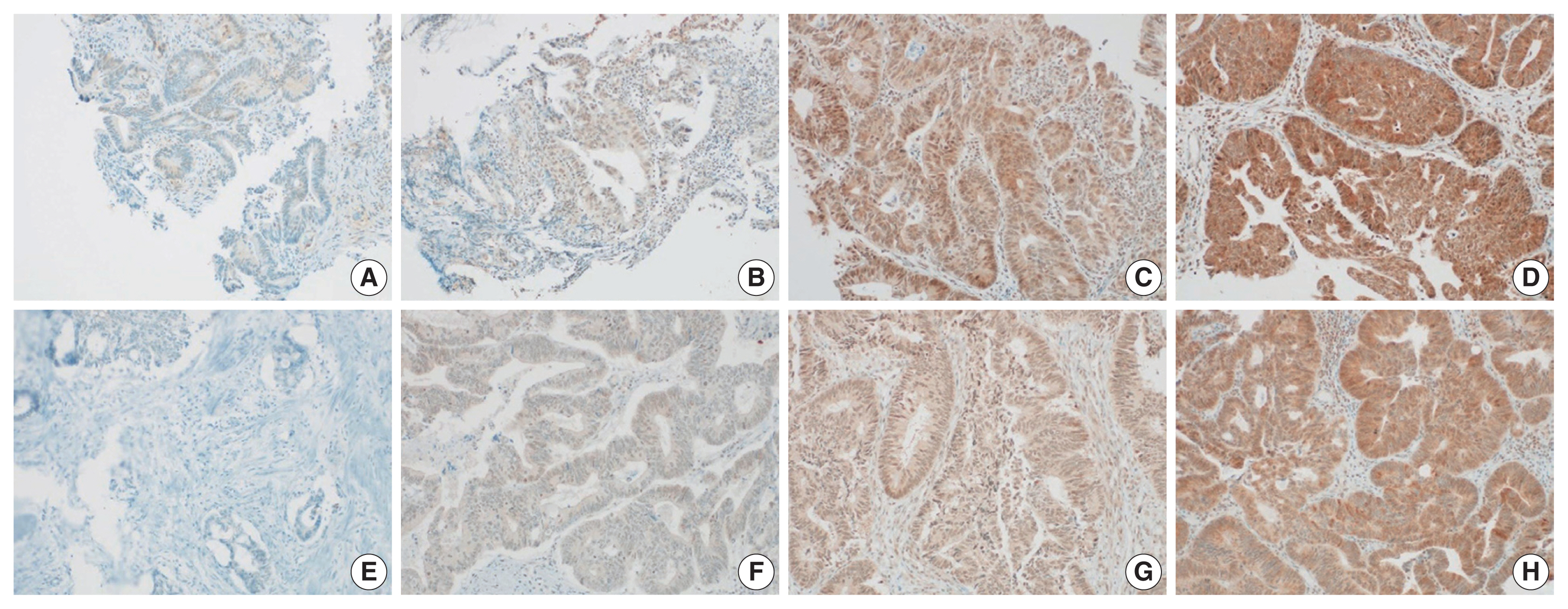
- Hyunseung Oh, Soon Gu Kim, Sung Uk Bae, Sang Jun Byun, Shin Kim, Jae-Ho Lee, Ilseon Hwang, Sun Young Kwon, Hye Won Lee
- J Pathol Transl Med. 2022;56(1):40-47. Published online November 16, 2021
- DOI: https://doi.org/10.4132/jptm.2021.10.07
- 6,808 View
- 163 Download
- 2 Web of Science
- 2 Crossref
-
Abstract
PDF
 Supplementary Material
 - Background
Polo-like kinase 4 (PLK4) is a serine/threonine protein kinase located in the centriole of the chromosome during the cell cycle. PLK4 overexpression has been described in a variety of many common human epithelial tumors. Conversely, PLK4 acts as a haploinsufficient tumor suppressor in some situations, highlighting the importance of strict regulation of PLK4 expression, activity, and function. Meanwhile, the importance of chemoradiation resistance in rectal cancer is being emphasized more than ever. We aimed to analyze PLK4 expression and the tumor regression grade (TRG) in patients with rectal cancer, treated with chemoradiotherapy (CRT).
Methods
A retrospective study was conducted on 102 patients with rectal cancer who received preoperative CRT. Immunohistochemistry for PLK4 in paraffin-embedded tissue was performed from the biopsy and surgical specimens.
Results
We found significant association between high expression of PLK4 and poor response to neoadjuvant CRT (according to both Mandard and The Korean Society of Pathologists TRG systems) in the pre-CRT specimens. Other clinicopathologic parameters did not reveal any correlation with PLK4 expression.
Conclusions
This study revealed an association between high expression of PLK4 in the pre-CRT specimens and TRG. Our results indicated that PLK4 could potentially be a new predictor for CRT effect in patients with rectal cancer. -
Citations
Citations to this article as recorded by- Polo-like kinase 4 (PLK4) as a therapeutic target in breast cancer
Armen Parsyan, Harjot Athwal, Vasudeva Bhat, Alison L Allan
Carcinogenesis.2025;[Epub] CrossRef - Polo-like kinase 4: A molecular linchpin in cancer and its management
Durdana Muntaqua, Gagan Chhabra, Karla B. Anaya Aldrete, Nihal Ahmad
iScience.2025; 28(12): 114186. CrossRef
- Polo-like kinase 4 (PLK4) as a therapeutic target in breast cancer
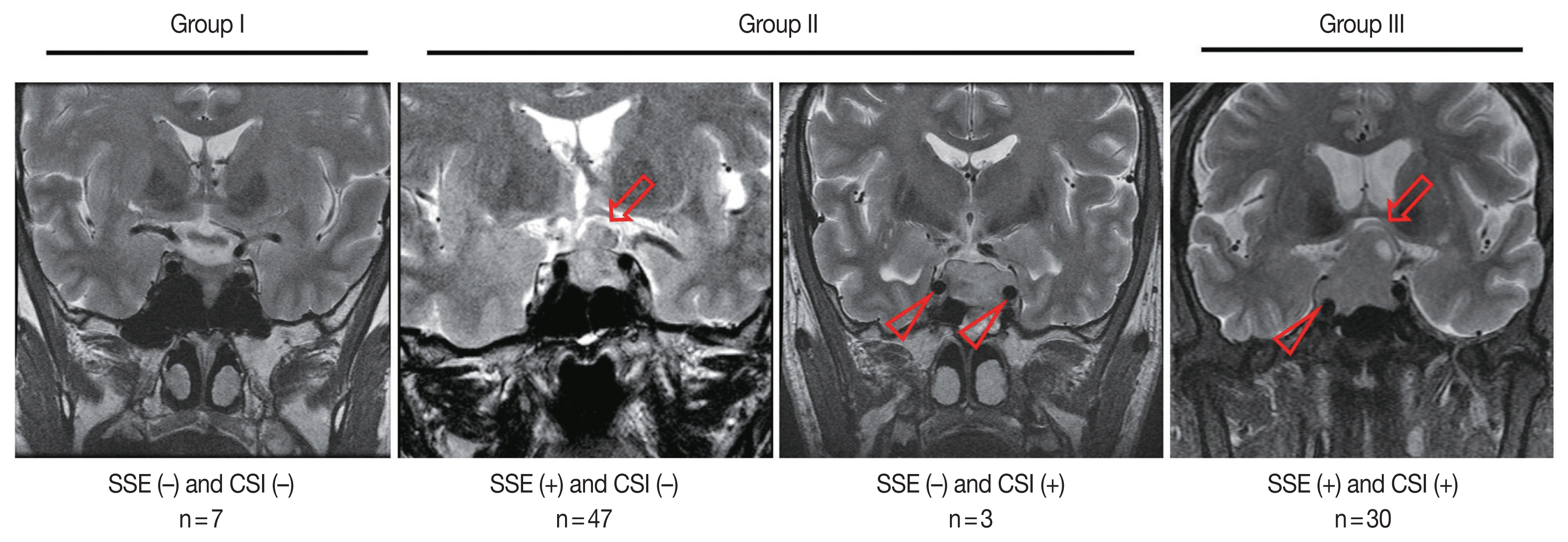
- Association of PTTG1 expression with invasiveness of non-functioning pituitary adenomas
- Su Jung Kum, Hye Won Lee, Soon Gu Kim, Hyungsik Park, Ilseon Hwang, Sang Pyo Kim
- J Pathol Transl Med. 2022;56(1):22-31. Published online October 15, 2021
- DOI: https://doi.org/10.4132/jptm.2021.08.31
- 8,321 View
- 212 Download
- 8 Web of Science
- 8 Crossref
-
Abstract
PDF
- Background
Pituitary tumor transforming gene 1 (PTTG1), paired-like homeodomain 2 (PITX2), and galectin-3 have been widely studied as predictive biomarkers for various tumors and are involved in tumorigenesis and tumor progression. We evaluated the usefulness of PTTG1, PITX2, and galectin-3 as predictive biomarkers for invasive non-functioning pituitary adenomas (NFPAs) by determining the relationship between the expressions of these three proteins and the invasiveness of the NFPAs. We also investigated whether PTTG1, E-cadherin, and Ki-67, which are known to be related to each other, show a correlation with NFPA features.
Methods
A retrospective study was conducted on 87 patients with NPFAs who underwent surgical removal. The NFPAs were classified into three groups based on magnetic resonance imaging findings of suprasellar extension and cavernous sinus invasion. Immunohistochemical staining for PTTG1, PITX2, galectin-3, E-cadherin, and Ki-67 was performed on tissue microarrays.
Results
PTTG1 expression showed a statistically significant correlation with the invasiveness of NFPAs, whereas PITX2 and galectin-3 did not have a relationship with the invasiveness of NFPAs. Moreover, there was no association among PTTG1, E-cadherin, and Ki-67 expression.
Conclusions
PTTG1 has the potential to serve as a predictive biomarker for invasive NFPA. Furthermore, this study may serve as a reference for the development of PTTG1-targeted therapeutic agents. -
Citations
Citations to this article as recorded by- Biomarkers Driving Precision Medicine in Nonfunctioning Pituitary Neuroendocrine Tumors: A Systematic Review of Recent Literature
Amalina Haydar Ali Tajuddin, Nur Firdaus Isa, Mohd Hamzah Mohd Nasir
The Journal of Clinical Endocrinology & Metabolism.2026; 111(4): e1195. CrossRef - The role of methylation in pituitary neuroendocrine tumors current insights and emerging perspectives
Yafei Wang, Tianlang Hu, Jingjing Jia, Chen Wang, Chenran Zhang
Molecular Biology Reports.2026;[Epub] CrossRef - The regulatory role of PTTG1 in proliferation and migration of thyroid cancer
Jianjun Wang, Chenjun Guo, Junyu Cao, Li Li
Discover Oncology.2025;[Epub] CrossRef - Pituitary tumor-transforming gene 1 and endocrine cancers: an up-to-date review through history, current insights and future perspectives
E Vergani, E Teveroni, F Mancini, F Di Nicuolo, S Raia, S Chiloiro, F Pierconti, A Bianchi, A M Isidori, A Pontecorvi, D Milardi
Endocrine-Related Cancer.2025;[Epub] CrossRef - High-throughput Screening for Cushing Disease: Therapeutic Potential of Thiostrepton via Cell Cycle Regulation
Takuro Hakata, Ichiro Yamauchi, Daisuke Kosugi, Taku Sugawa, Haruka Fujita, Kentaro Okamoto, Yohei Ueda, Toshihito Fujii, Daisuke Taura, Nobuya Inagaki
Endocrinology.2024;[Epub] CrossRef - Neoplasms and tumor-like lesions of the sellar region: imaging findings with correlation to pathology and 2021 WHO classification
Lorenzo Ugga, Raduan Ahmed Franca, Alessandra Scaravilli, Domenico Solari, Sirio Cocozza, Fabio Tortora, Luigi Maria Cavallo, Marialaura Del Basso De Caro, Andrea Elefante
Neuroradiology.2023; 65(4): 675. CrossRef - A comprehensive characterisation of phaeochromocytoma and paraganglioma tumours through histone protein profiling, DNA methylation and transcriptomic analysis genome wide
Prodromos Chatzikyriakou, Dimitria Brempou, Mark Quinn, Lauren Fishbein, Roberta Noberini, Ioannis N. Anastopoulos, Nicola Tufton, Eugenie S. Lim, Rupert Obholzer, Johnathan G. Hubbard, Mufaddal Moonim, Tiziana Bonaldi, Katherine L. Nathanson, Louise Izat
Clinical Epigenetics.2023;[Epub] CrossRef - Expression and clinical significance of Cathepsin K and MMPs in invasive non-functioning pituitary adenomas
Hongyan Liu, Saichun Zhang, Ting Wu, Zhaohui Lv, Jianming Ba, Weijun Gu, Yiming Mu
Frontiers in Oncology.2022;[Epub] CrossRef
- Biomarkers Driving Precision Medicine in Nonfunctioning Pituitary Neuroendocrine Tumors: A Systematic Review of Recent Literature
- Causes of necrotic features in fine-needle aspirates from cervical lymph nodes
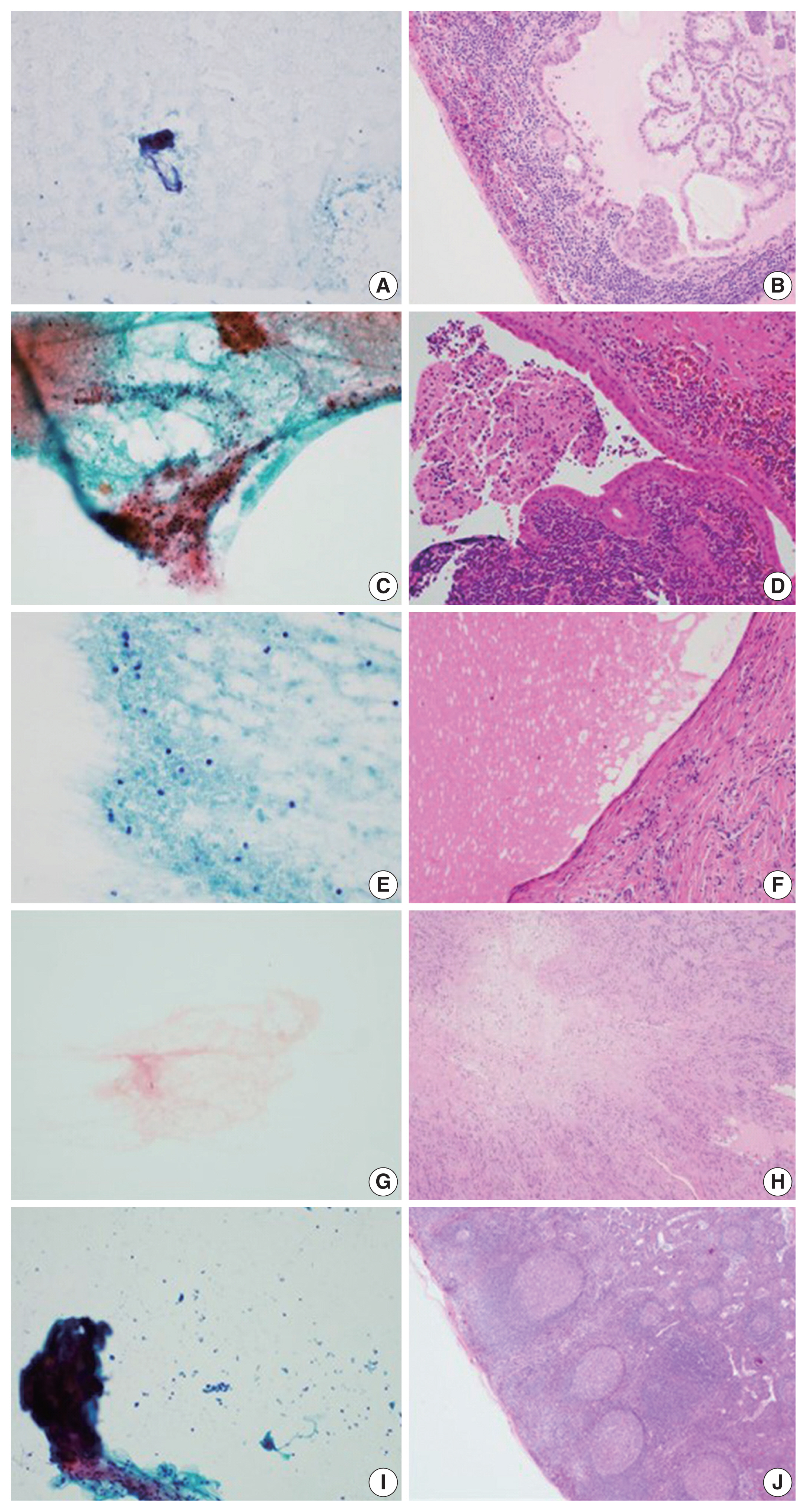
- Young Jin Seo, Hyeongchan Shin, Hye Won Lee, Hye Ra Jung
- J Pathol Transl Med. 2021;55(1):60-67. Published online November 27, 2020
- DOI: https://doi.org/10.4132/jptm.2020.09.28
- 21,832 View
- 212 Download
- 5 Web of Science
- 7 Crossref
-
Abstract
PDF
- Background
Lymph node fine-needle aspiration (LN FNA) cytology indicates necrosis in various diseases. Dominant necrotic features make the diagnosis of underlying conditions very difficult.
Methods
We retrospectively reviewed 460 patients who underwent cervical LN aspiration cytology that revealed necrotic findings at Keimyung University Dongsan Hospital in Daegu, Korea, from 2003–2017. Each specimen was evaluated and analyzed in association with the clinical findings, biopsy findings, and/or other ancillary tests, including acid-fast bacilli staining and molecular testing for Mycobacterium tuberculosis.
Results
When necrotic features were noted upon cervical LN FNA cytology, the most common pathologic LN FNA category was necrosis alone (31.5%). The second most common category was granulomatous inflammation (31.3%), followed by Kikuchi disease (20.0%) and malignant neoplasm (8.7%). In cases where the cervical LN FNA revealed necrosis alone, the most common final diagnosis was tuberculosis. In young patients, Kikuchi disease should be considered as one cervical LN FNA category, while metastatic carcinoma should be suspected in older patients.
Conclusions
Even when necrosis alone is observed in LN FNA cytology, it is important to determine the cause through further evaluation. -
Citations
Citations to this article as recorded by- Necrosis of a Metastatic Axillary Lymph Node in Breast Cancer Possibly Induced by Fine-Needle Aspiration Cytology: A Case Report
Yoshiko Masuda, Mikiko Aoki, Masumi Tanaka, Yukari Koga, Kyoko Eto, Yasuteru Yoshinaga, Toshihiko Sato
Case Reports in Oncology.2026; 19(1): 349. CrossRef - Telecytology in head and neck cytopathology: current applications and practical considerations
Yeongjoon Kim
Kosin Medical Journal.2026; 41(2): 126. CrossRef - The Potential Use of Peptides in the Fight against Chagas Disease and Leishmaniasis
Hayelom Berhe, Mahesh Kumar Cinthakunta Sridhar, Mulate Zerihun, Nir Qvit
Pharmaceutics.2024; 16(2): 227. CrossRef - Tips and tricks for a proper radiological assessment of abdominal and pelvic lymph nodes
Ana Laura Lopes Potente, Cynthia Lopes Pereira de Borborema, Iza Campos Pedra Vieira, Aley Talans, Eduardo Oliveira Pacheco, Lucas Rios Torres, Serli Kiyomi Nakao Ueda, Fernanda Lopez Mazzucato, Andrei Saraiva Purysko, Daniel Lahan Martins, Ulysses Santos
Abdominal Radiology.2024; 49(11): 4057. CrossRef - Does the Necrotic Portion of Metastatic Lymphadenopathy from Squamous Cell Carcinoma Still Have Tumoral Oncologic Information? Differential Diagnosis of Benign Necrotic Lymphadenopathy Using microRNA
Eun Shin, Seung Hoon Han, Il-Seok Park, Jee Hye Wee, Joong Seob Lee, Heejin Kim
Biomedicines.2023; 11(9): 2407. CrossRef - Impact of HPV status in T1–2 oropharyngeal squamous cell carcinoma with bulky N3 nodes: a multicenter GETTEC study
Charles Hurel, Florent Carsuzaa, Julia Salleron, Philippe Gorphe, Christian Righini, Maximilien Rogé, Erwan de Mones, Sylvain Morinière, Sébastien Vergez, Juliette Thariat, Xavier Dufour
European Archives of Oto-Rhino-Laryngology.2023; 280(2): 847. CrossRef - Lymph nodes in health and disease – A pathologist's perspective
N S Priya
Journal of Oral and Maxillofacial Pathology.2023; 27(1): 6. CrossRef
- Necrosis of a Metastatic Axillary Lymph Node in Breast Cancer Possibly Induced by Fine-Needle Aspiration Cytology: A Case Report
Case Studies
- Amoebic Encephalitis Caused by Balamuthia mandrillaris
- Su Jung Kum, Hye Won Lee, Hye Ra Jung, Misun Choe, Sang Pyo Kim
- J Pathol Transl Med. 2019;53(5):327-331. Published online May 24, 2019
- DOI: https://doi.org/10.4132/jptm.2019.05.14
- 22,777 View
- 196 Download
- 15 Web of Science
- 16 Crossref
-
Abstract
PDF
- We present the case of a 71-year-old man who was diagnosed with amoebic encephalitis caused by Balamuthia mandrillaris. He had rheumatic arthritis for 30 years and had undergone continuous treatment with immunosuppressants. First, he complained of partial spasm from the left thigh to the left upper limb. Magnetic resonance imaging revealed multifocal enhancing nodules in the cortical and subcortical area of both cerebral hemispheres, which were suggestive of brain metastases. However, the patient developed fever with stuporous mentality and an open biopsy was performed immediately. Microscopically, numerous amoebic trophozoites, measuring 20 to 25 µm in size, with nuclei containing one to four nucleoli and some scattered cysts having a double-layered wall were noted in the background of hemorrhagic necrosis. Based on the microscopic findings, amoebic encephalitis caused by Balamuthia mandrillaris was diagnosed. The patient died on the 10th day after being admitted at the hospital. The diagnosis of amoebic encephalitis in the early stage is difficult for clinicians. Moreover, most cases undergo rapid deterioration, resulting in fatal consequences. In this report, we present the first case of B. mandrillaris amoebic encephalitis with fatal progression in a Korean patient.
-
Citations
Citations to this article as recorded by- Prognostic Factors and Treatment Outcomes in Pediatric Balamuthia Amoebic Encephalitis: A Case Report and Literature Review
Juan-Juan Liu, Yong-Ping Xie, Lei Wang, Lv-Chang Zhu, Yi Shi, Wu-Jie Shi, Ao-Le Li, Wang Hua, Zhi-Peng Shen, Da-Qing Ma, Li-Su Huang
Infectious Microbes and Diseases.2026; 8(2): 129. CrossRef - The role of plasma metagenomic sequencing in identification of Balamuthia mandrillaris encephalitis
Sarah Y. Edminster, Ryan W. Rebbe, Christopher Khatchadourian, Kyle M. Hurth, Anna J. Mathew, Julie Huss-Bawab, Mark S. Shiroishi, Devin Clark, Andrew P. Norgan, Susan M. Butler-Wu, Annie Hiniker
Acta Neuropathologica Communications.2025;[Epub] CrossRef -
Understanding
Naegleria fowleri
: The Risks of Brain Infection
Pankti C. Balar, Savan Modi, Vivek P. Chavda
Outbreak Management and Response.2025;[Epub] CrossRef - Strain-dependent immune responses reveal a critical role of IL-17A in defense against Balamuthia mandrillaris
Yu Jin Jeong, Dong Gyu Han, Mi Kyung Park, Seo Yeong Choi, Shin Ae Kang, Hak Sun Yu
Parasites, Hosts and Diseases.2025; 63(4): 371. CrossRef - Molecular identification, phylogenetic analysis and histopathological study of pathogenic free-living amoebae isolated from discus fish (Symphysodon aequifasciatus) in Iran: 2020–2022
Hooman Rahmati-Holasoo, Maryam Niyyati, Marziye Fatemi, Fatemeh Mahdavi Abhari, Sara Shokrpoor, Alireza Nassiri, Amin Marandi
BMC Veterinary Research.2024;[Epub] CrossRef - Encephalomyelomeningitis Caused by Balamuthia mandrillaris: A Case Report and Literature Review
XueMei Fan, TianWen Chen, Hui Yang, Yue Gao, Yan Chen
Infection and Drug Resistance.2023; Volume 16: 727. CrossRef - Diagnosing Balamuthia mandrillaris amebic meningoencephalitis in a 64-year-old woman from the Southwest of China
Suhua Yao, Xiaoting Chen, Lian Qian, Shizheng Sun, Chunjing Zhao, Zongkai Bai, Zhaofang Chen, Youcong Wu
Parasites, Hosts and Diseases.2023; 61(2): 183. CrossRef - Diagnosis of Balamuthia mandrillaris Encephalitis by Thymine–Adenine Cloning Using Universal Eukaryotic Primers
Ju Yeong Kim, Myung-Hee Yi, Myungjun Kim, Joon-Sup Yeom, Hyun Dong Yoo, Seong Min Kim, Tai-Soon Yong
Annals of Laboratory Medicine.2022; 42(2): 196. CrossRef - Facial Balamuthia mandrillaris infection with neurological involvement in an immunocompetent child
Zhen Zhang, Jianying Liang, Ruoqu Wei, Xiaobo Feng, Lei Wang, Liuhui Wang, Piaoping Zhao, Hong Yu, Yan Gu, Zhirong Yao
The Lancet Infectious Diseases.2022; 22(3): e93. CrossRef - Subacute Balamuthia mandrillaris encephalitis in an immunocompetent patient diagnosed by next-generation sequencing
Changbo Xu, Xiaoyan Wu, Miaoqin Tan, Dongmei Wang, Shengnan Wang, Yongming Wu
Journal of International Medical Research.2022;[Epub] CrossRef - Distribution and Current State of Molecular Genetic Characterization in Pathogenic Free-Living Amoebae
Alejandro Otero-Ruiz, Leobardo Daniel Gonzalez-Zuñiga, Libia Zulema Rodriguez-Anaya, Luis Fernando Lares-Jiménez, Jose Reyes Gonzalez-Galaviz, Fernando Lares-Villa
Pathogens.2022; 11(10): 1199. CrossRef - Fulminant Disseminating Fatal Granulomatous Amebic Encephalitis: The First Case Report in an Immunocompetent Patient in South Korea
Ju Yeon Lee, In Kyu Yu, Seong Min Kim, Joo Heon Kim, Ha Youn Kim
Yonsei Medical Journal.2021; 62(6): 563. CrossRef - A Japanese case of amoebic meningoencephalitis initially diagnosed by cerebrospinal fluid cytology
Ryogo Aoki, Toshimasa Sakakima, Asuka Ohashi, Riyoko Niwa, Masashi Matsuyama, Fumimasa Etori, Naoki Watanabe, Kenji Yagita, Takuji Tanaka
Clinical Case Reports.2020; 8(9): 1728. CrossRef -
Balamuthia mandrillaris
infection in China: a retrospective report of 28 cases
Lei Wang, Wenjing Cheng, Bing Li, Zhe Jian, Xianlong Qi, Dongjie Sun, Jian Gao, Xuetao Lu, Yi Yang, Kun Lin, Chuanlong Lu, Jiaxi Chen, Chunying Li, Gang Wang, Tianwen Gao
Emerging Microbes & Infections.2020; 9(1): 2348. CrossRef - Methotrexate/nonsteroidal anti-inflammatory drugs/steroids
Reactions Weekly.2019; 1775(1): 307. CrossRef - Identification of plicamycin, TG02, panobinostat, lestaurtinib, and GDC-0084 as promising compounds for the treatment of central nervous system infections caused by the free-living amebae Naegleria, Acanthamoeba and Balamuthia
Monica M. Kangussu-Marcolino, Gretchen M. Ehrenkaufer, Emily Chen, Anjan Debnath, Upinder Singh
International Journal for Parasitology: Drugs and Drug Resistance.2019; 11: 80. CrossRef
- Prognostic Factors and Treatment Outcomes in Pediatric Balamuthia Amoebic Encephalitis: A Case Report and Literature Review
- Bile Granuloma Mimicking Peritoneal Seeding: A Case Report
- Hasong Jeong, Hye Won Lee, Hye Ra Jung, Ilseon Hwang, Sun Young Kwon, Yu Na Kang, Sang Pyo Kim, Misun Choe
- J Pathol Transl Med. 2018;52(5):339-343. Published online July 16, 2018
- DOI: https://doi.org/10.4132/jptm.2018.06.02
- 9,731 View
- 120 Download
- 7 Web of Science
- 9 Crossref
-
Abstract
PDF
- Laparoscopic cholecystectomy is a widely used treatment method for most cholelithiasis and is a relatively safe procedure. Foreign body granulomatous reaction to bile or gallstone spillage during laparoscopic cholecystectomy has rarely been reported. We report a case of bile granuloma after laparoscopic cholecystectomy, which mimicked peritoneal seeding. A 59-year-old Korean man presented with right upper quadrant pain. He underwent laparoscopic cholecystectomy for acute cholecystitis with cholelithiasis. Pathologic examination revealed an incidental adenocarcinoma invading the lamina propria with acute cholecystitis and cholelithiasis. After 3 months, follow-up abdominal computed tomography revealed a subhepatic nodule, which showed hypermetabolism on positron emission tomography–computed tomography. Suspecting localized peritoneal seeding, wedge resection of the liver, wedge resection of the transverse colon, and omentectomy were performed. Pathologic examination of the resected specimens revealed multiple bile granulomas. Awareness of bile granuloma mimicking malignancy is noteworthy for patient management to reduce unnecessary procedure during postoperative surveillance.
-
Citations
Citations to this article as recorded by- False Alarm: When Dropped Gallstones Mimic Malignant Recurrence. A Case Report and Literature Review
Nedaa Obeidi, Michael Egan, Michael E. Kelly, Feras Abu Saadeh, Karuna Sharma
Case Reports in Obstetrics and Gynecology.2026;[Epub] CrossRef - A mimic of peritoneal metastatic disease, multifocal intraabdominal foreign body granulomas secondary to feculent peritonitis
Damien Gibson, Christo Joseph, Diarmid P. Foulis, Christophe R. Berney
ANZ Journal of Surgery.2024; 94(4): 763. CrossRef - Practices and Attitudes of Surgeons With Regard to Spilled Gallstones During Laparoscopic Cholecystectomy: A Cross-Sectional Study From Saudi Arabia
Mohammed Alfehaid, Moath Aljohani, Sajad A Salati , Shoug Alaodah, Wejdan Alresheedi, Raghad Almarshud
Cureus.2024;[Epub] CrossRef - Spilled gallstone mimicking intra-abdominal seeding of gallbladder adenocarcinoma: A case report
Cheng-Ken Huang, Ruey-Hwa Lu, Chien-Cheng Chen, Po-Chun Chen, Wen-Chang Hsu, Meng-Jui Tsai, Chin-Tsung Ting
World Journal of Gastrointestinal Surgery.2024; 16(2): 622. CrossRef - Peritoneal bile granuloma formation at the site of caesarean surgical scar
Lila Marshall, Sharlin Varghese, Mary Ciranni-Callon
Journal of Case Reports and Images in Obstetrics and Gynecology.2024; 10(2): 6. CrossRef - Biliary Granulomatous Peritoneal Reaction as Consequence of Cholecystectomy: Case Report and Literature Review
Giuseppe Tarantino, Denise Menghini, Maria Eva Argenziano, Miriam Palmieri, Alessandra Mandolesi, Enrico Dalla Bona, Antonio Benedetti, Mario Guerrieri, Maria Giovanna Danieli
SN Comprehensive Clinical Medicine.2023;[Epub] CrossRef - Foreign body reaction mimicking local recurrence from polyactide adhesion barrier film after laparoscopic colorectal cancer surgery
Tien-Chan Hsieh, Chao-Wen Hsu
Medicine.2022; 101(5): e28692. CrossRef - Spilled gallstones after laparoscopic cholecystectomy: a systematic review
Sajad Ahmad Salati, Mohammed Alfehaid, Saleh Alsuwaydani, Lamees AlSulaim
Polish Journal of Surgery.2022; 94(4): 1. CrossRef - Foreign body granulomas mimic peritoneal dissemination caused by incarcerated femoral hernia perforation: A case report
Shinpei Ogino, Tatsuya Matsumoto, Yosuke Kamada, Noriaki Koizumi, Hiroshi Fujiki, Kenji Nakamura, Takeshi Yamano, Chouhei Sakakura
World Journal of Clinical Oncology.2021; 12(11): 1083. CrossRef
- False Alarm: When Dropped Gallstones Mimic Malignant Recurrence. A Case Report and Literature Review
Case Report
- Sporadic Cutaneous Keratocyst without Nevoid Basal Cell Carcinoma Syndrome: Report of 1 Case.
- Hye Won Lee, Ji Young Park, Sun Hee Kang, Misun Choe
- Korean J Pathol. 2011;45(3):322-325.
- DOI: https://doi.org/10.4132/KoreanJPathol.2011.45.3.322
- 5,946 View
- 65 Download
- 3 Crossref
-
Abstract
PDF
- A cutaneous keratocyst is very rare and is ordinarily associated with nevoid basal cell carcinoma syndrome (NBCCS), also known as Gorlin syndrome. NBCCS is a rare autosomal-dominant disorder that results from the mutation in the patched homologue 1 (PTCH1) gene located on chromosome 9q22.3, with high penetrance and variable expressivity. NBCCS demonstrates multisystem manifestations such as multiple basal cell carcinomas in early age, jaw cysts and pits of the hands and feet. Cutaneous keratocysts are characteristically lined by festooned keratinized squamous epithelium with parakeratosis. The cystic wall contains neither granular cell layer nor skin appendages. To the best of our knowledge, only two cases of cutaneous keratocysts not associated with NBCCS have been reported to date. We report one another case of a histologically confirmed cutaneous keratocyst in a 50-year-old female without a family history and clinical features of NBCCS.
-
Citations
Citations to this article as recorded by- Determining the Relationship Between Cutaneous Keratocysts and Basal Cell Nevus Syndrome
Madelyn M. Class, Claire Rose Kissinger, Sidra Ibad, Aspen Trautz, Lisa Zhai, Farhaan Hafeez
Journal of Cutaneous Pathology.2025; 52(6): 403. CrossRef - Sporadic Cutaneous Keratocyst of the Scalp: A Report of an Extremely Rare Lesion
Rana S AL-Zaidi, Eyad Tantawi, Rahaf AL-Radadi, Asrar Banjar
Cureus.2021;[Epub] CrossRef - A Large Extragnathic Keratocystic Odontogenic Tumour
Soumya Makarla, Radhika M. Bavle, Sudhakara Muniswamappa, Srinath Narasimhamurthy
Case Reports in Pathology.2015; 2015: 1. CrossRef
- Determining the Relationship Between Cutaneous Keratocysts and Basal Cell Nevus Syndrome
Original Article
- Expression of MUC1 and MUC4 and Its Prognostic Significance in Non-Small Cell Lung Carcinoma.
- Ji Min Jeon, Hye Won Lee, Ji Young Park, Hye Ra Jung, Ilseon Hwang, Sun Young Kwon, Mi Sun Choe, Yu Na Kang, Sang Pyo Kim, Sang Sook Lee, Won Il Choi, Kun Young Kwon
- Korean J Pathol. 2010;44(4):397-403.
- DOI: https://doi.org/10.4132/KoreanJPathol.2010.44.4.397
- 6,009 View
- 62 Download
- 6 Crossref
-
Abstract
PDF
- BACKGROUND
Mucin (MUC)1 and MUC4 (MUC1, 4) are high molecular weight glycoproteins expressed in normal and malignant epithelial cells, and these expressions are related to the prognosis of some carcinomas. In non-small cell lung carcinoma (NSCLC), the relationship between MUC1, 4 expressions and their prognostic significance is not well known. We evaluated these relationships in a series of NSCLC: 1) between MUC1, 4 expression levels and histologic subtypes, and 2) between high expression of MUC1, 4 and their prognostic significance.
METHODS
We performed immunohistochemical staining for MUC1, 4 in paraffin-embedded tissues from 165 NSCLC cases arranged in a tissue microarray.
RESULTS
We found a significant correlation between MUC1, 4 expressions and NSCLC histologic subtypes (p < 0.05). High MUC1 expression was characteristic of adenocarcinoma. Low MUC1, 4 expressions were characteristic of squamous cell carcinoma. In adenocarcinoma, we found significant association between diffuse MUC1 expression and short patient survival (p = 0.005). In squamous cell carcinoma, diffuse MUC4 expression showed long patient survival trend (p = 0.128).
CONCLUSIONS
MUC1, 4 expression levels were significantly correlated with NSCLC histologic subtypes. Diffuse MUC1 expression was significantly associated with shortened survival in NSCLC patients, especially in adenocarcinoma. -
Citations
Citations to this article as recorded by- Inhibition of MUC1-C Increases ROS and Cell Death in Mouse Embryonic Stem Cells
Jeong-A Park, Sangkyu Park, Jun-Kyu Choi, Myung-Kwan Han, Younghee Lee
International Journal of Stem Cells.2020;[Epub] CrossRef - Assessing the prognostic significance of MUC4β in mucoepidermoid carcinoma of the salivary glands: An immunohistochemical study
Poonam R. Sawant, Anita Spadigam, Anita Dhupar, Shaheen Syed, Karla Carvalho
Heliyon.2019; 5(11): e02753. CrossRef - Ultrasensitive cytosensing based on an aptamer modified nanobiosensor with a bioconjugate: Detection of human non-small-cell lung cancer cells
Tanveer A. Mir, Jang-Hee Yoon, N.G. Gurudatt, Mi-Sook Won, Yoon-Bo Shim
Biosensors and Bioelectronics.2015; 74: 594. CrossRef - Expression of MUC1 and MUC4 in Gallbladder Adenocarcinoma
Su-Mi Kim, Sun-Ju Oh, Bang Hur
Korean Journal of Pathology.2012; 46(5): 429. CrossRef - MUC4 and MUC1 Expression in Adenocarcinoma of the Stomach Correlates with Vessel Invasion and Lymph Node Metastasis: An Immunohistochemical Study of Early Gastric Cancer
Yukihiro Tamura, Michiyo Higashi, Sho Kitamoto, Seiya Yokoyama, Masahiko Osako, Michiko Horinouchi, Takeshi Shimizu, Mineo Tabata, Surinder K. Batra, Masamichi Goto, Suguru Yonezawa, Fazlul H. Sarkar
PLoS ONE.2012; 7(11): e49251. CrossRef - Prognostic significance of membrane-associated mucins 1 and 4 in gastric adenocarcinoma
ILSEON HWANG, YU NA KANG, JIN YOUNG KIM, YOUNG ROK DO, HONG SUK SONG, KEON UK PARK
Experimental and Therapeutic Medicine.2012; 4(2): 311. CrossRef
- Inhibition of MUC1-C Increases ROS and Cell Death in Mouse Embryonic Stem Cells
 First
First Prev
Prev


