 E-submission
Search
- Page Path
- HOME > Search
Case Studies
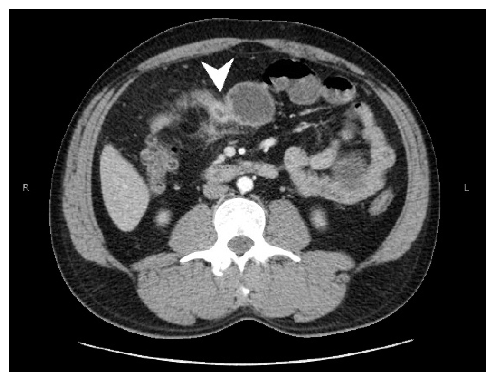
- Heterotopic mesenteric ossification: a report of two cases
- Hisham F. Bahmad, Olga Lopez, Tyson Sutherland, Marisa Vinas, Kfir Ben-David, Lydia Howard, Robert Poppiti, Sarah Alghamdi
- J Pathol Transl Med. 2022;56(5):294-300. Published online September 13, 2022
- DOI: https://doi.org/10.4132/jptm.2022.07.23
- 6,639 View
- 107 Download
- 3 Crossref
-
 Abstract
 PDF
PDF - Heterotopic mesenteric ossification (HMO) is abnormal bone formation in tissues which usually do not undergo ossification. There are approximately 75 cases reported worldwide. We present two cases of HMO. The first case is that of a 39-year-old man who presented with abdominal pain and a computerized tomography scan of the abdomen and pelvis revealed an apple core lesion resulting in small bowel obstruction. The second case is that of a 36-year-old woman who presented 2 months after undergoing robotic gastric sleeve resection complaining of weakness and emesis. An esophagogram revealed kinking at the distal esophagus. Surgical resection was performed in both, yielding the diagnosis of HMO. There are various theories as to the pathophysiology of HMO, but no clearly defined mechanism has been established. Management should be conservative whenever possible to prevent further ossification with subsequent surgical intervention.
-
Citations
Citations to this article as recorded by
- Bone fragility in a post-intestinal transplant patient with mesenteric heterotopic ossification
Husam Hamshary, Dania S Bacha, Leila Z Khan
JBMR Plus.2026;[Epub] CrossRef - Fatal Moans and Bones in Crohn's
Joyce Opara, Heather Jarrell, Nicole R. Jackson
American Journal of Forensic Medicine & Pathology.2025; 46(2): 151. CrossRef - Heterotopic mesenteric ossification caused by trauma: A case report
Bi-Fang Zhang, Jiang Liu, Shuai Zhang, Ling Chen, Jia-Zheng Lu, Ming-Qing Zhang
World Journal of Gastrointestinal Endoscopy.2024; 16(8): 494. CrossRef
- Bone fragility in a post-intestinal transplant patient with mesenteric heterotopic ossification
- Adrenal Cortical Neoplasm with Uncertain Malignant Potential Arising in the Heterotopic Adrenal Cortex in the Liver of a Patient with Beckwith-Wiedemann Syndrome
- Eun Na Kim, Dong Eun Song, Hee Mang Yoon, Beom Hee Lee, Chong Jai Kim
- J Pathol Transl Med. 2019;53(2):129-135. Published online November 26, 2018
- DOI: https://doi.org/10.4132/jptm.2018.11.13
- 8,802 View
- 110 Download
- 5 Web of Science
- 5 Crossref
-
Abstract
PDF
- Patients with Beckwith-Wiedemann syndrome (BWS) are predisposed to developing embryonal tumors, with hepatoblastoma being the most common type. Our patient showed hemihypertrophy, macroglossia, and paternal uniparental disomy in chromosome 11 and was diagnosed with BWS. When the patient was 9 months old, a 2.5×1.5 cm oval hypoechoic exophytic mass was detected in the inferior tip of his right liver. Preoperative imaging identified it as hepatoblastoma; however, histologic, immunohistochemistry, and electron microscopic findings were compatible with adrenal cortical neoplasm with uncertain malignant potential. The origin of the adrenal tissue seemed to be heterotopic. Here, we describe for the first time an adrenal cortical neoplasm with uncertain malignant potential arising in the heterotopic adrenal cortex located in the liver of a patient with BWS.
-
Citations
Citations to this article as recorded by- Adrenocortical tumors and hereditary syndromes
Kanakamani Jeyaraman, Paola Concolino, Henrik Falhammar
Expert Review of Endocrinology & Metabolism.2025; 20(1): 1. CrossRef - Functional adrenocortical carcinoma with adrenohepatic fusion: A case report
Pastor Escárcega-Fujigaki, Guillermo Hernández-Peredo Rezk, José de Jesús Loeza- Oliva, Anallely Luna-Hernández, Bethsaida Natali Arreguín-Cortés, Rafael López-Cruz
Journal of Pediatric Surgery Case Reports.2024; 107: 102841. CrossRef - Molecular and Clinical Features of Adrenocortical Tumors in Beckwith–Wiedemann Spectrum
Diana Carli, Federico Rondot, Maria Luca, Anna Campello, Stefano Gabriele Vallero, Elisa Tirtei, Andrea Gazzin, Simona Cardaropoli, Francesca Montanari, Claudio Graziano, Paola Quarello, Abu Saadat, Angela Sparago, Giovanni Battista Ferrero, Franca Fagiol
Cancers.2024; 16(23): 3967. CrossRef - Beckwith–Wiedemann syndrome: Clinical, histopathological and molecular study of two Tunisian patients and review of literature
Hela Sassi, Yasmina Elaribi, Houweyda Jilani, Imen Rejeb, Syrine Hizem, Molka Sebai, Nadia Kasdallah, Habib Bouthour, Samia Hannachi, Jasmin Beygo, Ali Saad, Karin Buiting, Dorra H’mida Ben‐Brahim, Lamia BenJemaa
Molecular Genetics & Genomic Medicine.2021;[Epub] CrossRef - Adrenocortical Tumors in Children With Constitutive Chromosome 11p15 Paternal Uniparental Disomy: Implications for Diagnosis and Treatment
Emilia Modolo Pinto, Carlos Rodriguez-Galindo, Catherine G. Lam, Robert E. Ruiz, Gerard P. Zambetti, Raul C. Ribeiro
Frontiers in Endocrinology.2021;[Epub] CrossRef
- Adrenocortical tumors and hereditary syndromes
Case Reports
- Intraocular Ossification: A Case Report.
- Ho Sung Park, Tae Shik Kong, Kyu Yun Jang, Myoung Ja Chung, Woo Sung Moon, Dong Geun Lee, Myoung Jae Kang
- Korean J Pathol. 2004;38(3):188-190.
- 2,609 View
- 29 Download
-
Abstract
PDF
- Heterotopic bone formation in the eyeball is a rare finding. Some etiologic factors, such as trauma, chronic inflammation, and long-standing retinal detachment have been associated with the onset of intraocular ossification. We report here on a case of a 21-year-old woman with a history of blunt trauma fifteen years ago, who complained of right eye blindness. When the right eyeball eviceration was done, a hard, grayish mass was found. On histopathologic examination, the mass showed lamellar bone with fatty marrow and hyalinized tissue with dystrophic calcification. We diagnosed her case as intraocular ossification.
- Adenocarcinoma Arising from Heterotopic Gastric Mucosa in Cervical Esophagus: A Case Report.
- Young Ok Hong, Jeong Eun Hwang, In Chul Lee, Jin Hyuk Lee, Seung Il Park, Kyung Ja Cho
- Korean J Pathol. 2008;42(1):33-36.
- 2,393 View
- 17 Download
-
Abstract
PDF
- Heterotopic gastric mucosa (HGM) of the upper esophagus, referred as "cervical inlet patch (CIP)", is a benign lesion that is present in 3.8-10% of the adult population. Adenocarcinomas arising from HGM of the upper esophagus are exceedingly rare. The authors report one additional case of histologically confirmed adenocarcinoma arising from a CIP. The patient had concomitant primary adenocarcinoma of the colon. The right hemicolectomy specimen and total esophagectomy specimen after preoperative chemoradiotherapy showed histologically different adenocarcinomas. The residual esophageal tumor was characterized by large mucin pools, fibrous septa, and floating tumor cells. HGM of both the fundic and antral types was seen on the surface and sides of the tumor. The independent origins of the two cancers were confirmed by immunohistochemical studies for cytokeratins 7 and 20. Without further treatment, the patient remained free of disease after 29 months of follow-up.
- Heterotopic Prostatic Tissue with Cystic Change in Retrovesical Space: A case report.
- Hyun Jin Son, Myoung Jae Kang, Dong Geun Lee
- Korean J Pathol. 2000;34(1):93-95.
- 2,138 View
- 16 Download
-
Abstract
PDF
- Heterotopic prostatic tissue has been reported in a variety of sites within and outside the urinary tract. Extra-urethral ectopic prostatic tissue is a distinct entity and may be more common than previously thought. We report a case of heterotopic prostatic tissue in 71-year-old man. Pelvic CT scan showed a well circumscribed cystic mass in the retrovesical space. Grossly, the tumor was 7.5 7.0 2.8 cm and revealed an ovoid unilocular cyst containing grayish amorphous granular materials. The prostatic origin of the tissue was confirmed by immunohistochemical staining for prostate specific antigen.
Original Article
- Heterotopic Glial Nodule in the Lung of an Anencephaly Patient : An autopsy case.
- Hye Joung Lee, Soo Min Kang, Gyung Hyuck Ko
- Korean J Pathol. 1991;25(5):457-461.
- 2,141 View
- 10 Download
-
Abstract
- The heterotopic and tissues may be divided into two categories: those that are found in the head and neck region, and those that arise elsewhere. The latter type is rare and most cases are found in the lungs of patients with neural tube defect, particularly anencephaly. Our report descrives anencephalic male infant with heterotopic glial nodules in both lungs. The largest nodule is 2x1.5x1 cm, locates in the lower lobe of the left lung, and has a round gray-white cut surface with cystic spaces. Microscopically, the nodules consist of irregularly arranged astrocytes and glial fibers, in which are embedded gland-like or cystically dilated bronchioles. The astrocytes and glial fibers are strongly positive for glial fibrillary acidic protein and show astrocytic filaments on electron microscopy. This will be an additional case supporting the amniotic fluid aspiration/implantation theory of pathogenetic mechanism of distal heerotopic glial tissue.
Case Report
- Heterotopic Mesenteric Ossification: A Case Report.
- Hoon Kyu Oh, Jong Yup Bae
- Korean J Pathol. 2006;40(1):70-72.
- 2,590 View
- 32 Download
-
Abstract
PDF
- Heterotopic mesenteric ossification is a very rare reactive lesion in the small bowel mesentery, and it is related with trauma or surgical operation. It is pathologically characterized by well formed bone trabeculae and prominent osteoblastic rimming and is clinically related to rapid and recurrent bowel obstruction symptoms. This unusual reactive process shares many clinical and pathologic features with myositis ossificans. We report here on a rare case of heterotopic mesenteric ossification in 28-year-old man who underwent a delayed small bowel resection 15 days after trauma.
Original Article
- Histologic Variations of Intramural Heterotopic Pancreas in Gastrointestinal Tract Analysis of 15 Cases.
- Seung Sook Lee, Yong Il Kim, Woo Ho Kim, Eun Sil Yu
- Korean J Pathol. 1991;25(6):520-527.
- 2,233 View
- 13 Download
-
Abstract
PDF
- We reviewed a total of 15 cases of heterotopic pancreatic tissue within the gastrointestinal wall(intramural type), and compared with 3 extramural ones. Intramural heterotopic pancreatic lesions were located in the antrum(33%), pylorus(20%), and body of stomach(7%), and the remainders in the duodenum(40%). Only two cases presented with chinical symptoms by their existence. Two of them were situated within the submucosa, 3 in the muscularis, 6 in submucosa-muscularis, 2 in the muscularis-subserosa, and 1 in the susbmucosa-subserosa. Intramural type was featured with their structural heterogeneity compared to the extramural ones; 10 cases showed participation of gastrointestinal mucosal elements, and some accompanied tissue elements that were indistinguishable from submucosal epithelial heterotopia or microduplication cyst of the stomach. Langerhans islets were found in 67%, and one developed islet cell tumor. The above results suggest that the initially engrafted heterotopic pancreatic tissue becomes modified and presents with heterogeneity of endodermal and mesodermal tissue-derived components by its intramural growth during the gastrointestinal organogenesis; failure of opening of its drainage system into the gastrointestinal lumen may result in the increase of intraductal pressure with subsequent atrophy of the acinar tissue and various metaplastic changes of ductal epithelium, aside from induction of smooth muscle coat around the heterotopic tissue.
Case Reports
- Heterotopic Enchondral Ossification in Metastatic Colonic Adenocarcinoma: A case report .
- So Yeon Park, Yong Il Kim, Woo Ho Kim
- Korean J Pathol. 2000;34(7):531-533.
- 2,201 View
- 12 Download
-
Abstract
PDF
- Calcification and ossification of colon cancer is frequently encountered, especially in the mucinous carcinoma. However, cartilage formation or enchondral ossification has rarely been described in human colon cancer. This report describes a case of a 59-year-old man with retroperitoneal metastasis of mucinous adenocarcinoma of colon, which showed a widespread heterotopic ossification through membranous or enchondral ossification. The ossification appeared in apposition to tumor cell nests and in the organized mucin pool. In our knowledge, this is the first case showing enchondral ossification in gastrointestinal carcinoma in Korea.
- Retroperitoneal Duplication Cyst Associated with Heterotopic Pancreas: A case report.
- So Yeong Oh, Myoung Ja Chung, Dong Geun Lee, Ho Yeul Choi
- Korean J Pathol. 1998;32(9):687-690.
- 2,286 View
- 10 Download
-
Abstract
- Occurrence of a retroperitoneal duplication cyst associated with a heterotopic pancreas is rare. We report a case of a retroperitoneal duplication cyst associated with a heterotopic pancreas. A 35-year-old male was admitted, presenting with back pain. A pelvic computed tomographic scan disclosed a 10 cm-sized cystic mass filling the lower pelvis and displacing the rectosigmoid colon anteriorly. Histologically, the cyst wall was lined partly by mucin-secreting columnar epithelium, showing atypical hyperplasia and partly by a gastric fundic-type and a colonic-like mucosa. Beneath the epithelium, organized bundles of smooth muscle were arranged in two layers analogous to smooth muscles layers of the bowel and a small piece of pancreatic tissue were present in the smooth muscle wall of the cyst.
- Intraabdominal Heterotopic Thymus: Report of an autopsy case.
- Hye Seung Han, Je Geun Chi
- Korean J Pathol. 1996;30(11):1057-1059.
- 2,277 View
- 13 Download
-
Abstract
PDF
- Ectopic thymus results from the aberrant migration of thymic tissue and is mostly present in the mediastinum, the base of the skull, the tracheal bifurcation and the cervical region. We report the first case of intraabdominal heterotopic thymus incidentally detected and attached to the liver without associated anomalies. This fetus was sent to the Department without any clinical information. The fetus was small for gestational age, but had no external abnormalities. Each organ showed normal development except for the liver. The liver weighed 6 gm(normal 17.064+/-4.143 gm). Gray white heterotopic thymus was attached to the superior surface of the liver in the subdiaphragmatic area. It measured 1.1x0.6x0.5 cm. There was no diaphragmatic defect. The cervical thymic tissue near the thyroid was small and measured 0.2 gm(normal 0.927+/-0.485 gm). There was no thymic tissue in the anterior superior mediastinum. The histologic features of the heterotopic thymus were identical to the orthotopic thymus showing features appropriate for the gestational age. The origin of this subdiaphragmatic heterotopic thymus is speculated.
 First
First Prev
Prev


