 E-submission
Search
- Page Path
- HOME > Search
- Diagnostic conundrums of schwannomas: two cases highlighting morphological extremes and diagnostic challenges in biopsy specimens of soft tissue tumors
- Chankyung Kim, Yang-Guk Chung, Chan Kwon Jung
- J Pathol Transl Med. 2023;57(5):278-283. Published online August 24, 2023
- DOI: https://doi.org/10.4132/jptm.2023.07.13
- 9,162 View
- 274 Download
- 3 Web of Science
- 4 Crossref
-
 Abstract
 PDF
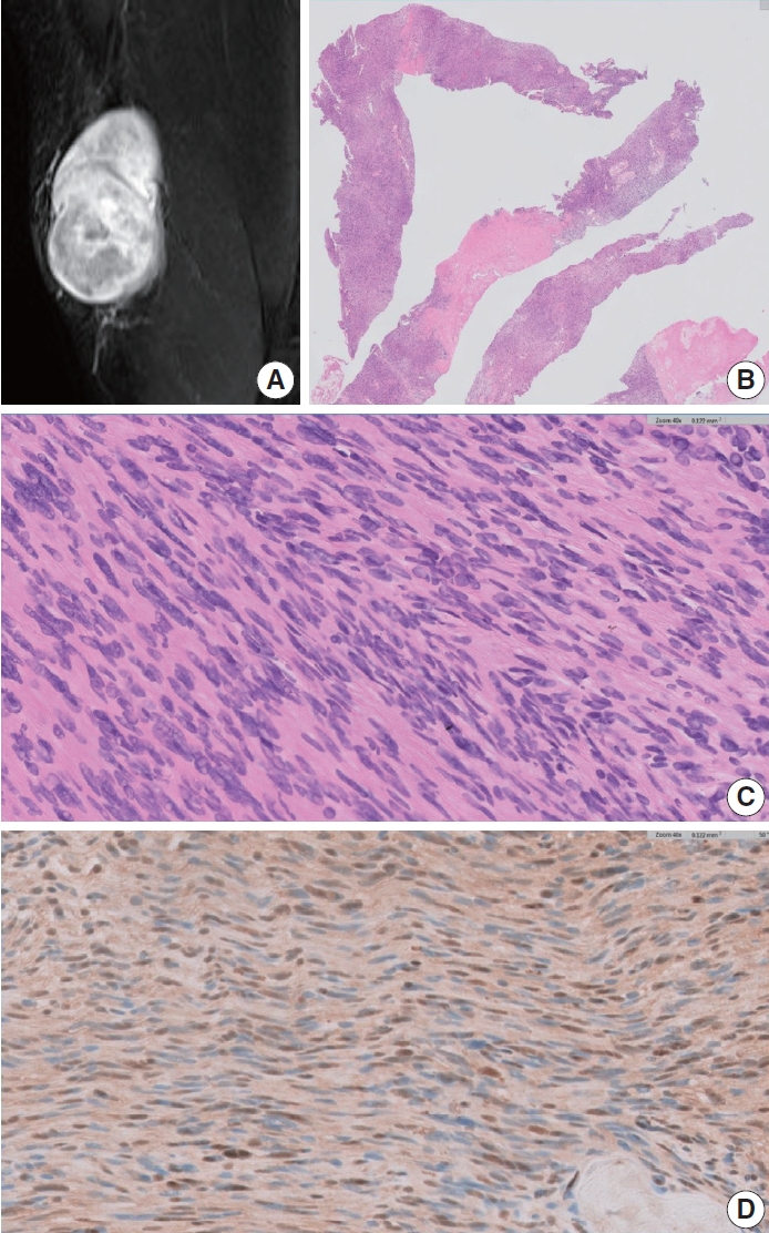
PDF - Schwannomas are benign, slow-growing peripheral nerve sheath tumors commonly occurring in the head, neck, and flexor regions of the extremities. Although most schwannomas are easily diagnosable, their variable morphology can occasionally create difficulty in diagnosis. Reporting pathologists should be aware that schwannomas can exhibit a broad spectrum of morphological patterns. Clinical and radiological examinations can show correlation and should be performed, in conjunction with ancillary tests, when appropriate. Furthermore, deferring a definitive diagnosis until excision may be necessary for small biopsy specimens and frozen sections. This report underscores these challenges through examination of two unique schwannoma cases, one predominantly cellular and the other myxoid, both of which posed significant challenges in histological interpretation.
-
Citations
Citations to this article as recorded by
- Oral and maxillofacial schwannoma (OMSCH): An institutional study of 102 patients
Lingli Huang, Wenya Zhu, Qicheng Ye, Shengwen Liu, Hao Lu, Wenjun Yang, Wanlin Xu
Journal of Stomatology Oral and Maxillofacial Surgery.2026; 127(3): 102678. CrossRef - Plexiform Schwannoma Over the Anterior Chest Wall: A Clinicopathological Review
Debojyoti Sasmal, Saswata Barenya, Hinglaj Saha, Pankaj Kumar Halder
Amrita Journal of Medicine.2025; 21(2): 95. CrossRef - Giant Retroperitoneal Schwannoma: Case Report and Review of the Literature
Magdalena Alexieva, Evgeni V Mekov, Silvia Ivanova, Alexandrina Vlahova, Georgi Yankov
Cureus.2025;[Epub] CrossRef - Breast schwannoma: review of entity and differential diagnosis
Sandra Ixchel Sanchez, Ashley Cimino-Mathews
Journal of Pathology and Translational Medicine.2025; 59(6): 353. CrossRef
- Oral and maxillofacial schwannoma (OMSCH): An institutional study of 102 patients
- Update on the Proposal for Creating a Guideline for Cancer Registration of the Gastrointestinal Tumors (I-2)
- Eun Sun Jung, Yun Kyung Kang, Mee-Yon Cho, Joon Mee Kim, Won Ae Lee, Hee Eun Lee, Sunhoo Park, Jin Hee Sohn, So-Young Jin
- Korean J Pathol. 2012;46(5):443-453. Published online October 25, 2012
- DOI: https://doi.org/10.4132/KoreanJPathol.2012.46.5.443
- 12,891 View
- 191 Download
- 18 Crossref
-
Abstract
PDF
Background Cancer registries play a fundamental role in cancer control and multicenter collaborative research. Recently, the need for reassessment of cancer registry criteria has arisen due to the newly released 2010 World Health Organization (WHO) classification. Accordingly, development of new coding guidelines for cancer is necessary to improve the quality of cancer registries, as well as to prevent conflicts that may arise when seeking medical insurance compensation.
Methods With funding from the Management Center for Health Promotion, 35 members of the Gastrointestinal Pathology Study Group and the Cancer Registration Committee of the Korean Society of Pathologists (KSP) participated in a second workshop for gastrointestinal tumor registration in Korea.
Results The topics of gastric epithelial tumor, colonic intramucosal carcinoma, neuroendocrine tumor (NET), gastrointestinal stromal tumor (GIST) and appendiceal mucinous tumor were discussed for new coding guidelines. A survey was then conducted among 208 members of the KSP for a consensus of the guidelines proposed in the workshop.
Conclusions Although a few issues were set aside for further discussion, such as coding for non-gastric GIST and some types of NET, the members agreed upon most of the proposed guidelines. Therefore, we suggest using the newly revised International Classification of Diseases for Oncology, 3rd edition (ICD-O-3) coding guidelines for registering gastrointestinal tumors in Korea.
-
Citations
Citations to this article as recorded by- Gastrointestinal Stromal Tumor: History, Molecular Subtypes, and Risk Stratification
In Hye Song, Soomin Ahn, Hyung-Don Kim, Jeong-Hyeon Jo, Jinho Shin, Min-Hee Ryu, Young Soo Park
Journal of Gastric Cancer.2026; 26(2): 202. CrossRef - Variation in mitotic counting and risk classification practices for gastrointestinal stromal tumors: a survey of pathologists in South Korea
In Hye Song, Soomin Ahn, Jeong-Hyeon Jo, Young Soo Park
Journal of Pathology and Translational Medicine.2025; 59(5): 348. CrossRef - Different miRNAs Related to FBXW7 Mutations or High Mitotic Indices Contribute to Rectal Neuroendocrine Tumors: A Pilot Study
Ho Suk Kang, Ha Young Park, Hyun Lim, Il Tae Son, Min-Jeong Kim, Nan Young Kim, Min Jeong Kim, Eun Sook Nam, Seong Jin Cho, Mi Jung Kwon
International Journal of Molecular Sciences.2023; 24(7): 6329. CrossRef - Clinicopathologic Impact of Peptide Hormonal Expression in Rectal Neuroendocrine Tumors
Jisup Kim, Dong-Hoon Yang, HaeSung Jung, HyungJun Cho, Hyeung-Jin Jang, Changhoon Yoo, In Ja Park, Baek-Yeol Ryoo, Jin-Sook Ryu, Seung-Mo Hong
Archives of Pathology & Laboratory Medicine.2023; 147(7): 797. CrossRef - Prognostic nomogram and novel risk-scoring system for small cell lung cancer with different patterns of metastases
Hongli Ruan, Huali Sun, Yu Guo, Yan Ding, Yanmei Liu, Shenpeng Ying, Peipei Lin
General Thoracic and Cardiovascular Surgery.2022; 70(12): 1022. CrossRef - Development of a nomogram model to predict survival outcomes in patients with primary hepatic neuroendocrine tumors based on SEER database
Ziteng Zhang, Xin Zhao, Zhiyan Li, Youchun Wu, Yao Liu, Zhiwei Li, Guobao Li
BMC Cancer.2021;[Epub] CrossRef - Standardization of the pathologic diagnosis of appendiceal mucinous neoplasms
Dong-Wook Kang, Baek-hui Kim, Joon Mee Kim, Jihun Kim, Hee Jin Chang, Mee Soo Chang, Jin-Hee Sohn, Mee-Yon Cho, So-Young Jin, Hee Kyung Chang, Hye Seung Han, Jung Yeon Kim, Hee Sung Kim, Do Youn Park, Ha Young Park, So Jeong Lee, Wonae Lee, Hye Seung Lee,
Journal of Pathology and Translational Medicine.2021; 55(4): 247. CrossRef - Analysis of the Incidence and Clinical Features of Colorectal Nonadenocarcinoma in Korea: A National Cancer Registry-Based Study
Soomin Nam, Dongwook Kim, Kyuwon Jung, Yoon Jung Choi, Jung Gu Kang
Annals of Coloproctology.2020; 36(6): 390. CrossRef - Novel Finding of Paired Box 5 (PAX5) Cytoplasmic Staining in Well-differentiated Rectal Neuroendocrine Tumors (Carcinoids) and Its Diagnostic and Potentially Prognostic Utility
Zhiyan Fu, Chunlai Zuo, Christine E. Sheehan, Deepa T. Patil, Jingmei Lin, Zhaohai Yang, Hwajeong Lee
Applied Immunohistochemistry & Molecular Morphology.2019; 27(6): 454. CrossRef - Lymphovascular invasion as a prognostic value in small rectal neuroendocrine tumor treated by local excision: A systematic review and meta-analysis
Ho Suk Kang, Mi Jung Kwon, Tae-Hwan Kim, Junhee Han, Young-Su Ju
Pathology - Research and Practice.2019; 215(11): 152642. CrossRef - Management Colorectal Gastrointestinal Stromal Tumors (Gists) in Surabaya
Yuda Handaya, Sutamto Wibowo, Iwan Kristian
Open Journal of Gastroenterology.2016; 06(04): 97. CrossRef - Non-L-cell Immunophenotype and Large Tumor Size in Rectal Neuroendocrine Tumors Are Associated With Aggressive Clinical Behavior and Worse Prognosis
Joo Young Kim, Ki-Suk Kim, Kyung-Jo Kim, In Ja Park, Jong Lyul Lee, Seung-Jae Myung, Yangsoon Park, Young Soo Park, Chang Sik Yu, Jin Cheon Kim, Eunsil Yu, Hyeung-Jin Jang, Seung-Mo Hong
American Journal of Surgical Pathology.2015; 39(5): 632. CrossRef - Diagnostic Coding for Intramucosal Carcinoma and Neuroendocrine Tumor in the Colorectum: Proposal for Avoiding Confusing Coding in Korea
Dong Soo Han, Jin Hee Sohn, Jeong-Sik Byeon, Hwang Choi, Joon Mee Kim
Clinical Endoscopy.2015; 48(3): 216. CrossRef - Prognostic Significance of Defining L-Cell Type on the Biologic Behavior of Rectal Neuroendocrine Tumors in Relation with Pathological Parameters
Jin Hee Sohn, Mee-Yon Cho, Yangsoon Park, Hyunki Kim, Woo Ho Kim, Joon Mee Kim, Eun Sun Jung, Kyoung-Mee Kim, Jae Hyuk Lee, Hee Kyung Chan, Do Youn Park, Mee Joo, Sujin Kim, Woo Sung Moon, Mi Seon Kang, So-Young Jin, Yun Kyung Kang, Sun Och Yoon, HyeSeung
Cancer Research and Treatment.2015; 47(4): 813. CrossRef - Diminutive and Small Colorectal Polyps: The Pathologist's Perspective
Yun Kyung Kang
Clinical Endoscopy.2014; 47(5): 404. CrossRef - Highlights from the 50th Seminar of the Korean Society of Gastrointestinal Endoscopy
Eun Young Kim, Il Ju Choi, Kwang An Kwon, Ji Kon Ryu, Seok Ho Dong, Ki Baik Hahm
Clinical Endoscopy.2014; 47(4): 285. CrossRef - Early Colorectal Epithelial Neoplasm in Korea: A Multicenter Survey of Pathologic Diagnosis
Yun Kyung Kang, So-Young Jin, Mee Soo Chang, Jung Yeon Kim, Gyeong Hoon Kang, Hye Seung Lee, Jin Hee Sohn, Ho Sung Park, Kye Won Kwon, Mi Jin Gu, Young Hee Maeng, Jong Eun Joo, Haeng Ji Kang, Hee Kyung Kim, Kee-Taek Jang, Mi Ja Lee, Hee Kyung Chang, Joon
Korean Journal of Pathology.2013; 47(3): 245. CrossRef - Expression of metallothionein‐1 and metallothionein‐2 as a prognostic marker in hepatocellular carcinoma
Yangsoon Park, Eunsil Yu
Journal of Gastroenterology and Hepatology.2013; 28(9): 1565. CrossRef
- Gastrointestinal Stromal Tumor: History, Molecular Subtypes, and Risk Stratification
- Proposal for Creating a Guideline for Cancer Registration of Microinvasive Tumors of the Breast and Ovary (II)
- Jin Hee Sohn, Gyungyub Gong, Kyu Rae Kim, Chang Suk Kang, Youn Soo Lee, Jin Man Kim, Woo Hee Jung, Kwang Sun Suh
- Korean J Pathol. 2012;46(3):226-232. Published online June 22, 2012
- DOI: https://doi.org/10.4132/KoreanJPathol.2012.46.3.226
- 12,597 View
- 63 Download
- 2 Crossref
-
Abstract
PDF
Background Cancer registration in Korea has a longer than 30-years of history, during which time cancer registration has improved and become well-organized. Cancer registries are fundamental for cancer control and multi-center collaborative research. However, there have been discrepancies in assigning behavior codes. Thus, we intend to propose appropriate behavior codes for the International Classification of Disease Oncology, 3rd edition (ICD-O-3) for microinvasive tumors of the ovary and breast not only to improve the quality of the cancer registry but also to prevent conflicts.
Methods As in series I, two pathology study groups and the Cancer Registration Committee of the Korean Society of Pathologists (KSP) participated. To prepare a questionnaire on provisional behavior code, the relevant subjects were discussed in the workshop, and consensus was obtained by convergence of opinion from members of KSP.
Results Microinvasive tumor of the breast should be designated as a microinvasive carcinoma which was proposed as malignant tumor (/3). Serous borderline tumor with microinvasion of the ovary was proposed as borderline tumor (/1), and mucinous borderline tumor with microinvasion of the ovary as either borderline (/1) or carcinoma (/3) according to the tumor cell nature.
Conclusions Some issues should be elucidated with the accumulation of more experience and knowledge. Here, however, we present our second proposal.
-
Citations
Citations to this article as recorded by- Update on the Proposal for Creating a Guideline for Cancer Registration of the Gastrointestinal Tumors (I-2)
Eun Sun Jung, Yun Kyung Kang, Mee-Yon Cho, Joon Mee Kim, Won Ae Lee, Hee Eun Lee, Sunhoo Park, Jin Hee Sohn, So-Young Jin
Korean Journal of Pathology.2012; 46(5): 443. CrossRef - A Proposal for Creating a Guideline for Cancer Registration of the Fibromatosis, PEComa Group, Malignant LymphomaIn Situand Dendritic Cell Tumors (III)
Changyoung Yoo, Chang Suk Kang, Yoon La Choi, Hye Yoon Kang, Jin Man Kim, Young Hye Koh, Joo Hee Lee, Seung Sook Lee, In Sun Kim, Dong Hoon Kim, Yong Ku Park, Jin Hee Sohn
Korean Journal of Pathology.2012; 46(5): 436. CrossRef
- Update on the Proposal for Creating a Guideline for Cancer Registration of the Gastrointestinal Tumors (I-2)
- The Analysis of Co-authorship and Networks among the Korean Pathologists.
- Jin Oh Kang, Seo Hyun Park, Yong Koo Park
- Korean J Pathol. 2011;45(3):227-236.
- DOI: https://doi.org/10.4132/KoreanJPathol.2011.45.3.227
- 5,088 View
- 27 Download
- 3 Crossref
-
Abstract
PDF
- BACKGROUND
To evaluate the characteristics of the co-authorship and its network within the Korean Pathologists' Society.
METHODS
In the KoreaMed database, 11,420 articles and 72,478 authors from 1991 to 2010 were searched. The patterns of co-authorship of the authors and institutions were analyzed to build a network matrix. The network centrality indices were measured with UCINET 6.0 and sociogram, and were drawn with Netdraw 5.0. KeyPlayer 1.44 was used for key player analysis.
RESULTS
The number of articles that pathologist participated in increased; however, the number of articles that the pathologists are the first author did not increase. The centrality degrees from 1991 to 2010 were 4.16% and 0.3% for the institutions and authors network, respectively. From 1991 to 2000, Seoul National University had the highest degree of centrality and was a key player. However, from 2001 to 2010, Ulsan replaced the position. For the authors, Chi, Je Geun was highest centrality author and key player during the 1991 to 2000 time period. From 2001 to 2010, Yoo, Jinyoung had the highest degree of centrality and Kim, Na Rae was a key player. Overall, most of the centrality indices were occupied by only a few institutions and authors.
CONCLUSIONS
The network among the pathologist society is a typical small world society. -
Citations
Citations to this article as recorded by- The therapeutic effect of Xuanbai Chengqi Decoction on chronic obstructive pulmonary disease with excessive heat in the lung and fu-organs based on gut and lung microbiota as well as metabolic profiles
Jiao Jiao, Qi Tang, Tie-jie Wang, Jin Fan, Tong-rui Zhang, Kai-shun Bi, Qing Li, Ran Liu
Journal of Chromatography B.2022; 1198: 123250. CrossRef - Generation of Collaboration Network and Analysis of Researcher's Role in National Cancer Center
Hae-Lan Jang
The Journal of the Korea Contents Association.2015; 15(10): 387. CrossRef - Co-authorship patterns and networks of Korean radiation oncologists
Jinhyun Choi, Jin Oh Kang, Seo Hyun Park, Sang Ki Kim
Radiation Oncology Journal.2011; 29(3): 164. CrossRef
- The therapeutic effect of Xuanbai Chengqi Decoction on chronic obstructive pulmonary disease with excessive heat in the lung and fu-organs based on gut and lung microbiota as well as metabolic profiles
- Porposal for Creating a Guideline for Cancer Registration of the Gastrointestinal Tumors (I).
- Mee Yon Cho, Yun Kyung Kang, Kyoung Mee Kim, Hee Kyung Chang, Hee Jin Chang, Mee Soo Chang, Joon Mee Kim, Dae Young Kang, Chanil Park, Jin Hee Sohn
- Korean J Pathol. 2008;42(3):140-150.
- 2,814 View
- 47 Download
-
Abstract
PDF
- BACKGROUND
Cancer registries are fundamental for cancer control and multicenter collaborative research. However, there have been discrepancies among pathologists in classifying cancer and assigning the codes according to the International Classification of Disease Oncology 3 (ICD-O3). To improve the quality of cancer registries as well as to prevent the conflict with medical insurance compensation, a guideline for the coding of cancer is mandatory.
METHODS
AND RESULTS: Funded by the Management Center for Health Promotion, 40 members of the Gastrointestinal Pathology Study Group and the Cancer Registration Committee of the Korean Society of Pathologists participated in the 1st workshop for gastrointestinal tumor registration. The subjects of gastric epithelial tumor, intramucosal carcinoma of the colon, carcinoid tumor, gastrointestinal stromal tumor and appendiceal mucinous tumor were discussed to create a guideline. A survey to obtain consensus for the guideline proposed by the workshop was carried out by the members of the Korean Society of Pathologists and 240 members completed the questionnaire.
CONCLUSION
Although there are some issues to be discussed further, such as coding of high grade dysplasia/adenoma and intramucosal carcinoma of stomach and colon, the members agreed upon most parts of the proposed guideline. Therefore, we suggest using the ICD-O3 coding guideline for gastrointestinal tumor.
- Perspective of The Korean Society of Pathologists.
- Je G Chi
- Korean J Pathol. 1997;31(10):902-908.
- 3,583 View
- 10 Download
-
Abstract
- Only since the introduction of western medicine by Japanese officials and American missionaries in the late 1890's, has the Pathology in its modern concept been considered a major part of basic science in medical schools in Korea, after its role as a hospital service had long been ignored. Limited service of tissue diagnosis on surgical material was the only service performed. Professor Inamoto was the first Japanese pathologist to come to Korea and set up a Pathology Department at the Chosun Chongdogbu Hospital in 1913, and Dr. Mills appears to be the first American hospital pathologist who worked at Severance Hospital in 1913 practicing bacteriology and parasitology as well as lecturing pathology at the medical school. Korea was annexed by Japan from 1910 to 1945. The Korean Society of Pathologists (The former Chosun Society of Pathology) was founded on October 1, 1946, during the turmoil after the end of the Second World War and liberation from Japanese occupation. Only a handful of pathologists gathered for the delivery of the Society. The purpose of the Society was to study, research and exchange information and knowledge in the field of Pathology among its members. Since 1947 the Society had held regular annual academic meetings. In 1950 the Korean War occurred and the Korean Society of Pathologists (KSP) had to restart after the war. The still existing Monthly Slide Conference started in 1959, and the Pathology specialist system was adopted in 1963. There had been a considerable confusion during the adoption period of the pathology specialist system in this country, mainly because of the confused concept of the term "clinical pathology". In its start three categories, i.e., anatomic pathology, clinical pathology, and combined anatomic and clinical pathology were opened. However, the combined training program was eliminated in 1975, which eventually resulted in the separation of clinical pathologists from the KSP to found a new society of Clinical Pathology in 1980 against the advice of the KSP. The first official Journal of the Society, The Korean Journal of Pathology was launched in 1967, marking the 20th anniversary of the Society. It started as a biannual Journal and became a quarterly in 1977. In 1991 the Journal became a bimonthly periodical, and since 1996 the Society issues 12 volumes a year. From 1976, academic activity of the Society was expanded by opening its Spring Meeting in addition to the conventional annual Fall Meeting. In 1992 the Society adopted board of trustee system, providing a fresh blood transfusion. In 1996, the Society commemorated its 50th Anniversary, and published a record book, "The First Fifty Years of The Korean Society of Pathologists". As of December 1996, the Society has a total membership of 500 and 7 special study groups The Society holds 2 annual meetings, monthly slide conferences, several long and short courses, and workshops every year. Approximately 400 papers have been presented each year at the annual meetings. Approximately 350 anatomic pathologists work at hospitals, and a additional 50 pathologists are engaged in full time research at the Department of Pathology in medical schools and other research institutes. As we turn the first half century of founding the Korean Society of Pathologists we realize that we have to be well prepared for various expected and unexpected situations in the future. Enforcement of research pathology at medical schools appears to be the most urgent and important issue. For this purpose, the concept of basic pathology, research pathology, and hospital pathology (surgical pathology) should be clearly established. We also have to clearly define the differnece between anatomic pathology and clinical pathology in this country. At present, the clinical pathology stands alone without any collaboration with the KSP in terms of training program, specialty qualification and hospital practice. Undergraduate pathology education is another issue that we have to pay special attention. The number of full time research pathologists should be increased, and their active and dominant participation in the Society are needed. As the demand for the knowledge and promotions of special field of pathology increases, establishment of additional study group should be encouraged. And if the requirements are met, founding a new Special Pathology Society could also be encouraged. However, the basic skeleton and executive power of the KSP in training residents, qualifying specialty or subspecialty, and in representing the entire pathology field should be maintained and strictly enforced. Hospital pathology has been a dominant drive of the KSP for the last 35 years since the adoption of specialty system. The term, "Diagnostic Pathology" appears to be a term that can replace "Anatomic Pathology", "Surgical Pathology", or "Tissue Pathology" in this country. In future the demand of diagnostic pathology particularly endoscopy biopsy diagnosis, cytological diagnosis and evaluation of surgical operation would be greatly increased. Therefore, we have to be ready for the requirements of professional diagnostician in various fields of pathology as well as overall general diagnostic pathologist. Subspecialty qualification could be expected around the year 2005, when the membership of the Society is expected to be 700. The Korean Journal of Pathology has yet to be improved. It should contain more basic research articles produced by full-time basic pathology researchers. Papers related to hospital pathology (diagnostic pathology) including cytopathology should pursure not only originality but also its practical importance in our situation in this country. The Korean Journal of Pathology should aim for its acception and inclusion in international indexing system in near future.
- Immunohistochemical Study of Gastrointestinal Stromal Tumors.
- Jung Weon Shim, Hye Jae Cho, Ill Hyang Ko, Ok Kyung Kim
- Korean J Pathol. 1991;25(2):93-103.
- 2,002 View
- 10 Download
-
Abstract
PDF
- Historically, gastrointestinal stroma tumors (GIST) have been considered as smooth muscle tumors, but the controversy over this histogenesis is provoked due to various results with utilizing immunohistochemical methods. In andeffort to further clarify the histogenesis of GIST, we performed the immunohistochemical study, as well as histopathologic reexamination, of 24 cases, all diagnosed as smooth muscle tumors of gastrointestinal tract, from Seoul Paik Hospital and Ewha University Hospital between 1980 and 1989, and the main results were as follows; 1) In the histopathologic features by light microscopic study, 11 benign and 13 malignant lesions (including one high grade malignancy and 12 low-grade malignant lesions) were disclosed. 2) In the immunohistochemical study, all tumors showed Vimentin positivity (100%), but no tumor showed S-100 protein positivity (0%), and 7 cases (29.1%) showed Desmin positivity. Positive reaction for Desmin made it possible to suggest that the histogenesis of GIST be in smooth muscle, and neurogenic origin would be excluded by all negativity for S-100 protein. In summary, we would like to conclude that GIST would be smooth muscle tumors on account of their morphological characteristics and their intramural location, but most of them appear poorly differentiated by immunohistochemical method.
 First
First Prev
Prev


