
 E-submission
Search
- Page Path
- HOME > Search
Case Study
- Fibrosarcoma of bone masquerading as fibrous dysplasia: diagnostic pitfall and molecular correlates of aggressive behavior
- Wangpan J. Shi, Brady K. Huang, Li Lei
- J Pathol Transl Med. 2026;60(5):578-586. Published online August 3, 2026
- DOI: https://doi.org/10.4132/jptm.2026.05.29
- 950 View
- 21 Download
-
 Abstract
 PDF
PDF Supplementary Material
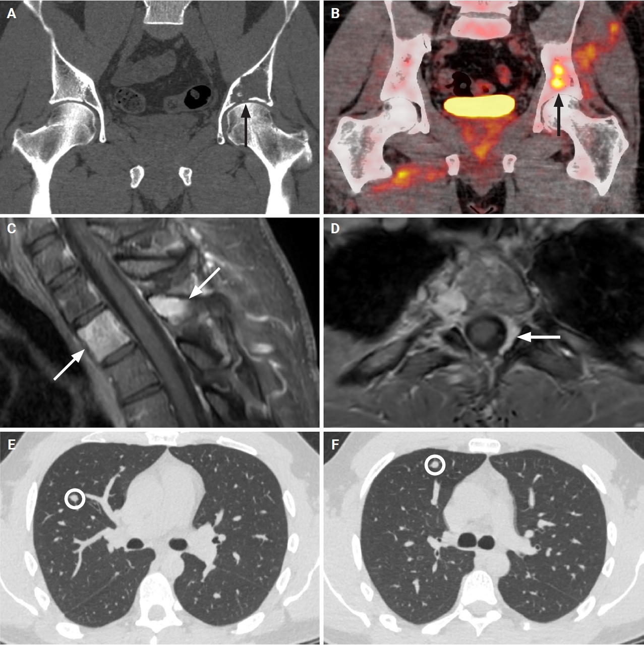
 - Low-grade fibro-osseous lesions can be challenging to classify. A 33-year-old man presented with synchronous lesions involving the ilium and T2 vertebra. Because of an impending pathologic fracture, he received several doses of denosumab. Initial biopsies showed bland fibro-osseous lesions lacking GNAS or MDM2 alterations and were favored to represent polyostotic fibrous dysplasia. Subsequent iliac curettage revealed a fascicular spindle cell proliferation with subtle atypia and focal ossification, leading to a revised diagnosis of low-grade fibrosarcoma. Re-biopsy of the T2 lesion demonstrated a similar spindle cell proliferation without ossification. Sequencing identified copy number gains involving KIT, PDGFRA, and TERT. At 16-month follow-up, two metastatic pulmonary nodules developed, one responsive to chemotherapy. The patient remains alive with disease at 27 months after presentation. This case highlights a diagnostic pitfall in which low-grade fibrosarcoma with denosumab-associated ossification may mimic fibrous dysplasia and underscores the prognostic value of molecular profiling beyond histologic grading.
Case Report
- Liposclerosing Myxofibrous Tumor in Tibia: A Case Report and Review of the Literature.
- Jung Woo Choi, Young Seok Lee, Ju Han Lee, Han Kyeom Kim, Bom Woo Yeom, Jong Sang Choi, Hong Chul Lim, Chul Hwan Kim
- Korean J Pathol. 2005;39(3):207-210.
- 2,982 View
- 51 Download
-
Abstract
PDF
- Liposclerosing myxofibrous tumor (LSMFT) is a benign fibro-osseous lesion that is characterized by a complex mixture of histologic elements, including its fibrous dysplasia-like features and its lipoma, myxofibroma, xanthoma and pseudo-Paget's bone patterns. However, this lesion is considered by some researchers as a variant of fibrous dysplasia or as the non-specific end result of degenerative change, while it is considered by others as a definite clinicopathologic entity. Here, we report on a case of LSMFT occurring in tibia, which is a very uncommon location for this tumor, and we review the related literatures. The case presented here shares features with those described for LSMFT, except for the location of this tumor. We believe that more studies on a larger scale that compare LSMFT with other benign bone lesions, including fibrous dysplasia, are required to clarify the origin and behavior of this lesion.
 First
First Prev
Prev



