 E-submission
Search
- Page Path
- HOME > Search
Case Study
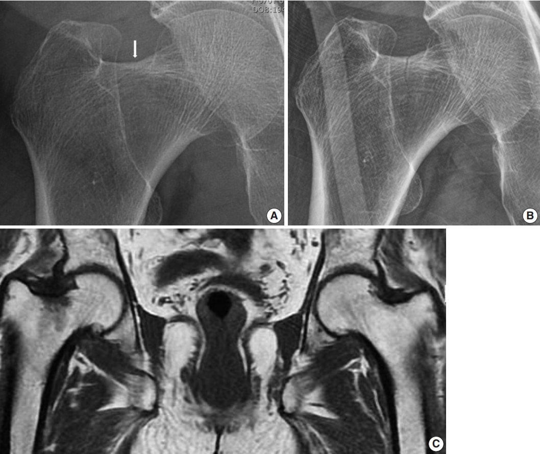
- Atypical femoral neck fracture after prolonged bisphosphonate therapy
- Kwang-kyoun Kim, Young-wook Park, Tae-hyeong Kim, Kyung-deok Seo
- J Pathol Transl Med. 2020;54(4):346-350. Published online June 29, 2020
- DOI: https://doi.org/10.4132/jptm.2020.05.14
- Correction in: J Pathol Transl Med 2020;54(5):435
- 6,763 View
- 140 Download
- 10 Web of Science
- 11 Crossref
-
 Abstract
 PDF
PDF - Of the drugs developed to prevent and treat osteoporosis, bisphosphonate has played a very important role in preventing osteoporotic fractures. However, case reports describing atypical femoral fractures in patients using long-term bisphosphonates have emerged. The majority of atypical femur fractures occurs in the lateral aspect of the subtrochanteric or femur diaphysis, which is explained by accumulation of tensile stress in these areas. Although the superior cortex of the femur neck withstands maximum tensile stress, to our knowledge, there have been only two reports (three cases) of atypical femoral neck fracture. In addition, none of those case reports revealed detailed pathology related to suppressed bone turnover rate. We encountered an incomplete femoral neck fracture and diagnosed it as “atypical” on the basis of the patient’s lack of trauma and medication history and pathological findings. For patients with groin pain, minimal or no trauma, and a history of long-term bisphosphonate use, an atypical femoral neck fracture should be considered.
-
Citations
Citations to this article as recorded by
- Frequency and Classification of Femoral Neck Fractures: A Cross-Sectional Study From a Tertiary Care Hospital in Peshawar, Pakistan
Muhammad Mannan, Zawar Ahmad, Syed Muneeb Ali Shah, Muhammad Tayyab, Faisal Karim, Rehan Raza Shan, Muhammad Zeeshan Akram
Cureus.2025;[Epub] CrossRef - Asian Federation of Osteoporosis Societies 2025 consensus on atypical femoral fractures in patients with osteoporosis
Thanut Valleenukul, Thawee Songpatanasilp, Unnop Jaisamrarn, Surapong Anuraklekha, Varalak Srinonprasert, Sumapa Chaiamnuay, Aasis Unnanuntana, Lalita Wattanachanya, Hataikarn Nimitphong, Noratep Kulachote, Ong-art Phruetthiphat, Rahat Jarayabhand, Tanawa
Osteoporosis and Sarcopenia.2025; 11(4): 111. CrossRef - Improvement of osteoblast adhesion, viability, and mineralization by restoring the cell cytoskeleton after bisphosphonate discontinuation in vitro
Somying PATNTIRAPONG, Chunya CHAMPAKERDSAP, Pichaya MATHAVEECHOTIKUL, Apichaya VATANASILP
Journal of Applied Oral Science.2024;[Epub] CrossRef - Is there genetic susceptibility for atypical femoral fractures?
Maximilian Ellacott, Hüseyin Bilgehan Çevik, Peter V. Giannoudis
Injury.2024; 55(2): 111312. CrossRef - Incomplete femoral neck fracture with characteristics of atypical femoral fracture: A case report and literature review
Yasuhiro Higai, Yuji Kanaya, Hiroyoshi Hagiwara, Yuichirou Yano, Takashi Fukushima, Norihiro Akazawa, Takahiro Shimizu, Sueo Nakama, Katsushi Takeshita
Trauma Case Reports.2024; 53: 101091. CrossRef - Clinical value of quantitative parameters of MSCT in the diagnosis of occult femoral neck fractures
Yongzhong Xu, Shan Gao
Biotechnology and Genetic Engineering Reviews.2023; : 1. CrossRef - Femoral neck stress fracture return to activity and the effect of metabolic dysfunction on recovery: A systematic review
Kristine Yang, Senthil Sambandam, Matthew J. Yan, Michael Huo
Journal of Orthopaedics.2023; 43: 79. CrossRef - Association of atypical femoral fracture location and lower limb mechanical axis: a computed tomography-based finite element analysis
Donghwan Hwang, Chul-Ho Kim, Yongkoo Lee, Ji Wan Kim
Osteoporosis International.2022; 33(6): 1285. CrossRef - Bilateral Bisphosphonate-related Atypical Femoral Neck Fracture in a Patient with Familial Mediterranean Fever: A Case Report
Sibel Balıkçı, Bilinç Doğruöz Karatekin
Turkish Journal of Osteoporosis.2022; 28(1): 74. CrossRef - Clinical Outcomes and Radiologic Characteristics of Insufficiency Femoral Neck Fracture in Elderly Patients
Hee-Uk Ye, Kyung-Jae Lee, Byung-Woo Min, Kyung-Hwan Lim, Beom-Soo Kim, Young-Hoon Kim
Journal of the Korean Fracture Society.2021; 34(1): 1. CrossRef - Ibandronic acid
Reactions Weekly.2020; 1827(1): 165. CrossRef
- Frequency and Classification of Femoral Neck Fractures: A Cross-Sectional Study From a Tertiary Care Hospital in Peshawar, Pakistan
Case Report
- Intraosseous Well Differentiated Osteosarcoma: A case report.
- Mee Hye Oh, So Young Park, Yeon Lim Suh, Shin Khang Kang
- Korean J Pathol. 1992;26(6):627-631.
- 2,105 View
- 16 Download
-
Abstract
PDF
- Well differentiated osteosarcomas are variants of osteosarcoma composed mainly of fibrous and osseous tissue with minimal cystologic atypia. This tumor may be misinterpretated as a benign lesion if the radiologic and clinical features are not taken into account. We report a typical case of intraosseous well differentiated osteosarcoma occuring in the left distal femur of a 58-year-old woman. Radiologically, it appered as an ill-defined lesion with a mixture of sclerotic and osteolytic ares. But there was a lack of highly destructive appearance of conventional osteosarcoma. Grossly, the mass occupied a metaphysis of the distal femur with extension into the diaphysis and epiphysis. Multifocal cortical destruction and sclerosis were also associated. Histologically, the mass showed typical features of intraosseous well differentiated osteosarcoma. There were various patterns of osteoid deposits and bone formation mimicking those of fibrous dysplasia, nonossifying fibroma or parosteal osteosarcoma.
 First
First Prev
Prev


