 E-submission
Search
- Page Path
- HOME > Search
Review Article
- Ganglioglioma and gangliocytoma: a review for pathologists
- Gianfranco E. Umeres-Francia, Melissa Mejia-Bautista, Pouya Jamshidi, Jared T. Ahrendsen
- J Pathol Transl Med. 2026;60(4):379-387. Published online July 15, 2026
- DOI: https://doi.org/10.4132/jptm.2026.06.06
- 537 View
- 18 Download
-
 Abstract
 PDF
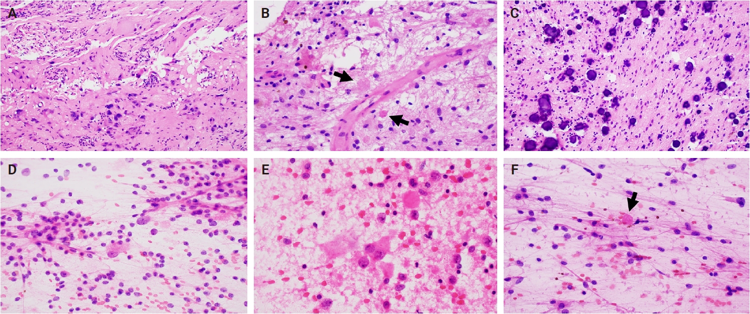
PDF - Ganglioglioma and gangliocytoma are rare, predominantly low-grade neuroepithelial tumors that commonly present with epilepsy in children and young adults. Advances in molecular profiling have improved understanding of their pathogenesis, highlighting key roles for the mitogen-activated protein kinase/ERK signaling pathway. Diagnosis relies on a combination of clinical, radiologic, and histopathologic features, with complete surgical resection offering the best clinical outcomes. This review summarizes current knowledge on their epidemiology, etiology, clinical presentation, imaging characteristics, pathology, treatment strategies, and prognosis.
Review
- Dysembryoplastic Neuroepithelial Tumors
- Yeon-Lim Suh
- J Pathol Transl Med. 2015;49(6):438-449. Published online October 23, 2015
- DOI: https://doi.org/10.4132/jptm.2015.10.05
- 19,640 View
- 305 Download
- 29 Web of Science
- 35 Crossref
-
Abstract
PDF
- Dysembryoplastic neuroepithelial tumor (DNT) is a benign glioneuronal neoplasm that most commonly occurs in children and young adults and may present with medically intractable, chronic seizures. Radiologically, this tumor is characterized by a cortical topography and lack of mass effect or perilesional edema. Partial complex seizures are the most common presentation. Three histologic subtypes of DNTs have been described. Histologically, the recognition of a unique, specific glioneuronal element in brain tumor samples from patients with medically intractable, chronic epilepsy serves as a diagnostic feature for complex or simple DNT types. However, nonspecific DNT has diagnostic difficulty because its histology is indistinguishable from conventional gliomas and because a specific glioneuronal element and/or multinodularity are absent. This review will focus on the clinical, radiographic, histopathological, and immunohistochemical features as well as the molecular genetics of all three variants of DNTs. The histological and cytological differential diagnoses for this lesion, especially the nonspecific variant, will be discussed.
-
Citations
Citations to this article as recorded by
- A Case of Dysembryoplastic Neuroepithelial Tumor in an HIV-Positive Adult: Diagnostic Lessons for Clinicians
Mariana Lobo, Susana Viana, Andreia Sá Lima, Isabel Monteiro, Carolina M Cerqueira, Frederico Duarte, Luís M Ribeiro, Sara Camões
Cureus.2026;[Epub] CrossRef - Magnetic resonance imaging findings of dysembryoplastic neuroepithelial tumors and low-grade astrocytomas
Kai-Wei Yu, Shih-Chieh Lin, Hsin-Hung Chen, Chia-Hung Wu, Wei-An Tai, Chung-Han Yang, Te-Ming Lin, Feng-Chi Chang
Journal of the Chinese Medical Association.2026; 89(3): 228. CrossRef - Imaging diagnosis of cystic intraparenchymal brain neoplasms
Sonoko Oshima, Yasutaka Fushimi, Sachi Okuchi, Satoshi Nakajima, Akihiko Sakata, Takayuki Yamamoto, Yuji Nakamoto, Noriko Salamon
Japanese Journal of Radiology.2026;[Epub] CrossRef - Histopathological and molecular heterogeneity of dysembryoplastic neuroepithelial tumors
Yuxiu Wang, Sarra Belakhoua, Yiying Yang, Jonathan Serrano, Craig Horbinski, Daniel R Boué, John C DeWitt, Benjamin Liechty, Declan McGuone, Qinwen Mao, Olga Krasnozhen-Ratush, Stephen Yip, Christopher Dunham, Melissa Umphlett, Seema Shroff, Matija Snuder
Journal of Neuropathology & Experimental Neurology.2026;[Epub] CrossRef - An Imaging Review of Common Pediatric Brain Tumors
Joseph Yang, Brandon Collins, Matthew Beniuk, Alexandra Hodder, Dani Bahnam, Angela Pickles
Roentgen Ray Review.2025;[Epub] CrossRef - Ruptured intratumoral arteriovenous malformation in a patient with dysembryoplastic neuroepithelial tumor: A case report
Takashi Aoka, Masaaki Nishimoto, Hideki Ogiwara
Surgical Neurology International.2025; 16: 375. CrossRef - Pediatric Neuroglial Tumors: A Review of Ependymoma and Dysembryoplastic Neuroepithelial Tumor
Melissa Arfuso, Sandeepkumar Kuril, Harshal Shah, Derek Hanson
Pediatric Neurology.2024; 156: 139. CrossRef - From bedside to bench: New insights in epilepsy‐associated tumors based on recent classification updates and animal models on brain tumor networks
Silvia Cases‐Cunillera, Lea L. Friker, Philipp Müller, Albert J. Becker, Gerrit H. Gielen
Molecular Oncology.2024; 18(12): 2951. CrossRef - Imaging of pediatric glioneuronal and neuronal tumors
Vivek Pai, Suzanne Laughlin, Birgit Ertl-Wagner
Child's Nervous System.2024; 40(10): 3007. CrossRef - Dysembryoplastic Neuroepithelial Tumor: A Case Report of A Benign Intracranial Lesion Masquerading as Seizure Disorder
Garima S Agarwal, Anil K Agrawal, Daksh Singhal, Jayashree Bhawani
Cureus.2024;[Epub] CrossRef - Super T2-FLAIR mismatch sign: a prognostic imaging biomarker for non-enhancing astrocytoma, IDH-mutant
Iori Ozono, Shumpei Onishi, Ushio Yonezawa, Akira Taguchi, Novita Ikbar Khairunnisa, Vishwa Jeet Amatya, Fumiyuki Yamasaki, Yukio Takeshima, Nobutaka Horie
Journal of Neuro-Oncology.2024; 169(3): 571. CrossRef - Genotype-relevant neuroimaging features in low-grade epilepsy-associated tumors
Keiya Iijima, Hiroyuki Fujii, Fumio Suzuki, Kumiko Murayama, Yu-ichi Goto, Yuko Saito, Terunori Sano, Hiroyoshi Suzuki, Hajime Miyata, Yukio Kimura, Takuma Nakashima, Hiromichi Suzuki, Masaki Iwasaki, Noriko Sato
Frontiers in Neurology.2024;[Epub] CrossRef - Extra-temporal pediatric low-grade gliomas and epilepsy
José Hinojosa, Victoria Becerra, Santiago Candela-Cantó, Mariana Alamar, Diego Culebras, Carlos Valencia, Carlos Valera, Jordi Rumiá, Jordi Muchart, Javier Aparicio
Child's Nervous System.2024; 40(10): 3309. CrossRef - Atypical Presentation of Dysembryoplastic Neuroepithelial Tumor
Varis S. Khalilov, Aleksey N. Kislyakov, Natalia A. Medvedeva, Natalia S. Serova
Annals of Clinical and Experimental Neurology.2024; 18(3): 109. CrossRef - Unusual low-grade neuroepithelial tumour in a child
Leia Salongo, Ali Nael, Pournima Navalkele, John Ross Crawford
BMJ Case Reports.2024; 17(10): e262692. CrossRef - Glioneuronal and Neuronal Tumors: Who? When? Where? An Update Based on the 2021 World
Health Organization Classification
A.S. Ayres, G.A. Bandeira, S.F. Ferraciolli, J.T. Takahashi, R.A. Moreno, L.F. de Souza Godoy, Y.R. Casal, L.G.C.A. de Lima, F.P. Frasseto, L.T. Lucato
Neurographics.2023; 13(1): 1. CrossRef - Biological functions of the Olig gene family in brain cancer and therapeutic targeting
Jenny I. Szu, Igor F. Tsigelny, Alexander Wojcinski, Santosh Kesari
Frontiers in Neuroscience.2023;[Epub] CrossRef - Aspekte der Bildgebung des Hippokampus
Isabela S. Alves, Artur M. N. Coutinho, Ana Vieira, Bruno P. Rocha, Ula L. Passos, Vinicius T. Gonçalves, Paulo D. S. Silva, Malia X. Zhan, Paula C. Pinho, Daniel S. Delgado, Marcos F. L. Docema, Hae W. Lee, Bruno A. Policeni, Claudia C. Leite, Maria G. M
Neuroradiologie Scan.2023; 13(03): 197. CrossRef - T2-FLAIR Mismatch Sign in Pediatric Low-Grade Glioma
M.W. Wagner, L. Nobre, K. Namdar, F. Khalvati, U. Tabori, C. Hawkins, B.B. Ertl-Wagner
American Journal of Neuroradiology.2023; 44(7): 841. CrossRef - Clinicopathological features of dysembryoplastic neuroepithelial tumor: a case series
Shabina Rahim, Nasir Ud Din, Jamshid Abdul-Ghafar, Qurratulain Chundriger, Poonum Khan, Zubair Ahmad
Journal of Medical Case Reports.2023;[Epub] CrossRef - Imaging Aspects of the Hippocampus
Isabela S. Alves, Artur M. N. Coutinho, Ana P. F. Vieira, Bruno P. Rocha, Ula L. Passos, Vinicius T. Gonçalves, Paulo D. S. Silva, Malia X. Zhan, Paula C. Pinho, Daniel S. Delgado, Marcos F. L. Docema, Hae W. Lee, Bruno A. Policeni, Claudia C. Leite, Mari
RadioGraphics.2022; 42(3): 822. CrossRef - Dysembryoplastic neuroepithelial tumors: A single-institutional series with special reference to glutamine synthetase expression
Chiara Caporalini, Mirko Scagnet, Selene Moscardi, Gioia Di Stefano, Gianna Baroni, Flavio Giordano, Federico Mussa, Carmen Barba, Iacopo Sardi, Lorenzo Genitori, Anna Maria Buccoliero
Annals of Diagnostic Pathology.2021; 54: 151774. CrossRef - Unusual case of occipital lobe dysembryoplastic neuroepithelial tumour with GNAi1-BRAF fusion
Jennifer H Yang, Denise M Malicki, Michael L Levy, John Ross Crawford
BMJ Case Reports.2021; 14(1): e241440. CrossRef - Malformations of Cortical Development, Cognitive Involvementand Epilepsy: A Single Institution Experience in 19 Young Patients
Valeria Venti, Maria Chiara Consentino, Pierluigi Smilari, Filippo Greco, Claudia Francesca Oliva, Agata Fiumara, Raffaele Falsaperla, Martino Ruggieri, Piero Pavone
Children.2021; 8(8): 637. CrossRef - A Case of Dysembryoplastic Neuroepithelial Tumor in an Adolescent Male
Marcel Yibirin, Diana De Oliveira, Isabella Suarez, Gabriela Lombardo, Carlos Perez
Cureus.2021;[Epub] CrossRef - Clinical and histopathological profile of dysembryoplastic neuroepithelial tumor
Pooja Gupta, Fouzia Siraj, Akanksha Malik, K. B. Shankar
Journal of Cancer Research and Therapeutics.2021; 17(4): 912. CrossRef - Neuroradiological and pathomorphological features of epilepsy associated brain tumors
V. S. Khalilov, A. A. Kholin, A. N. Kisyakov, N. A. Medvedeva, B. R. Bakaeva
Diagnostic radiology and radiotherapy.2021; 12(2): 7. CrossRef - Evaluación prequirúrgica mediante resonancia magnética funcional en pacientes con tumores neuroepiteliales disembrioplásicos: una serie de casos
Natalia García-Casares, Francisco Alfaro-Rubio, José Ramón Ramos-Rodríguez, Álvaro Ocaña-Ledesma, Bernarda Márquez-Márquez, Victoria E. Fernández-Sánchez, Guillermo Ibáñez-Botella, Miguel Ángel Arráez-Sánchez, Pedro J. Serrano-Castro
Neurocirugía.2020; 31(4): 158. CrossRef - Features of the neuroradiological picture of ganglioglioma on the example of 20 clinical cases
V.S. Khalilov, A.A. Kholin, Kh.Sh. Gazdieva, A.N. Kislyakov, N.N. Zavadenko
Zhurnal nevrologii i psikhiatrii im. S.S. Korsakova.2020; 120(11): 90. CrossRef - Malignant Glial Neuronal Tumors After West Nile Virus Neuroinvasive Disease: A Coincidence or a Clue?
Akanksha Sharma, Marie F. Grill, Scott Spritzer, A. Arturo Leis, Mark Anderson, Parminder Vig, Alyx B. Porter
The Neurohospitalist.2019; 9(3): 160. CrossRef - Particularities in differential diagnostics of epileptogenic brain malformations on the low-field MRI-device
V. S. Khalilov, A. A. Kholin, B. R. Bakaeva, M. Yu. Bobylova, Kh. Sh. Gazdieva
Russian Journal of Child Neurology.2019; 13(4): 23. CrossRef - Dysembryoplastic Neuroepithelial Tumors: What You Need to Know
Sabino Luzzi, Angela Elia, Mattia Del Maestro, Samer K. Elbabaa, Sergio Carnevale, Francesco Guerrini, Massimo Caulo, Patrizia Morbini, Renato Galzio
World Neurosurgery.2019; 127: 255. CrossRef - The miR‐139‐5p regulates proliferation of supratentorial paediatric low‐grade gliomas by targeting the PI3K/AKT/mTORC1 signalling
G. Catanzaro, Z. M. Besharat, E. Miele, M. Chiacchiarini, A. Po, A. Carai, C. E. Marras, M. Antonelli, M. Badiali, A. Raso, S. Mascelli, D. Schrimpf, D. Stichel, M. Tartaglia, D. Capper, A. von Deimling, F. Giangaspero, A. Mastronuzzi, F. Locatelli, E. Fe
Neuropathology and Applied Neurobiology.2018; 44(7): 687. CrossRef - Dysembryoplastic Neuroectodermal Tumor: An Analysis from the Surveillance, Epidemiology, and End Results Program, 2004–2013
Ha Son Nguyen, Ninh Doan, Michael Gelsomino, Saman Shabani
World Neurosurgery.2017; 103: 380. CrossRef - Common Histologically Benign Tumors of the Brain
Roy E. Strowd, Jaishri O. Blakeley
Continuum.2017; 23(6): 1680. CrossRef
- A Case of Dysembryoplastic Neuroepithelial Tumor in an HIV-Positive Adult: Diagnostic Lessons for Clinicians
Original Articles
- Non-neoplastic Lesions in Temporal Lobe Epilepsy: A Pathologic Review of 64 cases.
- Sang Pyo Kim, Kun Young Kwon, Eun Sook Chang, Kwan Kyu Park, Sang Do Yi, Eun Ik Son
- Korean J Pathol. 1996;30(4):281-292.
- 2,441 View
- 20 Download
-
Abstract
PDF
- Temporal lobe epilepsy is characterized by complex partial seizures with either primary intracranial neoplasms or other non-neoplastic lesions. We reviewed 64 cases of surgically resected temporal lobes and amygdalo-hippocampal regions for temporal lobe epilepsy ansed by non-neoplastic lesions to elucidate the incidence and histologic features of each histologic group for a period of 2 years. The patient's age ranged from 12 to 49 years and the ratio of male to female was 42:22. There were 37 cases(57.8%) with single pathology and an additional 20 cases(31.3%) with dual pathology. The emaining 7 cases(10.9%) had no structural alternations. The most common temporal lobe pathology was hippocampal sclerosis in 41 cases(64.1%), diagnosed alone in 21 cases and as dual lesions in 20 cases. The hippocampal neuron loss was most pro,omemt in CA1, followed by CA4, CA3, and CA2. Amygdaloid sclerosis was present in 28 cases(43.8%), lases had 13 dual lesions, 25 cases also had hippocampal sclerosis. The 20 dual lesions showed that 6 cortical dysplasia, 10 microdysgenesis, 1 chronic non-specific inflammatory lesion, and 3 cysticercosis were associated with the various degree of mesial temporal sclerosis. Neuronoglial malformative lesions were identified in 21 cases(32.8%) including 16 dual lesion cases, which composed of 15 microdysgenesis and 6 cortical dysplasia. Neurofilament immunostain for cortical dysplasia revealed abnormally beaded disarray of axons in dysplastic pyramidal cells. The remaining pathologic lesions observed were 1 cysticercosis, 1 chronic non-specific inflammatory lesion, 3 arteriovenous malformation, 2 fibrous nodule, and 1 fibrous adhesions of the arachnoid.
- Pathologic Analysis of 39 Cases of Epilepsy Surgery.
- Young Mee Cho, Joong Koo Kang, Youn Mee Hwang, Jung Kyo Lee, Ghee Young Choe
- Korean J Pathol. 1996;30(5):388-395.
- 2,136 View
- 17 Download
-
Abstract
PDF
- Pharmacologic therapy is still the primary management for epilpsy; however, surgical treatment is a reasonable therapeutic option for patients suffering from medically intractable seizures, especially temporal lobe epilepsy having a documented unilateral epileptogenic area. Thirty nine patients with pharmaco-resistant complex partial seizures underwent anterior temporal lobectomy and hippocampectomy in 38 cases and frontal cortisectomy in one case. On pathological examination, hippocampal sclerosis was a predominent pathologic finding and was identified in 18 cases. Other non-neoplastic lesions consisted of 5 cases of vascular lesions(2 cavernous angiomas, 2 arteriovenous malformations and 1 angiomatosis), 3 cases of fibrous nodule, 2 cases of cicatrical changes of cerebral cortex, and 1 case of parasitic infection. Neoplasms including two cases of oligodendroglioma and one case of anaplastic astrocytoma were also noted. In seven cases, there was no detectable lesion on gross and microscopic examination. On post-operative follow-up, seizures were completely terminated in most cases(31 cases, 79%). The rest of the patients also displayed marked alleviation of symptoms. The seizures tended to recur more aften among the patients with neoplasm or no pathologically detectable lesion. In order to detect any minute pathological lesion, thorough gross and microcsopic examinations are considered to be essential.
- Comparison of Pathologic Findings of Cortical Lobectomy for Intractable Seizures between Children and Adults: An Analysis of 164 Cases.
- Na Rae Kim, Yeon Lim Suh
- Korean J Pathol. 1999;33(12):1175-1181.
- 2,072 View
- 14 Download
-
Abstract
PDF
- Many pathological surveys of brain tissue in patients with intractable epilepsy have been reported. There have been, however, few studies focused on the differences between childhood and adults in pathological alterations of brain. We retrospectively analyzed histopathology of 164 lobectomy specimens for intractable epilepsy in view of the differences between children and adults. Among 164 cases, 28 cases were children (less than 15 years) and 136 cases adults. We compared frequency of histopathologic features, distribution of involved cortex (temporal or extratemporal lobe), previous injury histories, such as brain trauma, encephalitis or febrile seizure, and coexistence of other lesions (dual pathology) between two groups. Pathologic alterations were encountered in 92% of 164 patients. In children focal cortical dysplasia (n=16, 57.1%), neoplasm (n=8, 28.6%), hippocampal sclerosis (n=6, 21.4%), cortical tuber (n=1, 3.6%), leukomalacia (n=1, 3.6%), and Rasmussen's encephalitis (n=1, 3.6%) were observed, whereas focal cortical dysplasia (n=81, 59.6%), hippocampal sclerosis (n=80, 58.8%), neoplasm (n=19, 14%), and cerebral cysticercosis (n=3, 2.2%) were found in adults. Pediatric patients had a higher proportion of severe focal cortical dysplasia (17.9% in children, 0.7% in adults). Neoplasia and extratemporal lobe involvement were more commonly found in children (28.6%, 50%) than in adults (14.0%, 24.3%), whereas hippocampal sclerosis and dual pathology were more common in adults (58.8%, 44.9%) than in children (21.4%, 17.9%). Previous injury history was statistically significant in patients with hippocampal sclerosis, and lent support to the hypothesis that hippocampal sclerosis is related with acquired lesions. Incidence of focal cortical dysplasia was nearly similar in both adult (59.6%) and pediatric groups (57.1%), and supported the hypothesis that focal cortical dysplasia is developmental abnormality occurring during a prenatal period.
Case Report
- Suprasellar Endodermal Sinus Tumor Presenting with Tonic-Clonic Seizure: An Autopsy Case Report.
- Min Jin Lee, Hea Soo Koo, Woon Sup Han, Sun Hee Sung, Yong Jae Kim, Hye Young Choi
- Korean J Pathol. 2002;36(6):433-439.
- 2,211 View
- 13 Download
-
Abstract
PDF
- We report the clinical course and autopsy findings of a 19-year-old girl with endodermal sinus tumor involving the thalamus, hypothalamus, and basal ganglia. The patient initially had tonic-clonic seizures with abnormal signal involving the right hippocampus, amygdala, basal ganglia, putamen, and dentate gyrus. The signal intensity of the posterior pituitary on T1-weighted images was decreased at the time of admission, which was not associated with clinical symptoms of diabetes insipidus (DI). A huge tumor mass as well as central DI developed within 10 months. The postmortem examination showed gliosis with calcification involving the right basal ganglia, internal capsule, and white matter, in addition to a tumor mass involving the thalamus, hypothalamus, and basal ganglia. Dissemination of tumor cells in the leptomeninges and the gliotic area and hydrocephalus were also noted.
Original Article
- Synaptic Reorganization of Dentate Mossy Fibers and Expression of Calcium Binding Proteins in Hippocampal Sclerosis of Temporal Lobe Epilepsy.
- Sang Pyo Kim, Seung Pil Kim, Seung Che Cho, Young Rok Cho, Ji Min Jeon, Yu Na Kang, Kun Young Kwon, Eun Sook Chang
- Korean J Pathol. 1998;32(5):328-336.
- 2,301 View
- 10 Download
-
Abstract
- This study was designed to identify expression of calcium-binding proteins and synaptic reorganizations of dentate mossy fibers in hippocampal sclerosis of human temporal lobe epilepsy. Hippocampal neuronal density was quantitively analyzed in temporal lobe epilepsy group (n=50) to investigate the degree of hippocampal sclerosis and it was compared with that of autopsy control (n=3). To verify the distribution of calcium-binding proteins in neurons of epileptic hippocampi, the parvalbumin (PV)-immunoreactive and calbindin-D28K (CB)-immunoreactive neurons were quantitively analyzed in each area of Ammon's horn by immunohistochemical stain. Also, to clarify synaptic reorganizations of the dentate mossy fibers, a part of each hippocampus was examined under light microscopy and transmission electron microscopy using Timm sulphide silver method. In epileptic hippocampi, severity of hippocampal sclerosis (HS) was graded four, which consisted of 3 cases with no HS, 6 mild HS, 12 moderate HS, and 29 severe HS. The hippocampal neuronal loss was most prominent in CA1, followed by CA4 and CA2. Expression of calcium-binding proteins was more prevalent in CA2 of all groups. The proportion of PV-immunoreactive neurons in CA1 and CA4 significantly increased in the moderate and severe HS group, whereas the proportion of CB-immunoreactive neurons did not correlated with the severity of HS. Timm granules were noted in inner molecular supragranular layer of dentate gyrus of epileptic hippocampi and they tended to increase in proportion along with the severity of hippocampal sclerosis. Transmission electron microscopy showed that supragranular Timm granules corresponded to synaptic terminals of mossy fibers. These results suggest that parvalbumin appears to have more protective effect against neuronal loss and that mossy fiber synaptic reorganization seems to play a major role in pathogenesis of hippocampal sclerosis of human temporal lobe epilepsy.
 First
First Prev
Prev


