 E-submission
Search
- Page Path
- HOME > Search
- Frequent apocrine changes in pleomorphic adenoma with malignant transformation: a possible pre-malignant step in ductal carcinoma ex pleomorphic adenoma
- Joon Seon Song, Yeseul Kim, Yoon-Se Lee, Seung-Ho Choi, Soon Yuhl Nam, Sang Yoon Kim, Kyung-Ja Cho
- J Pathol Transl Med. 2023;57(3):158-165. Published online May 10, 2023
- DOI: https://doi.org/10.4132/jptm.2023.03.13
- 7,350 View
- 196 Download
- 4 Web of Science
- 4 Crossref
-
 Abstract
 PDF
PDF - Background
The most common type of carcinoma ex pleomorphic adenoma (CPA) is histologically equivalent to salivary duct carcinoma, which has an apocrine phenotype. Invasive CPA is often accompanied by non-invasive or in situ carcinoma, an observation that suggests the presence of precursor lesions. The aim of this study was to identify candidate precursor lesions of CPA within pleomorphic adenoma (PA).
Methods
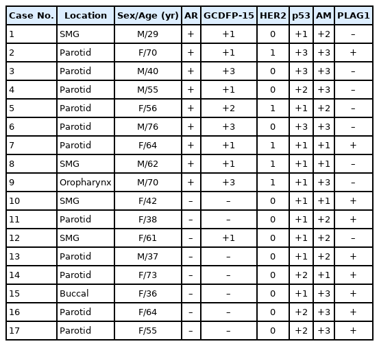
Eleven resected cases of CPA with residual PA and 17 cases of PA with atypical changes were subjected to immunohistochemistry (IHC) for p53, human epidermal growth factor receptor 2 (HER2), androgen receptor (AR), pleomorphic adenoma gene 1, gross cystic disease fluid protein-15 (GCDFP-15), and anti-mitochondrial antibody.
Results
Invasive or in situ carcinoma cells in all CPAs were positive for AR, GCDFP-15, and HER2. Atypical foci in PAs corresponded to either apocrine or oncocytic changes on the basis of their reactivity to AR, GCDFP-15, and anti-mitochondrial antibody. Atypical cells in PAs surrounding CPAs had an apocrine phenotype without HER2 expression.
Conclusions
Our study identified frequent apocrine changes in residual PAs in CPA cases, suggesting a possible precursor role of apocrine changes. We recommend the use of HER2 IHC in atypical PAs, and that clinicians take HER2 positivity into serious consideration. -
Citations
Citations to this article as recorded by
- Pleomorphic Adenoma with Epithelial Atypia, Apocrine Metaplasia, and/or In situ/Intracapsular Salivary Duct Carcinoma Are Indolent Lesions with Good Prognosis: A Proposal for Unified Nomenclature and Clinical Observation
Grayson G. Cole, Matt Levin, David Ferber, Spencer C. Roark, Peter M. Sadow, Daniel Lubin, Julie Guilmette, Jason R. Pettus, Adam S. Fisch, Dipti P. Sajed, Fouad R. Zakka, Mark W. Lingen, Nicole A. Cipriani
Head and Neck Pathology.2025;[Epub] CrossRef - Progression of Nasopharyngeal Pleomorphic Adenoma to Carcinoma Ex Pleomorphic Adenoma With Metastases: A Case Report
Krystsina Zhukovich, Alisher Tashbayev, Vladimir Osipov
Cureus.2025;[Epub] CrossRef - Carcinoma Ex Pleomorphic Adenoma of the Palate in a Young Female: A Rare and Aggressive Malignant Transformation
Sakshi Akolkar, Alka Hande, Archana Sonone, Husna Tehzeeb
Journal of Datta Meghe Institute of Medical Sciences University.2025; 20(4): 919. CrossRef - Characterization of a Molecularly Distinct Subset of Oncocytic Pleomorphic Adenomas/Myoepitheliomas Harboring Recurrent ZBTB47-AS1::PLAG1 Gene Fusion
Ziyad Alsugair, Jimmy Perrot, Françoise Descotes, Jonathan Lopez, Anne Champagnac, Daniel Pissaloux, Claire Castain, Mihaela Onea, Philippe Céruse, Pierre Philouze, Charles Lépine, Marie-Delphine Lanic, Marick Laé, Valérie Costes-Martineau, Nazim Benzerdj
American Journal of Surgical Pathology.2024; 48(5): 551. CrossRef
- Pleomorphic Adenoma with Epithelial Atypia, Apocrine Metaplasia, and/or In situ/Intracapsular Salivary Duct Carcinoma Are Indolent Lesions with Good Prognosis: A Proposal for Unified Nomenclature and Clinical Observation
- Squamous Cell Carcinoma of the Extrahepatic Common Hepatic Duct
- Myunghee Kang, Na Rae Kim, Dong Hae Chung, Hyun Yee Cho, Yeon Ho Park
- J Pathol Transl Med. 2019;53(2):112-118. Published online October 1, 2018
- DOI: https://doi.org/10.4132/jptm.2018.09.03
- 9,315 View
- 178 Download
- 9 Web of Science
- 11 Crossref
-
Abstract
PDF
- We report a rare case of hilar squamous cell carcinoma. A 62-year-old Korean woman complaining of nausea was referred to our hospital. Her biliary computed tomography revealed a 28 mm-sized protruding solid mass in the proximal common bile duct. The patient underwent left hemihepatectomy with S1 segmentectomy and segmental excision of the common bile duct. Microscopically, the tumor was a moderately differentiated squamous cell carcinoma of the extrahepatic bile duct, without any component of adenocarcinoma or metaplastic portion in the biliary epithelium. Immunohistochemically, the tumor was positive for cytokeratin (CK) 5/6, CK19, p40, and p63. Squamous cell carcinoma of the extrahepatic bile duct is rare. To date, only 24 cases of biliary squamous cell carcinomas have been reported. Here, we provide a clinicopathologic review of previously reported extrahepatic bile duct squamous cell carcinomas.
-
Citations
Citations to this article as recorded by- Deciphering cholangiocarcinoma heterogeneity and specific progenitor cell niche of extrahepatic cholangiocarcinoma at single-cell resolution
Chunliang Liu, Xiang Wang, Erdong Liu, Yali Zong, Wenlong Yu, Youhai Jiang, Jianan Chen, Mingye Gu, Zhengyuan Meng, Jingfeng Li, Yang Liu, Yongjie Zhang, Jing Tang, Hongyang Wang, Jing Fu
Journal of Hematology & Oncology.2025;[Epub] CrossRef - Extrahepatic cholangiocarcinoma: Current concepts in histopathology, immunohistochemistry, and molecular diagnostics
Jared Beyersdorf, M. Lisa Zhang
Seminars in Diagnostic Pathology.2025; 42(6): 150949. CrossRef - Cholangiocarcinoma With Liver Metastasis in Squamous Cell Carcinoma Type: A Case Report
Jane Chiang
Journal of Diagnostic Medical Sonography.2024; 40(6): 609. CrossRef - A Rare Case of Squamous Cell Carcinoma of the Bile Duct
Julianna Tantum, Rachael Schneider, Stefanie Gallagher, Kyley Leroy, Jared Lander, Patricia Wong
ACG Case Reports Journal.2023; 10(8): e01119. CrossRef - Metastatic Anal Squamous Cell Carcinoma Presenting as an Indeterminate Biliary Stricture Diagnosed By Cholangioscopy
Ritu Nahar, Ian Holmes, Jeffrey Baliff, Austin Chiang, Thomas Kowalski
ACG Case Reports Journal.2022; 9(6): e00785. CrossRef - Temporal Changes in Cholangiocarcinoma Incidence and Mortality in the United States from 2001 to 2017
Milind Javle, Sunyoung Lee, Nilofer S Azad, Mitesh J Borad, Robin Kate Kelley, Smitha Sivaraman, Anna Teschemaker, Ishveen Chopra, Nora Janjan, Shreekant Parasuraman, Tanios S Bekaii-Saab
The Oncologist.2022; 27(10): 874. CrossRef - PRIMARY SQUAMOUS CELL CARCINOMA OF THE COMMON BILE DUCT WITH LIVER METASTASES
Dhouha BACHA, Mohamed HAJRI, Wael FERJAOUI, Ghofrane TALBI, Lasaad GHARBI, Mohamed Taher KHALFALLAH, Sana ben SLAMA, Ahlem LAHMAR
ABCD. Arquivos Brasileiros de Cirurgia Digestiva (São Paulo).2021;[Epub] CrossRef - S1510 A Rare Case of Squamous Cell Carcinoma of the Bile Duct
Stefanie Gallagher, Kyley Leroy, Julianna Tantum, Babak Etemad
American Journal of Gastroenterology.2021; 116(1): S688. CrossRef - Heparin
Reactions Weekly.2019; 1752(1): 184. CrossRef - Carcinoma primario de células escamosas del conducto hepático común: a propósito de un caso
Ana Delgado Maroto, Andrés Barrientos Delgado, Marta Lázaro Sáez, Samia Hallouch Toutouh, Enrique Práxedes González
Gastroenterología y Hepatología.2019; 42(7): 436. CrossRef - Primary squamous cell carcinoma of the extrahepatic bile duct: A case report
Ana Delgado Maroto, Andrés Barrientos Delgado, Marta Lázaro Sáez, Samia Hallouch Toutouh, Enrique Práxedes González
Gastroenterología y Hepatología (English Edition).2019; 42(7): 436. CrossRef
- Deciphering cholangiocarcinoma heterogeneity and specific progenitor cell niche of extrahepatic cholangiocarcinoma at single-cell resolution
- Diverse Immunoprofile of Ductal Adenocarcinoma of the Prostate with an Emphasis on the Prognostic Factors
- Se Un Jeong, Anuja Kashikar Kekatpure, Ja-Min Park, Minkyu Han, Hee Sang Hwang, Hui Jeong Jeong, Heounjeong Go, Yong Mee Cho
- J Pathol Transl Med. 2017;51(5):471-481. Published online August 9, 2017
- DOI: https://doi.org/10.4132/jptm.2017.06.02
- 11,201 View
- 208 Download
- 14 Web of Science
- 14 Crossref
-
Abstract
PDF
- Background
Ductal adenocarcinoma (DAC) of the prostate is an uncommon histologic subtype whose prognostic factors and immunoprofile have not been fully defined. Methods: To define its prognostic factors and immunoprofile, the clinicopathological features, including biochemical recurrence (BCR), of 61 cases of DAC were analyzed. Immunohistochemistry was performed on tissue microarray constructs to assess the expression of prostate cancer-related and mammalian target of rapamycin (mTOR) signaling-related proteins. Results: During the median follow-up period of 19.3 months, BCR occurred in 26 cases (42.6%). DAC demonstrated a wide expression range of prostate cancer-related proteins, including nine cases (14.8%) that were totally negative for pan-cytokeratin (PanCK) immunostaining. The mTOR signaling-related proteins also showed diverse expression. On univariate analysis, BCR was associated with high preoperative serum levels of prostate-specific antigen (PSA), large tumor volume, predominant ductal component, high Gleason score (GS), comedo-necrosis, high tumor stage (pT), lymphovascular invasion, and positive surgical margin. High expressions of phospho-mTOR (p-mTOR) as well as low expressions of PSA, phospho-S6 ribosomal protein (pS6) and PanCK were associated with BCR. On multivariable analysis, GS, pT, and immunohistochemical expressions of PanCK and p-mTOR remained independent prognostic factors for BCR. Conclusions: These results suggest GS, pT, and immunohistochemical expressions of PanCK and p-mTOR as independent prognostic factors for BCR in DAC. Since DAC showed diverse expression of prostate cancer–related proteins, this should be recognized in interpreting the immunoprofile of DAC. The diverse expression of mTOR-related proteins implicates their potential utility as predictive markers for mTOR targeted therapy. -
Citations
Citations to this article as recorded by- Intermediate risk prostate tumors contain lethal subtypes
William L. Harryman, James P. Hinton, Rafael Sainz, Jaime M. C. Gard, John M. Ryniawec, Gregory C. Rogers, Noel A. Warfel, Beatrice S. Knudsen, Raymond B. Nagle, Juan J. Chipollini, Benjamin R. Lee, Belinda L. Sun, Anne E. Cress
Frontiers in Urology.2025;[Epub] CrossRef - High GLUT1 membrane expression and low PSMA membrane expression in Ductal Adenocarcinoma and Intraductal Carcinoma of the prostate
Xingming Wang, Li Zhou, Lin Qi, Ye Zhang, Hongling Yin, Yu Gan, Xiaomei Gao, Yi Cai
Prostate Cancer and Prostatic Diseases.2024; 27(4): 720. CrossRef - Association of Lymphovascular Invasion with Biochemical Recurrence and Adverse Pathological Characteristics of Prostate Cancer: A Systematic Review and Meta-analysis
Jakub Karwacki, Marcel Stodolak, Andrzej Dłubak, Łukasz Nowak, Adam Gurwin, Kamil Kowalczyk, Paweł Kiełb, Nazar Holdun, Wojciech Szlasa, Wojciech Krajewski, Agnieszka Hałoń, Anna Karwacka, Tomasz Szydełko, Bartosz Małkiewicz
European Urology Open Science.2024; 69: 112. CrossRef - Impact of Epithelial Histological Types, Subtypes, and Growth Patterns on Oncological Outcomes for Patients with Nonmetastatic Prostate Cancer Treated with Curative Intent: A Systematic Review
Giancarlo Marra, Geert J.L.H. van Leenders, Fabio Zattoni, Claudia Kesch, Pawel Rajwa, Philip Cornford, Theodorus van der Kwast, Roderick C.N. van den Bergh, Erik Briers, Thomas Van den Broeck, Gert De Meerleer, Maria De Santis, Daniel Eberli, Andrea Faro
European Urology.2023; 84(1): 65. CrossRef - Impact of comedonecrosis on prostate cancer outcome: a systematic review
Kaveri T S Aiyer, Lisa J Kroon, Geert J L H van Leenders
Histopathology.2023; 83(3): 339. CrossRef - Survival after radical prostatectomy vs. radiation therapy in ductal carcinoma of the prostate
Francesco Chierigo, Marco Borghesi, Christoph Würnschimmel, Rocco Simone Flammia, Benedikt Horlemann, Gabriele Sorce, Benedikt Höh, Zhe Tian, Fred Saad, Markus Graefen, Michele Gallucci, Alberto Briganti, Francesco Montorsi, Felix K. H. Chun, Shahrokh F.
International Urology and Nephrology.2022; 54(1): 89. CrossRef - Defining Diagnostic Criteria for Prostatic Ductal Adenocarcinoma at Multiparametric MRI
Weranja K. B. Ranasinghe, Patricia Troncoso, Devaki Shilpa Surasi, Juan José Ibarra Rovira, Priya Bhosale, Janio Szklaruk, Andrea Kokorovic, Xuemei Wang, Mohamed Elsheshtawi, Miao Zhang, Ana Aparicio, Brian F. Chapin, Tharakeswara K. Bathala
Radiology.2022; 303(1): 110. CrossRef - Oncological outcomes of patients with ductal adenocarcinoma of the prostate receiving radical prostatectomy or radiotherapy
Mengzhu Liu, Kun Jin, Shi Qiu, Pengyong Xu, Mingming Zhang, Wufeng Cai, Xiaonan Zheng, Lu Yang, Qiang Wei
Asian Journal of Urology.2021; 8(2): 227. CrossRef - Ductal Prostate Cancers Demonstrate Poor Outcomes with Conventional Therapies
Weranja Ranasinghe, Daniel D. Shapiro, Hyunsoo Hwang, Xuemei Wang, Chad A. Reichard, Mohamed Elsheshtawi, Mary F. Achim, Tharakeswara Bathala, Chad Tang, Ana Aparicio, Shi-Ming Tu, Nora Navone, Timothy C. Thompson, Louis Pisters, Patricia Troncoso, John W
European Urology.2021; 79(2): 298. CrossRef - Optimizing the diagnosis and management of ductal prostate cancer
Weranja Ranasinghe, Daniel D. Shapiro, Miao Zhang, Tharakeswara Bathala, Nora Navone, Timothy C. Thompson, Bradley Broom, Ana Aparicio, Shi-Ming Tu, Chad Tang, John W. Davis, Louis Pisters, Brian F. Chapin
Nature Reviews Urology.2021; 18(6): 337. CrossRef - A first case of ductal adenocarcinoma of the prostate having characteristics of neuroendocrine phenotype with PTEN, RB1 and TP53 alterations
Hiroaki Kobayashi, Takeo Kosaka, Kohei Nakamura, Kazunori Shojo, Hiroshi Hongo, Shuji Mikami, Hiroshi Nishihara, Mototsugu Oya
BMC Medical Genomics.2021;[Epub] CrossRef - Knowing what’s growing: Why ductal and intraductal prostate cancer matter
Mitchell G. Lawrence, Laura H. Porter, David Clouston, Declan G. Murphy, Mark Frydenberg, Renea A. Taylor, Gail P. Risbridger
Science Translational Medicine.2020;[Epub] CrossRef - Integrative Genomic Analysis of Coincident Cancer Foci Implicates CTNNB1 and PTEN Alterations in Ductal Prostate Cancer
Marc Gillard, Justin Lack, Andrea Pontier, Divya Gandla, David Hatcher, Adam G. Sowalsky, Jose Rodriguez-Nieves, Donald Vander Griend, Gladell Paner, David VanderWeele
European Urology Focus.2019; 5(3): 433. CrossRef - Genomic Characterization of Prostatic Ductal Adenocarcinoma Identifies a High Prevalence of DNA Repair Gene Mutations
Michael T. Schweizer, Emmanuel S. Antonarakis, Tarek A. Bismar, Liana B. Guedes, Heather H. Cheng, Maria S. Tretiakova, Funda Vakar-Lopez, Nola Klemfuss, Eric Q. Konnick, Elahe A. Mostaghel, Andrew C. Hsieh, Peter S. Nelson, Evan Y. Yu, R. Bruce Montgomer
JCO Precision Oncology.2019; (3): 1. CrossRef
- Intermediate risk prostate tumors contain lethal subtypes
- Isolated Mass-Forming IgG4-Related Cholangitis as an Initial Clinical Presentation of Systemic IgG4-Related Disease
- Seokhwi Kim, Hyunsik Bae, Misun Choi, Binnari Kim, Jin Seok Heo, Ho Seong Kim, Seung Hee Choi, Kee-Taek Jang
- J Pathol Transl Med. 2016;50(4):300-305. Published online January 11, 2016
- DOI: https://doi.org/10.4132/jptm.2015.12.01
- 11,654 View
- 97 Download
- 5 Web of Science
- 3 Crossref
-
Abstract
PDF
- IgG4-related disease (IgG4-RD) may involve multiple organs. Although it usually presents as diffuse organ involvement, localized mass-forming lesions have been occasionally encountered in pancreas. However, the same pattern has been seldom reported in biliary tract. A 61-year-old male showed a hilar bile duct mass with multiple enlarged lymph nodes in imaging studies and he underwent trisectionectomy under impression of cholangiocarcinoma. Gross examination revealed a mass-like lesion around hilar bile duct. Histopathologically, dense lymphoplasmacytic infiltration and storiform fibrosis were identified without evidence of malignancy. Immunohistochemical stain demonstrated rich IgG4-positive plasma cell infiltration. Follow-up imaging studies disclosed multiple enlarged lymph nodes with involvement of pancreas and perisplenic soft tissue. The lesions have been significantly reduced after steroid treatment, which suggests multi-organ involvement of systemic IgG4-RD. Here, we report an unusual localized mass-forming IgG4-related cholangitis as an initial presentation of IgG4-RD, which was biliary manifestation of systemic IgG4-related autoimmune disease.
-
Citations
Citations to this article as recorded by- Pathologic interpretation of endoscopic ultrasound–guided fine needle aspiration cytology/biopsy for pancreatic lesions
Haeryoung Kim, Kee-Taek Jang
Journal of Pathology and Translational Medicine.2020; 54(5): 367. CrossRef - Multivisceral IgG4-related disease presenting as recurrent massive gastrointestinal bleeding: a case report and literature review
Xuexue Deng, Ronghua Fang, Jianshu Zhang, Rongqiong Li
BMC Gastroenterology.2018;[Epub] CrossRef - Recent advances in understanding and managing IgG4-related disease
Anna R. Wolfson, Daniel L. Hamilos
F1000Research.2017; 6: 185. CrossRef
- Pathologic interpretation of endoscopic ultrasound–guided fine needle aspiration cytology/biopsy for pancreatic lesions
- Neuroendocrine Tumors of the Female Reproductive Tract: A Literature Review
- Yi Kyeong Chun
- J Pathol Transl Med. 2015;49(6):450-461. Published online October 13, 2015
- DOI: https://doi.org/10.4132/jptm.2015.09.20
- 19,852 View
- 282 Download
- 28 Web of Science
- 29 Crossref
-
Abstract
PDF
- Neuroendocrine tumors of the female reproductive tract are a heterogeneous group of neoplasms that display various histologic findings and biologic behaviors. In this review, the classification and clinicopathologic characteristics of neuroendocrine tumors of the female reproductive tract are described. Differential diagnoses are discussed, especially for non-neuroendocrine tumors showing high-grade nuclei with neuroendocrine differentiation. This review also discusses recent advances in our pathogenetic understanding of these disorders.
-
Citations
Citations to this article as recorded by- Mixed neuroendocrine-non-neuroendocrine neoplasm (MiNEN) of the cervix in a 38-year-old female: a case report and review of literature
Josh Matthew B. Chen, Denise B. Andal, Benedict Jose P. Canora, Claire Anne Therese M. Hemedez
Human Pathology Reports.2026; 43: 300815. CrossRef - Neuroendocrine Neoplasms of the Gastrointestinal Tract: Morphology, WHO 2022 Grading, and Prognostic Perspectives
Hussein Qasim, Shaima' Dibian, Mohammad Abu Shugaer, Karis Khattab, Mudhaffer Touqan, Matteo Luigi Giuseppe Leoni , Giustino Varrassi
Cureus.2026;[Epub] CrossRef - A rare case report of primary ovarian carcinoid presenting with constipation
Xiaofeng Deng, Qian Huang, Bangfang Xie, Hailong Huang, Jianguo Chen
Frontiers in Oncology.2025;[Epub] CrossRef - Clinical, pathological characteristics, and therapeutic outcomes of primary ovarian carcinoid tumors: a case series of 15 cases
Xinyue Dai, Suidan Chen, Simeng Yang
World Journal of Surgical Oncology.2025;[Epub] CrossRef - Smart Red Blood Cell Carriers: A Nanotechnological Approach to Cancer Drug Delivery
Ioannis Tsamesidis, Georgios Dryllis, Sotirios P. Fortis, Andreas Sphicas, Vasiliki Konstantinidou, Maria Chatzidimitriou, Stella Mitka, Maria Trapali, Petros Skepastianos, Anastasios G. Kriebardis, Ilias Pessach
Current Issues in Molecular Biology.2025; 47(9): 711. CrossRef - Imaging of Gynecologic Neuroendocrine Tumors: A Case-Based Pictorial Essay
Ana Paula Bavaresco, Ulysses S. Torres, Mayara S. Cruz, Vitor V.C. Machado, Cynthia L.P. Borborema, Giovanna S. Torre, Jhonata Soares Da Silva, Tulio A. Kawai, Gustavo R.A. Focchi, Eduardo O. Pacheco, Aley Talans, Daniel Bekhor, Ana Paula C. Moura, Lucas
Seminars in Ultrasound, CT and MRI.2025;[Epub] CrossRef - Challenges in Diagnosis and Management of Ovarian Neuroendocrine Carcinoma: A Case of Aggressive Disease With Multimodal Treatment Approach
Javeria Haider, Humera Mahmood, Muhammad Faheem, Shaista Khurshid, Abdullah, Biruk Demisse Ayalew, Humza Saeed
Clinical Case Reports.2025;[Epub] CrossRef - Neuroendocrine Marker Expression in Primary Non-neuroendocrine Epithelial Tumors of the Ovary: A Study of 551 Cases
Michaela Kendall Bártů, Kristýna Němejcová, Romana Michálková, Quang Hiep Bui, Jana Drozenová, Pavel Fabian, Oluwole Fadare, Jitka Hausnerová, Jan Laco, Radoslav Matěj, Gábor Méhes, Adam Šafanda, Naveena Singh, Petr Škapa, Zuzana Špůrková, Simona Stolnicu
International Journal of Gynecological Pathology.2024; 43(2): 123. CrossRef - Diagnostic and therapeutic challenge of neuroendocrine endometrial carcinoma: a case report
Hariyono Winarto, David Calvin, Fitriyadi Kusuma, Kartiwa Hadi Nuryanto, Yuri Feharsal, Dewita Nilasari, Hartono Tjahjadi
The Pan African Medical Journal.2024;[Epub] CrossRef - Neuroendocrine carcinoma of ovary: Hitherto rare entity in primary ovarian tumors
Md A. Osama, Seema Rao, Punita Bhardwaj, Geeta Mediratta, Sunita Bhalla, Sonia Badwal
Indian Journal of Pathology and Microbiology.2023; 66(4): 855. CrossRef - Mixed neuroendocrine–non-neuroendocrine neoplasm with mucinous adenocarcinoma and amphicrine carcinoma components in the bile duct: an autopsy case
Toji Murabayashi, Yoshihide Kanno, Takashi Odaira, Shinsuke Koshita, Takahisa Ogawa, Hiroaki Kusunose, Toshitaka Sakai, Keisuke Yonamine, Kazuaki Miyamoto, Fumisato Kozakai, Kazuki Endo, Yutaka Noda, Takashi Sawai, Kei Ito
Clinical Journal of Gastroenterology.2023; 16(2): 310. CrossRef - Coexistence of Papillary Thyroid Carcinoma and Strumal Carcinoid Arising from Struma Ovarii in Pregnant Women: a Case Report and Review
Myungsoo Im, Doohwa Kim, Soree Ryang, Bo Hyun Kim
International Journal of Thyroidology.2023; 16(1): 134. CrossRef - Role of radiotherapy in the management of rare gynaecological cancers
R. Morcet-Delattre, S. Espenel, P. Tas, C. Chargari, A. Escande
Cancer/Radiothérapie.2023; 27(8): 778. CrossRef - Small cell carcinoma of the ovary, pulmonary type: A role for adjuvant radiotherapy after carboplatin and etoposide?
Anase S. Asom, Ricardo R. Lastra, Yasmin Hasan, Lori Weinberg, Gini F. Fleming, Katherine C. Kurnit
Gynecologic Oncology Reports.2022; 39: 100925. CrossRef - MicroRNA and Metabolic Profiling of a Primary Ovarian Neuroendocrine Carcinoma Pulmonary-Type Reveals a High Degree of Similarity with Small Cell Lung Cancer
Stefano Miglietta, Giulia Girolimetti, Lorena Marchio, Manuela Sollazzo, Noemi Laprovitera, Sara Coluccelli, Dario De Biase, Antonio De Leo, Donatella Santini, Ivana Kurelac, Luisa Iommarini, Anna Ghelli, Davide Campana, Manuela Ferracin, Anna Myriam Perr
Non-Coding RNA.2022; 8(5): 64. CrossRef - Neuroendocrine Carcinomas of the Uterine Cervix, Endometrium, and Ovary Show Higher Tendencies for Bone, Brain, and Liver Organotrophic Metastases
Hyung Kyu Park
Current Oncology.2022; 29(10): 7461. CrossRef - Uterine carcinoma admixed with neuroendocrine carcinoma
Maria Victoria Olinca, Anca Potecă, Elvira Brătilă, Mihai Mitran
Ginecologia.ro.2022; 4(38): 32. CrossRef - The puzzle of gynecologic neuroendocrine carcinomas: State of the art and future directions
Giuseppe Caruso, Carolina Maria Sassu, Federica Tomao, Violante Di Donato, Giorgia Perniola, Margherita Fischetti, Pierluigi Benedetti Panici, Innocenza Palaia
Critical Reviews in Oncology/Hematology.2021; 162: 103344. CrossRef - Pitfalls and challenges in managing neuroendocrine carcinoma of gynecological origin: A case series and brief review
Lauren E. Farmer, Rutmi U. Goradia, Nisha A. Lakhi
Clinical Case Reports.2021;[Epub] CrossRef - Primary mixed large cell neuroendocrine and high grade serous carcinoma of the endometrium
Liesel Elisabeth Hardy, Zia Chaudry, King Wan, Chloe Ayres
BMJ Case Reports.2020; 13(9): e234977. CrossRef - Neuroendocrine carcinoma of the endometrium: Disease course, treatment, and outcomes
Kathryn Schlechtweg, Ling Chen, Caryn M. St. Clair, Ana I. Tergas, Fady Khoury-Collado, June Y. Hou, Alexander Melamed, Alfred I. Neugut, Dawn L. Hershman, Jason D. Wright
Gynecologic Oncology.2019; 155(2): 254. CrossRef - Peritoneal Fluid Cytology of Disseminated Large Cell Neuroendocrine Carcinoma Combined with Endometrioid Adenocarcinoma of the Endometrium
Yong-Moon Lee, Min-Kyung Yeo, Song-Yi Choi, Kyung-Hee Kim, Kwang-Sun Suh
Journal of Pathology and Translational Medicine.2019; 53(6): 407. CrossRef - Pro-Gastrin Releasing Peptide: A New Serum Marker for Endometrioid Adenocarcinoma
Mine Kiseli, Gamze Sinem Caglar, Asli Yarci Gursoy, Tolga Tasci, Tuba Candar, Egemen Akincioglu, Emre Goksan Pabuccu, Nurettin Boran, Gokhan Tulunay, Haldun Umudum
Gynecologic and Obstetric Investigation.2018; 83(6): 540. CrossRef - Tumeur neuroendocrine à petite cellule de l’endomètre : prise en charge originale
E. Galmiche, N. Hudry, P. Sagot, P. Ginod, S. Douvier
Gynécologie Obstétrique Fertilité & Sénologie .2017; 45(6): 381. CrossRef - Twist on a classic: vitamin D and hypercalcaemia of malignancy
Juan C Osorio, Masha G Jones, Nina Schatz-Siemers, Stephanie J Tang
BMJ Case Reports.2017; 2017: bcr-2017-220819. CrossRef - Mixed Neuroendocrine-Nonneuroendocrine Neoplasms (MiNENs): Unifying the Concept of a Heterogeneous Group of Neoplasms
Stefano La Rosa, Fausto Sessa, Silvia Uccella
Endocrine Pathology.2016; 27(4): 284. CrossRef - Neuroendocrine tumours in rare sites: differences in nomenclature and diagnostics—a rare and ubiquitous histotype
Elia Guadagno, Gaetano De Rosa, Marialaura Del Basso De Caro
Journal of Clinical Pathology.2016; 69(7): 563. CrossRef - Primary ovarian neuroendocrine tumor arising in association with a mature cystic teratoma: A case report
Nicolas M. Orsi, Mini Menon
Gynecologic Oncology Reports.2016; 17: 83. CrossRef - Benign Endometrial Polyp and Primary Endometrial Small Cell Neuroendocrine Carcinoma Confined to the Polyp: A Rare Association
Pembe Oltulu, Ceyhan Uğurluoğlu, Ayşenur Uğur, Sıdıka Fındık, Lema Tavlı
Journal of Clinical and Experimental Investigations.2016;[Epub] CrossRef
- Mixed neuroendocrine-non-neuroendocrine neoplasm (MiNEN) of the cervix in a 38-year-old female: a case report and review of literature
- Intraductal Carcinoma of Prostate: A Comprehensive and Concise Review
- Jordan A. Roberts, Ming Zhou, Yong Wok Park, Jae Y. Ro
- Korean J Pathol. 2013;47(4):307-315. Published online August 26, 2013
- DOI: https://doi.org/10.4132/KoreanJPathol.2013.47.4.307
- 15,721 View
- 151 Download
- 11 Crossref
-
Abstract
PDF
Intraductal carcinoma of the prostate (IDC-P) is defined as a proliferation of prostate adenocarcinoma cells distending and spanning the lumen of pre-existing benign prostatic ducts and acini, with at least focal preservation of basal cells. Studies demonstrate that IDC-P is strongly associated with high-grade (Gleason grades 4/5), large-volume invasive prostate cancers. In addition, recent genetic studies indicate that IDC-P represents intraductal spread of invasive carcinoma, rather than a precursor lesion. Some of the architectural patterns in IDC-P exhibit architectural overlap with one of the main differential diagnoses, high-grade prostatic intraepithelial neoplasia (HGPIN). In these instances, additional diagnostic criteria for IDC-P, including marked nuclear pleomorphism, non-focal comedonecrosis (>1 duct showing comedonecrosis), markedly distended normal ducts/acini, positive nuclear staining for ERG, and cytoplasmic loss of PTEN by immunohistochemistry, can help make the distinction. This distinction between IDC-P and HGPIN is of critical importance because IDC-P has an almost constant association with invasive carcinoma and has negative clinical implications, including shorter relapse-free survival, early biochemical relapse, and metastatic failure rate after radiotherapy. Therefore, IDC-P should be reported in prostate biopsies and radical prostatectomies, regardless of the presence of an invasive component. This article will review the history, diagnostic criteria, molecular genetics, and clinical significance of IDC-P.
-
Citations
Citations to this article as recorded by- Microfluidic Applications in Prostate Cancer Research
Kailie Szewczyk, Linan Jiang, Hunain Khawaja, Cindy K. Miranti, Yitshak Zohar
Micromachines.2024; 15(10): 1195. CrossRef - Detection limits of significant prostate cancer using multiparametric MR and digital rectal examination in men with low serum PSA: Up-date of the Italian Society of Integrated Diagnostic in Urology
Andrea B. Galosi, Erika Palagonia, Simone Scarcella, Alessia Cimadamore, Vito Lacetera, Rocco F. Delle Fave, Angelo Antezza, Lucio Dell'Atti
Archivio Italiano di Urologia e Andrologia.2021; 93(1): 92. CrossRef - Prostate cancer with comedonecrosis is frequently, but not exclusively, intraductal carcinoma: a need for reappraisal of grading criteria
Raghav Madan, Mustafa Deebajah, Shaheen Alanee, Nilesh S Gupta, Shannon Carskadon, Nallasivam Palanisamy, Sean R Williamson
Histopathology.2019; 74(7): 1081. CrossRef - The impact of intraductal carcinoma of the prostate on the site and timing of recurrence and cancer‐specific survival
Vincent Q. Trinh, Jennifer Sirois, Nazim Benzerdjeb, Babak K. Mansoori, Andrée‐Anne Grosset, Roula Albadine, Mathieu Latour, Anne‐Marie Mes‐Masson, Hélène Hovington, Alain Bergeron, Martin Ladouceur, Yves Fradet, Fred Saad, Dominique Trudel
The Prostate.2018; 78(10): 697. CrossRef - Comedonecrosis Revisited
Samson W. Fine, Hikmat A. Al-Ahmadie, Ying-Bei Chen, Anuradha Gopalan, Satish K. Tickoo, Victor E. Reuter
American Journal of Surgical Pathology.2018; 42(8): 1036. CrossRef - Focal Signet Ring Cell High-Grade Prostatic Intraepithelial Neoplasia on Needle Biopsy
Guang-Qian Xiao, Pamela D. Unger
International Journal of Surgical Pathology.2017; 25(4): 344. CrossRef - Exposure to maternal obesogenic diet worsens some but not all pre-cancer phenotypes in a murine genetic model of prostate cancer
Theresa Okeyo-Owuor, Emily Benesh, Scott Bibbey, Michaela Reid, Jacques Halabi, Siobhan Sutcliffe, Kelle Moley, Shree Ram Singh
PLOS ONE.2017; 12(5): e0175764. CrossRef - Histopathological features of intra-ductal carcinoma of prostatic and high grade prostatic intraepithelialneoplasia and correlation with PTEN and P63
Simin Torabi-Nezhad, Leila Malekmakan, Mohadese Mashayekhi, Arghavan Daneshian
The Prostate.2016; 76(4): 394. CrossRef - Intraduktales Karzinom der Prostata
G. Kristiansen, M. Varma, G. Seitz
Der Pathologe.2016; 37(1): 27. CrossRef - A Better Understating of the Morphological Features and Molecular Characteristics of Intraductal Carcinoma Helps Clinicians Further Explain Prostate Cancer Aggressiveness
Rodolfo Montironi, Liang Cheng, Antonio Lopez-Beltran, Marina Scarpelli, Francesco Montorsi
European Urology.2015; 67(3): 504. CrossRef - Clinicopathological analysis of intraductal proliferative lesions of prostate: intraductal carcinoma of prostate, high-grade prostatic intraepithelial neoplasia, and atypical cribriform lesion
Kosuke Miyai, Mukul K. Divatia, Steven S. Shen, Brian J. Miles, Alberto G. Ayala, Jae Y. Ro
Human Pathology.2014; 45(8): 1572. CrossRef
- Microfluidic Applications in Prostate Cancer Research
- HDAC1 Expression in Invasive Ductal Carcinoma of the Breast and Its Value as a Good Prognostic Factor
- Minseob Eom, Sung Soo Oh, Sayamaa Lkhagvadorj, Airi Han, Kwang Hwa Park
- Korean J Pathol. 2012;46(4):311-317. Published online August 23, 2012
- DOI: https://doi.org/10.4132/KoreanJPathol.2012.46.4.311
- 9,108 View
- 60 Download
- 7 Crossref
-
Abstract
PDF
Background Histone deacetylase 1 (HDAC1) is associated with the expression and function of estrogen receptors and the proliferation of tumor cells, and has been considered a very important factor in breast tumor progression and prognosis. Several studies have reported an association between HDAC1 expression and poorer prognosis in cancers including breast cancer, with a few exceptions. However, because of the dearth of studies on HDAC1 expression in breast cancer, its significance for breast cancer prognosis has not been well defined. Therefore, we examined HDAC1 expression in invasive ductal carcinoma (IDC), the most common breast cancer, and investigated its potential prognostic significance.
Methods We used 203 IDC tissue samples. Immunohistochemical stains for HDAC1 and real-time polymerase chain reaction for HDAC1 mRNA were performed and the results were compared to generally well-established prognostic factors in breast cancer and patient survival rates.
Results HDAC1 expression was significantly reduced in proportion to higher histologic grade, higher nuclear pleomorphism score, and higher mitotic counts, and with lower estrogen receptor expression. Furthermore, it was significantly associated with the survival rate.
Conclusions HDAC1 expression is a good prognostic indicator in IDC.
-
Citations
Citations to this article as recorded by- Are HDAC and Glutamine Synthetase Expression Levels Associated with Ga68-DOTATATE PET/CT Data and Prognosis in Gastroenteropancreatic Neuroendocrine Tumours?
Ozge Ulas, Ramazan Oguz Yuceer, Zekiye Hasbek, Hatice Ozer, Kerim Seker, Mukaddes Yılmaz, Mahmut Uçar
Medicina.2025; 61(11): 1952. CrossRef - SNP rs4971059 predisposes to breast carcinogenesis and chemoresistance via TRIM46‐mediated HDAC1 degradation
Zihan Zhang, Xiaoping Liu, Lei Li, Yang Yang, Jianguo Yang, Yue Wang, Jiajing Wu, Xiaodi Wu, Lin Shan, Fei Pei, Jianying Liu, Shu Wang, Wei Li, Luyang Sun, Jing Liang, Yongfeng Shang
The EMBO Journal.2021;[Epub] CrossRef - The Impact of Androgen Receptor and Histone Deacetylase 1 Expression on the Prognosis of Ductal Carcinoma In Situ
Choong Man Lee, Il Yong Chung, Yangsoon Park, Keong Won Yun, Hwi Gyeong Jo, Hye Jin Park, Hee Jin Lee, Sae Byul Lee, Hee Jeong Kim, Beom Seok Ko, Jong Won Lee, Byung Ho Son, Sei Hyun Ahn, Jisun Kim
Journal of Breast Cancer.2020; 23(6): 610. CrossRef - Prognostic and clinical significance of histone deacetylase 1 expression in breast cancer: A meta-analysis
Weiqiang Qiao, Heyang Liu, Ruidong Liu, Qipeng Liu, Ting Zhang, Wanying Guo, Peng Li, Miao Deng
Clinica Chimica Acta.2018; 483: 209. CrossRef - HDAC1 triggers the proliferation and migration of breast cancer cells via upregulation of interleukin-8
Zhaohui Tang, Sijuan Ding, Honglin Huang, Pengfei Luo, Bohua Qing, Siyuan Zhang, Ruoting Tang
Biological Chemistry.2017; 398(12): 1347. CrossRef - Identification of novel histone deacetylase 1 inhibitors by combined pharmacophore modeling, 3D-QSAR analysis, in silico screening and Density Functional Theory (DFT) approaches
Sanjay K. Choubey, Richard Mariadasse, Santhosh Rajendran, Jeyakanthan Jeyaraman
Journal of Molecular Structure.2016; 1125: 391. CrossRef - The Potential of Histone Deacetylase Inhibitors in Breast Cancer Therapy
Namita Chatterjee, Martin Tenniswood
Breast Cancer Management.2015; 4(2): 85. CrossRef
- Are HDAC and Glutamine Synthetase Expression Levels Associated with Ga68-DOTATATE PET/CT Data and Prognosis in Gastroenteropancreatic Neuroendocrine Tumours?
- The Ratio of Atypical Ductal Hyperplasia Foci to Core Numbers in Needle Biopsy: A Practical Index Predicting Breast Cancer in Subsequent Excision
- Jeong-Ju Lee, Hee Jin Lee, Jun Kang, Jeong-Hyeon Jo, Gyungyub Gong
- Korean J Pathol. 2012;46(1):15-21. Published online February 23, 2012
- DOI: https://doi.org/10.4132/KoreanJPathol.2012.46.1.15
- 13,067 View
- 50 Download
- 1 Crossref
-
Abstract
PDF
Background Although core needle biopsy (CNB) is considered to be the standard technique for histological diagnosis of breast lesions, it is less reliable for diagnosing atypical ductal hyperplasia (ADH). We therefore assessed the characteristics of CNB-diagnosed ADH that are more likely to be associated with more advanced lesions on subsequent surgical excision.
Methods We retrospectively examined 239 consecutive CNBs, 127 of which were diagnosed as ADH following surgical excision, performed at Asan Medical Center between 1995 and 2010. Archival slides were analyzed for the number of cores per specimen, the number of ADH foci, and the ratio of ADH foci to number of cores (FC ratio).
Results We found that ADH foci in 3 or more cores (p=0.003) and the presence of ADH in 3 or more foci (p=0.002) were correlated with malignancy following excision lesion. Moreover, an FC>1.1 was significantly associated with malignancy in the subsequent excision (p=0.000).
Conclusions Including the number of ADH foci, the number of cores involved according to ADH, FC ratio, and histologic type in a pathology report of CNB may help in making clinical decisions about surgical excision.
-
Citations
Citations to this article as recorded by- Active Surveillance for Atypical Ductal Hyperplasia and Ductal Carcinoma In Situ
Rachel Miceli, Cecilia L Mercado, Osvaldo Hernandez, Chloe Chhor
Journal of Breast Imaging.2023; 5(4): 396. CrossRef
- Active Surveillance for Atypical Ductal Hyperplasia and Ductal Carcinoma In Situ
- Esophageal Gland Duct Adenoma.
- Yoonjung Kim, Yang Soon Park, Jei So Bang, Ji Yeon Kim, Young Hyeh Ko, Cheol Keun Park, Kyoung Mee Kim
- Korean J Pathol. 2011;45:S45-S47.
- DOI: https://doi.org/10.4132/KoreanJPathol.2011.45.S1.S45
- 3,801 View
- 48 Download
-
Abstract
PDF
- Benign ductal or glandular neoplasms of the esophagus unrelated to Barrett esophagus are extremely rare. Only 9 cases have been reported in the English language literature. We now report a case of esophageal gland duct adenoma incidentally found in a 73-year-old man. A 0.8 cm-sized, polypoid submucosal lesion in the distal esophagus was removed. Histologically, the lesion was well circumscribed and consisted of several ducts or cysts with focal papillary configurations. Interstitial lymphocytic infiltration with germinal centers was also observed. The lining cells of ducts or cysts were composed of two layers: an inner intensely eosinophilic luminal duct cell layer and an outer myoepithelial cell layer that was accentuated by alpha-smooth muscle actin. There was no significant nuclear atypia or mitosis. Mucin production was occasionally observed in a few goblet cells. To the best of our knowledge, this is the first case of benign ductal or glandular neoplasm of the esophagus among Koreans.
- Simultaneous Pancreatic Serous Microcystic Adenoma and Intraductal Papillary Mucinous Tumor of the Pancreas: A Case Report.
- Hyoung Jong Kwak, Young Kon Kim, Baik Hwan Cho, Woo Sung Moon
- Korean J Pathol. 2011;45:S29-S31.
- DOI: https://doi.org/10.4132/KoreanJPathol.2011.45.S1.S29
- 3,634 View
- 24 Download
-
Abstract
PDF
- Serous cystadenomas of the pancreas account for approximately a third of pancreatic cystic neoplasms. Their coexistence with a second tumor is extremely rare. We now report a case of a serous microcystic adenoma combined with an intraductal papillary mucinous tumor of the pancreas in a 69-year-old man. Abdominal computed tomography scans demonstrated an incidental cystic mass in the body with cystic dilatation of the duct in the head of the pancreas. Central pancreatectomy with pancreatico-jejunostomy, and cyst excision of the pancreatic head were performed. Histologic examination demonstrated a serous microcystic cystadenoma in the body coexisting with an intraductal papillary mucinous adenoma in the head of the pancreas. This case study highlights the importance of careful intra-operative and pathologic examination for synchronous pancreatic tumors.
- Ectopic Epididymis in Testicular Appendices: Report of Two Cases.
- Hyun Soo Kim, Gou Young Kim, Hyung Lae Lee, Youn Wha Kim, Sung Jig Lim
- Korean J Pathol. 2011;45:S11-S14.
- DOI: https://doi.org/10.4132/KoreanJPathol.2011.45.S1.S11
- 3,877 View
- 25 Download
-
Abstract
PDF
- We report two cases of ectopic epididymal ducts and efferent ductules in the testicular appendices (TAs) of adult men with normally descended testes. In both cases, a sessile TA was incidentally found at the upper pole of the right testis during the scrotal hydrocelectomy. Microscopically, a few closely arranged tubules were detected within the TA. In the first case, the tubules were lined with a pseudostratified columnar epithelium with numerous, long microvilli, and were surrounded by a smooth muscle coat. In contrast, in the second case, the tubules had a wavy luminal surface, because ciliated columnar cells alternated with groups of cuboidal cells. In both cases, strong CD10 immunoreactivity was observed in the luminal border of the lining epithelium. Surgical pathologists should be aware of the presence of both ectopic epididymal ducts and efferent ductules that can occur in TAs, in order to avoid misinterpretation as transected, functional reproductive structures.
- Idiopathic Duct Centric Pancreatitis in Korea: A Clinicopathological Study of 14 Cases.
- Hyo Jeong Kang, Tae Jun Song, Eunsil Yu, Jihun Kim
- Korean J Pathol. 2011;45(5):491-497.
- DOI: https://doi.org/10.4132/KoreanJPathol.2011.45.5.491
- 4,285 View
- 20 Download
-
Abstract
PDF
- BACKGROUND
Idiopathic duct centric pancreatitis (IDCP) is a subtype of autoimmune pancreatitis (AIP) that is histologically characterized by granulocytic epithelial lesion and scarce IgG4-positive cells. This subtype of AIP has not been documented in Asian countries.
METHODS
We reviewed 38 histologically confirmed AIP cases and classified them into lymphoplasmacytic sclerosing pancreatitis (LPSP) and IDCP. Then, clinicopathological characteristics were compared between LPSP and IDCP.
RESULTS
Fourteen cases (36.8%) were IDCP. IDCP affected younger patients more than LPSP. IDCP was associated with ulcerative colitis in 35.7% of cases, whereas LPSP was associated with IgG4-related sclerosing diseases such as cholangitis, retroperitoneal fibrosis or sialadenitis in 41.7% of cases. IDCP was microscopically characterized by neutrophilic ductoacinitis with occasional granulocytic epithelial lesions, whereas LPSP was characterized by storiform inflammatory cell-rich fibrosis and obliterative phlebitis. IgG4-positive cells were not detected in any IDCP case but more than 20 IgG4-positive cells per high-power-field were invariably detected in LPSP cases. All patients with IDCP responded dramatically to steroids without recurrence, whereas 33.3% of patients with LPSP developed recurrences.
CONCLUSIONS
IDCP is clinicopathologically distinct from LPSP and can be diagnosed when neutrophilic ductoacinitis or granulocytic epithelial lesions are observed in a pancreatic biopsy under the appropriate clinical setting.
- Uncoupling Protein 2 (UCP2) and p53 Expression in Invasive Ductal Carcinoma of Breast.
- Kyu Yeoun Won, Gou Young Kim, Youn Wha Kim, Sung Jig Lim, Jeong Yoon Song
- Korean J Pathol. 2010;44(6):565-570.
- DOI: https://doi.org/10.4132/KoreanJPathol.2010.44.6.565
- 4,987 View
- 29 Download
- 2 Crossref
-
Abstract
PDF
- BACKGROUND
Uncoupling protein 2 (UCP2) is a recently identified mitochondrial inner membrane anion carrier and a negative regulator of reactive oxygen species production. In this study, we evaluated the characteristics and relationships of UCP2 and p53 expression in breast cancer tissues.
METHODS
Tissue microarray slides from 107 cases of invasive ductal carcinoma of the breast were constructed, UCP2 and p53 immunohistochemical staining was conducted, and clinicopathological correlations were investigated.
RESULTS
UCP2 expression in invasive ductal carcinoma was high in 53 cases (49.5%), while p53 expression in invasive ductal carcinoma was high in 37 cases (34.6%). UCP2 expression was correlated significantly with histological grade (p = 0.038) and mitotic count (p = 0.050). UCP2 expression was correlated significantly with p53 expression in invasive ductal carcinoma of the breast (p = 0.045). UCP2 expression (p = 0.8308) and p53 expression (p = 0.3292) showed no significant difference for the overall survival rate in patients with invasive ductal carcinoma.
CONCLUSIONS
UCP2 expression in invasive ductal carcinoma increased proportionally with histological grade and mitotic count. High UCP2 expression in invasive ductal carcinoma was observed in conjunction with high p53 expression. -
Citations
Citations to this article as recorded by- Forkhead box protein A1 inhibits the expression of uncoupling protein 2 in hydrogen peroxide-induced A549 cell line
Lan Song, Zhaojun Xu, Ling Li, Mei Hu, Lijuan Cheng, Lingli Chen, Bo Zhang
Cell Stress and Chaperones.2014; 19(1): 53. CrossRef - New Aspects of Mitochondrial Uncoupling Proteins (UCPs) and Their Roles in Tumorigenesis
Delira Robbins, Yunfeng Zhao
International Journal of Molecular Sciences.2011; 12(8): 5285. CrossRef
- Forkhead box protein A1 inhibits the expression of uncoupling protein 2 in hydrogen peroxide-induced A549 cell line
- Clinical Outcome of Surgically Resected Pancreatic Intraductal Papillary Mucinous Neoplasm According to the Marginal Status: A Single Center Experience.
- Sun A Kim, Eunsil Yu, Song Cheol Kim, Jihun Kim
- Korean J Pathol. 2010;44(4):410-419.
- DOI: https://doi.org/10.4132/KoreanJPathol.2010.44.4.410
- 4,518 View
- 26 Download
- 3 Crossref
-
Abstract
PDF
- BACKGROUND
Surgical resection is the treatment of choice of intraductal papillary mucinous neoplasm (IPMN) of the pancreas. However, the benefit of clearing resection margin is still controversial.
METHODS
We reviewed 281 surgically resected cases of IPMN. The recurrences were compared according to the histologic grade (benign or borderline IPMN, malignant noninvasive IPMN, invasive carcinoma) and size (pancreatic intraepithelial neoplasia, PanIN, less than 0.5 cm in the long axis; and IPMN, greater than or equal to 0.5 cm) of the residual lesions at the resection margin.
RESULTS
Sixty cases (21.4%) were invasive carcinoma, and 221 (78.6%) noninvasive cases included 87 (31.0%) benign, 107 (38.1%) borderline and 11 (3.9%) malignant noninvasive IPMN cases. In noninvasive IPMN, increased recurrence in patients with five or more years of follow-up was only related to the involvement of resection margin by severe dysplasia. The recurrence of invasive carcinoma was high (27.3%) even when the resection margin was clear, and was not related to the grade or size of residual tumors at the resection margin.
CONCLUSIONS
Invasiveness is a strong risk factor for recurrence in IPMN regardless of the status of the resection margin. However, in noninvasive IPMN, histologic grading of residual lesions at the resection margin predicts local recurrence. -
Citations
Citations to this article as recorded by- Systematic review of challenging issues in pathology of intraductal papillary mucinous neoplasms
Laura D. Wood, N. Volkan Adsay, Olca Basturk, Lodewijk A.A. Brosens, Noriyoshi Fukushima, Seung-Mo Hong, Sung-Joo Kim, Jae W. Lee, Claudio Luchini, Michaël Noë, Martha B. Pitman, Aldo Scarpa, Aatur D. Singhi, Mariko Tanaka, Toru Furukawa
Pancreatology.2023; 23(7): 878. CrossRef - The Use of Intraoperative Frozen Sections in Guiding the Extent of Pancreatic Resections for Intraductal Papillary Mucinous Neoplasms
Zhikai Chi, Deepti Dhall, Richard Mertens
Pancreas.2022; 51(1): 63. CrossRef - Recurrence of non-invasive intraductal papillary municious neoplasm seven years following total pancreatectomy
Nayima M. Clermont Dejean, Sinziana Dumitra, Jeffrey S. Barkun
International Journal of Surgery Case Reports.2013; 4(9): 789. CrossRef
- Systematic review of challenging issues in pathology of intraductal papillary mucinous neoplasms
- Clinicopathological Significance of Invasive Ductal Carcinoma with High Prevalence of CD44(+)/CD24(-/low) Tumor Cells in Breast Cancer.
- Ji Youn Sung, Gou Young Kim, Yong Koo Park, Juhie Lee, Youn Wha Kim, Sung Jig Lim
- Korean J Pathol. 2010;44(4):390-396.
- DOI: https://doi.org/10.4132/KoreanJPathol.2010.44.4.390
- 4,689 View
- 36 Download
- 3 Crossref
-
Abstract
PDF
- BACKGROUND
Epithelial tumor cells with a CD44(+)/CD24(-/low) immunoprofile may have the ability to cause breast cancer. We studied these cells and their clinicopathological significance.
METHODS
The clinicopathologic findings of 100 invasive ductal carcinoma (IDC) cases and 45 ductal carcinoma in situ (DCIS) cases were reviewed. CD44(+)/CD24(-/low) tumor cells were identified by immunohistochemistry, and their clinicopathological implications in IDC and DCIS were analyzed.
RESULTS
IDC with a high prevalence of CD44(+)/CD24(-/low) tumor cells was significantly associated with larger mass, higher grade, estrogen receptor (ER) negativity, and tumor cells with a higher frequency of metastasis. The proportion of CD44(+)/CD24(-/low) tumor cells in IDC, and its DCIS components was not significantly different, whereas the proportion of CD44(+)/CD24(-/low) tumor cells was higher in DCIS than in the DCIS component of IDC (p < 0.001).
CONCLUSIONS
IDC with a high prevalence of CD44(+)/CD24(-/low) tumor cells might correlate with aggressive features, such as ER and higher grades. Moreover, the proportion of CD44(+)/CD24(-/low) tumor cells in the DCIS components of IDC and DCIS might harbor different biology, which may lead to differences in cancer progression and early carcinogenesis. -
Citations
Citations to this article as recorded by- CD44 Marks Dormant Tumor Cells After HER2 Inhibition in Breast Cancer Cells
Carla Vargas, Adam Aguirre-Ducler, Karina Cereceda, Sebastián Quijada, Nicolás Escobar-Gómez, Rodrigo L. Castillo, Matías Escobar-Aguirre
International Journal of Molecular Sciences.2025; 26(10): 4907. CrossRef - Clinicopathologic Characteristics of Breast Cancer Stem Cells Identified on the Basis of Aldehyde Dehydrogenase 1 Expression
Yoon Seok Kim, Min Jung Jung, Dong Won Ryu, Chung Han Lee
Journal of Breast Cancer.2014; 17(2): 121. CrossRef - CD44/CD24 as potential prognostic markers in node-positive invasive ductal breast cancer patients treated with adjuvant chemotherapy
Agnieszka Adamczyk, Joanna A. Niemiec, Aleksandra Ambicka, Anna Mucha-Małecka, Jerzy Mituś, Janusz Ryś
Journal of Molecular Histology.2014; 45(1): 35. CrossRef
- CD44 Marks Dormant Tumor Cells After HER2 Inhibition in Breast Cancer Cells
- Expression of Raf-1 Kinase Inhibitory Protein in Extrahepatic Bile Duct Carcinoma.
- Hyun Soo Kim, Gou Young Kim, Sung Jig Lim, Youn Wha Kim
- Korean J Pathol. 2010;44(3):234-242.
- DOI: https://doi.org/10.4132/KoreanJPathol.2010.44.3.234
- 4,720 View
- 21 Download
- 6 Crossref
-
Abstract
PDF
- BACKGROUND
Raf-1 kinase inhibitory protein (RKIP) recently has been identified as a metastasis suppressor in a variety of human carcinomas. The prognostic significance of RKIP expression in extrahepatic bile duct (EBD) carcinoma has not been studied. The aims of the current study were to evaluate RKIP expression and to determine the prognostic significance of RKIP expression in EBD carcinoma.
METHODS
Immunohistochemical staining for RKIP was performed for 131 cases of EBD carcinoma. The associations of RKIP expression with clinicopathologic parameters and patient outcomes were examined. Multivariate logistic regression analysis was used to identify independent predictive parameters for lymphovascular invasion and nodal and distant metastases.
RESULTS
Loss of RKIP expression was observed in 55.0% (72/131) of cases. EBD carcinoma had significantly lower RKIP immunoreactivity than normal EBD (p < 0.001). Loss of RKIP expression was significantly associated with lymphatic invasion (p = 0.030) and nodal metastasis (p = 0.036), but it was not found to be a significant prognostic predictor for overall, disease-free or distant metastasis-free survival. In addition, loss of RKIP expression was an independent predictor for lymphatic invasion (p = 0.027).
CONCLUSIONS
These results suggest that RKIP may play a role in the suppression of lymphatic invasion and nodal metastasis in EBD carcinoma. -
Citations
Citations to this article as recorded by- Down-regulation of osteoprotegerin expression as a novel biomarker for colorectal carcinoma
Hyun-Soo Kim, Gun Yoon, Sung-Im Do, Sung-Joo Kim, Youn-Wha Kim
Oncotarget.2016; 7(12): 15187. CrossRef - Expression of phosphorylated extracellular signal-regulated kinase at the invasive front of hepatic colorectal metastasis
HYUN-SOO KIM, SUNG-IM DO, BYEONG-JOO NOH, YOUNG IN JEONG, SUN JIN PARK, YOUN WHA KIM
Oncology Letters.2015; 9(3): 1261. CrossRef - Reduced expression of Raf-1 kinase inhibitory protein predicts regional lymph node metastasis and shorter survival in esophageal squamous cell carcinoma
Hyun-Soo Kim, Kyu Yeoun Won, Gou Young Kim, Soo Cheol Kim, Yong-Koo Park, Youn Wha Kim
Pathology - Research and Practice.2012; 208(5): 292. CrossRef - Expression of Raf-1 kinase inhibitory protein in carcinoma of the ampulla of Vater
Hyun-Soo Kim, Sun Ho Lee, Kyu Yeoun Won, Gou Young Kim, Yong-Koo Park, Youn Wha Kim
Virchows Archiv.2012; 460(1): 61. CrossRef - Raf-1 Kinase Inhibitory Protein Expression in Thyroid Carcinomas
Hyun-Soo Kim, Gou Young Kim, Sung-Jig Lim, Youn Wha Kim
Endocrine Pathology.2010; 21(4): 253. CrossRef - Loss of Raf-1 kinase inhibitory protein in pancreatic ductal adenocarcinoma
Hyun-Soo Kim, Gou Young Kim, Sung-Jig Lim, Youn Wha Kim
Pathology.2010; 42(7): 655. CrossRef
- Down-regulation of osteoprotegerin expression as a novel biomarker for colorectal carcinoma
- A Case of Paraduodenal Pancreatitis and Immunohistochemical Analysis.
- Mi Jung Kwon, Eun Sook Nam, Seong Jin Cho, Hyung Sik Shin, Joo Seop Kim, Doo Jin Kim
- Korean J Pathol. 2010;44(2):199-203.
- DOI: https://doi.org/10.4132/KoreanJPathol.2010.44.2.199
- 3,505 View
- 30 Download
-
Abstract
PDF
- Paraduodenal pancreatitis (PP) is a rare, distinct form of chronic pancreatitis, and it is related to alcohol abuse in middle-aged men. A 36-year-old man with a history of chronic recurrent pancreatitis for 4 years and alcohol abuse for 15 years presented with abdominal pain. Computed tomography revealed a multilocular cystic mass 3.2 x 3 x 3 cm in size and it was located within the muscular layer of the duodenal wall. The cysts were lined by a single layer of eosinophilic cuboidal epithelial cells that stained positively for mucin (MUC)1, MUC6, cytokeratin (CK)7 and CK19 and they stained negatively for MUC2, MUC5AC and CK5/6. Mild, chronic inflammatory reaction around the cystic wall, Brunner's gland hyperplasia and several clusters of heterotopic pancreatic tissue were noted. We report here on a case of PP and we demonstrated that the pancreatitis was of pancreatic ductal cell origin according to the MUC and CK expression patterns we observed on the immunohistochemical analysis.
- Intraductal Papillary Mucinous Tumor Simultaneously Involving the Liver and Pancreas: A Case Report.
- Bong Hee Park, Jae Hee Suh, Hee Jeong Cha, Young Min Kim, Hye Jeong Choi
- Korean J Pathol. 2010;44(1):83-86.
- DOI: https://doi.org/10.4132/KoreanJPathol.2010.44.1.83
- 4,335 View
- 31 Download
- 6 Crossref
-
Abstract
PDF
- We describe here a 67-year-old man who was diagnosed with a rare case of intraductal papillary mucinous tumors that occurred simultaneously in the liver and pancreas. Abdominal computed tomography showed a tubular and cystic dilatation of the pancreatic duct in the pancreas tail, which suggested an intraductal papillary mucinous tumor (IPMT), and multiple intrahepatic duct stones. The patient underwent a distal pancreatectomy with splenectomy and a lateral segmentectomy of the liver. Microscopic examination showed an intraductal papillary mucinous neoplasms of borderline malignancy in the pancreas and a non-invasive intraductal papillary mucinous tumor with moderate dysplasia of the bile duct. Although several cases of intraductal papillary mucinous neoplasm of the liver (IPNL) without any pancreatic association have been described, the simultaneous presentation of both IPMT of the pancreas and IPNL is very rare. The patient has been doing well for 10 months postoperatively.
-
Citations
Citations to this article as recorded by- Surgical resection for simultaneous intraductal papillary mucinous neoplasm of the bile duct and pancreatic duct: A case report
Xiao-Rui Huang, Deng-Sheng Zhu, Ya-Hong Yu
World Journal of Gastrointestinal Surgery.2025;[Epub] CrossRef - Reoperation for heterochronic intraductal papillary mucinous neoplasm of the pancreas after bile duct neoplasm resection: A case report
Gang Xiao, Tao Xia, Yi-Ping Mou, Yu-Cheng Zhou
World Journal of Gastrointestinal Surgery.2023; 15(7): 1542. CrossRef - Intraductal papillary neoplasm of the bile duct: The new frontier of biliary pathology
Federico Mocchegiani, Paolo Vincenzi, Grazia Conte, Daniele Nicolini, Roberta Rossi, Andrea Benedetti Cacciaguerra, Marco Vivarelli
World Journal of Gastroenterology.2023; 29(38): 5361. CrossRef - Multicentric recurrence of intraductal papillary neoplasm of bile duct after spontaneous detachment of primary tumor: A case report
Hiroki Fukuya, Akifumi Kuwano, Shigehiro Nagasawa, Yusuke Morita, Kosuke Tanaka, Masayoshi Yada, Akihide Masumoto, Kenta Motomura
World Journal of Clinical Cases.2022; 10(3): 1000. CrossRef - Co-occurrence of IPMN and malignant IPNB complicated by a pancreatobiliary fistula: A case report and review of the literature
Xu Ren, Chun-Lan Zhu, Xu-Fu Qin, Hong Jiang, Tian Xia, Yong-Ping Qu
World Journal of Clinical Cases.2019; 7(1): 102. CrossRef - Synchronous pancreatic adenocarcinoma and intrahepatic cholangiocarcinoma arising in the context of intraductal papillary neoplasms
Anmol Bansal, Swan N. Thung, Hongfa Zhu, Myron Schwartz, Sara Lewis
Clinical Imaging.2016; 40(5): 897. CrossRef
- Surgical resection for simultaneous intraductal papillary mucinous neoplasm of the bile duct and pancreatic duct: A case report
- Immunohistochemical Study about the Origin of Bile Ductules Proliferation in Obstructive Liver Disease.
- Hyun Jung Sung, Byung Chul Ann, Jae Tae Lee, Yoon Seup Kum, Jae Bok Park, Kwan Kyu Park
- Korean J Pathol. 2009;43(2):126-132.
- DOI: https://doi.org/10.4132/KoreanJPathol.2009.43.2.126
- 3,702 View
- 30 Download
-
Abstract
PDF
- BACKGROUND
The relationship between bile duct proliferation and portal fibrosis in obstructive liver diseases remains unclear. The purpose of this study is to analyze the relationship between hepatic stellate cells (HSC), hepatocytes and bile ductule proliferation in obstructive liver disease using immunoreactivity for alpha-SMA (alpha-smooth muscle actin), CK7, and CK19.
METHODS
We used 20 human tissue samples with hepatic fibrosis due to intrahepatic stones and liver cirrhosis. Immunohistochemical staining was performed using the streptavidin-biotin method.
RESULTS
Proliferations of bile ductules at the periphery of the hepatic lobules, and diffuse HSC activation in the perisinusoidal spaces were observed in all cases. Immunoreactivity of the hepatocytes for CK7 and CK19 suggested a possible phenotypic transformation into bile duct epithelium during fibrogenesis. Immunohistochemical-analyses of alpha-SMA expression profiles showed that intralobular HSCs and some hepatocytes underwent early phenotypic changes, and that the accumulation of collagen coincides with that of alpha-SMA-labeled myofibroblasts around portal/septal ductular structures.
CONCLUSIONS
Our results showed the possibility of a phenotypic transformation of hepatocytes into bile ductular epithelium. It is suggested that hepatocytes might play a role in bile ductule proliferation in obstructive liver disease.
- The Clinicopathological Parameters for Making the Differential Diagnosis of Neonatal Cholestasis.
- Heejin Lee, Jun Kang, Kyung Mo Kim, Joo Young Jang, Se Jin Jang, Eunsil Yu
- Korean J Pathol. 2009;43(1):43-47.
- DOI: https://doi.org/10.4132/KoreanJPathol.2009.43.1.43
- 5,594 View
- 46 Download
- 5 Crossref
-
Abstract
PDF
- BACKGROUND
The diseases that cause neonatal cholestasis display several overlapping clinical feature. Making the differential diagnosis using liver biopsy specimens from infants with neonatal cholestasis is important for delivering the proper treatment.
METHODS
We assessed the clinical manifestations, laboratory data, and histopathologic features of the pretreatment liver biopsy specimens from patients suffering with biliary atresia (n=66), intrahepatic bile duct paucity (n=15), and neonatal hepatitis (n=21).
RESULTS
The gender distribution was nearly equal for the patients with biliary atresia and intrahepatic bile duct paucity, whereas males predominated for the cases of neonatal hepatitis. Only the gamma-glutamyl transferase level differed significantly amongst the groups. The diagnostic features for making the differential diagnosis of bile duct lesions included marked bile ductular proliferation, severe fibrosis, and bile duct loss. The difference of the average percentage of portal tracts with bile duct loss was statistically significant between the patients with intrahepatic bile duct paucity (73.9%) and those patients with neonatal hepatitis (39.1%) (p<0.001).
CONCLUSIONS
Bile ductular proliferation, bile duct loss, and advanced fibrosis are useful for the differential diagnosis of neonatal cholestasis. Moreover, stricter diagnostic criteria for bile duct loss (more than 2/3 of bile ducts) should be applied for the definitive diagnosis of intrahepatic bile duct paucity, because bile duct loss also frequently occurs in infants suffering with neonatal hepatitis. -
Citations
Citations to this article as recorded by- False-negative Hepatobiliary Scintigraphy for Biliary Atresia
Hyunji Kim, Sujin Park, Sejin Ha, Jae Seung Kim, Dae Yeon Kim, Minyoung Oh
Nuclear Medicine and Molecular Imaging.2019; 53(5): 356. CrossRef - Morphometric assessment of liver fibrosis may enhance early diagnosis of biliary atresia
Ahmed F. Abdalla, Abeer Fathy, Khaled R. Zalata, Ahmed Megahed, Ahmed Abo-Alyazeed, Mohammed Ezz El regal
World Journal of Pediatrics.2013; 9(4): 330. CrossRef - Differential hepatic expression of CD56 can discriminate biliary atresia from other neonatal cholestatic disorders
Mostafa Mohamed Sira, Mohamed Abdel-Salam El-Guindi, Magdy Anwar Saber, Nermin Ahmad Ehsan, Marwa Sabry Rizk
European Journal of Gastroenterology & Hepatology.2012; 24(10): 1227. CrossRef - Biliary Atresia: A Multidisciplinary Approach to Diagnosis and Management
Roger Klein Moreira, Rodrigo Cabral, Robert A. Cowles, Steven J. Lobritto
Archives of Pathology & Laboratory Medicine.2012; 136(7): 746. CrossRef - Tentative Proposal of Optimal Timing of Kasai Operation for Biliary Atresia Based on Fibroscan Results
Hwa Young Lee, Young A Park, Seok Joo Han, Hong Koh
Korean Journal of Pediatric Gastroenterology and Nutrition.2011; 14(1): 74. CrossRef
- False-negative Hepatobiliary Scintigraphy for Biliary Atresia
- The Relationship between the Methylenetetrahydrofolate Reductase Genotypes and the Methylation Status of the CpG Island Loci, LINE-1 and Alu in Prostate Adenocarcinoma.
- Jung Ho Kim, Nam Yun Cho, Baek Hee Kim, Wook Youn Kim, Bo Sung Kim, Kyung Chul Moon, Gyeong Hoon Kang
- Korean J Pathol. 2009;43(1):26-35.
- DOI: https://doi.org/10.4132/KoreanJPathol.2009.43.1.26
- 4,973 View
- 34 Download
- 2 Crossref
-
Abstract
PDF
- BACKGROUND
Genetic polymorphism of methylenetetrahydrofolate reductase (MTHFR), in association with the influence of MTHFR upon DNA methylation, may cause differences of the methylation profile of cancer. Thus, we investigated the relationship between the methylation status of prostate adenocarcinoma and the genetic polymorphism of MTHFR.
METHODS
We examined 179 cases of prostate adenocarcinoma for determining the genotypes of MTHFR 677 and 1298, the methylation status of 16 CpG island loci and the methylation levels of the LINE-1 and Alu repeats with using polymerase chain reaction/restriction fragment length polymorphism, methylation-specific polymerase chain reaction and combined bisulphite restriction analysis, respectively.
RESULTS
There was a higher proportion of the CT genotype of MTHFR 677 in the prostate adenocarcinoma than that in the normal control. The TT genotype of MTHFR 677 showed the highest frequency of methylation in six out of nine major CpG island loci, and these were which were frequently hypermethylated in prostate adenocarcinoma. The CT type showed the lowest methylation levels of LINE-1 and Alu among the MTHFR 677 genotypes. Interestingly, the CC type of MTHFR 1298 demonstrated favorable prognostic factors.
CONCLUSIONS
Our study is the first to examine the methylation profile of prostate adenocarcinoma according to the MTHFR genotypes. The differences of the cancer risk, the genomic hypomethylation and the prognosis between the MTHFR genotypes in prostate adenocarcinoma should be further explored. -
Citations
Citations to this article as recorded by- Association Between MTHFR 1298A>C Polymorphism and Spontaneous Abortion with Fetal Chromosomal Aneuploidy
Shin Young Kim, So Yeon Park, Ji Won Choi, Do Jin Kim, Shin Yeong Lee, Ji Hyae Lim, Jung Yeol Han, Hyun Mee Ryu, Min Hyoung Kim
American Journal of Reproductive Immunology.2011; 66(4): 252. CrossRef - Distinctive patterns of age-dependent hypomethylation in interspersed repetitive sequences
Pornrutsami Jintaridth, Apiwat Mutirangura
Physiological Genomics.2010; 41(2): 194. CrossRef
- Association Between MTHFR 1298A>C Polymorphism and Spontaneous Abortion with Fetal Chromosomal Aneuploidy
- Immunohistochemical Analysis of nm23 Protein in Infiltrating Ductal Carcinoma of the Breast.
- Min Hee Jung, Seung Cheol Lee, Yoon Kyung Sohn, In Soo Suh
- Korean J Pathol. 1997;31(2):145-151.
- 1,981 View
- 12 Download
-
Abstract
PDF
- The nm23 gene was originally identified from murine melanoma cell lines of varying metastatic potential. A strong association has been observed between reduced expression of nm23 gene and acquisition of metastatic behavior in some tumor cells including breast cancer and melanoma, but not in others such as colon cancer, neuroblastoma, and cervical cancer. It was proposed that nm23 may function as a suppressor gene for tumor metastasis. It has recently been found that the sequence of nm23 and NDP-kinase(NDP-K) was identical. Mortality associated with human breast carcinoma is almost entirely due to subsequent metastasis, but the molecular basis of this metastasis is not understood. Elucidation of the genetic control of metastatic propensity of a tumor is important in determining prognosis and choice of therapy. The purpose of this study was to investigate the relationship of nm23 protein expression with axillary lymph node metastasis and other prognostic factors. Using an immunohistochemical technique and employing a polyclonal antibody to nm23 protein, we have determined nm23 expression in a series of 72 infiltrating ductal carcinomas of the breast. Immunostaining for the nm23 gene product have heterogenous cytoplasmic and nuclear staining in 61 patients(84.7%). Sections were scored according to relative abundance(1 = less than 25% of the cells, 2 = 26-75%, 3 = 76-100%). In 61 patients with positive immunostaining, the staining was scored as 1 in 41.6%, 2 in 18.0%, and 3 in 40.2%. The staining of tumor cells was greater than that in normal epithelial cells and stromal cells. No relationship was found between nm23 expression and lymph node metastasis, histologic grade, tumor size, estrogen receptors or progesterone receptors. Therefore, nm23 protein is increased in neoplastic tissues but no correlation with metastatic potential could be demonstrated. The biological mechanism of over-expression of nm23 in malignant cells and its role in tumor progression remain to be determined.
- Expression of Matrix Metalloproteinase-2 (MMP-2) and Tissue Inhibitor of Metalloproteinase-2 (TIMP-2) in Pancreatic Ductal Adenocarcinoma.
- Mi Jin Gu, Young Kyung Bae, Joon Hyuk Choi
- Korean J Pathol. 2004;38(2):73-78.
- 2,372 View
- 14 Download
-
Abstract
PDF
- BACKGROUND
Matrix metalloproteinase-2 (MMP-2) is known to be one of the key molecules for tumor invasion and metastasis. MMP-2 activity is modulated through interaction with the tissue inhibitor of metalloproteinase-2 (TIMP-2). The purpose of this study was to evaluate the expression of MMP-2 and TIMP-2 in pancreatic ductal adenocarcinoma.
METHODS
Using immunohistochemical staining, we investigated the expression of MMP-2 and TIMP-2 in 30 pancreatic ductal adenocarcinomas and 10 normal pancreas.
RESULTS
MMP-2 expression was present in tumor cells in 11 cases, and in stromal cells in 24 cases, out of 30 carcinomas. MMP-2 expression of tumor cells was significantly higher in poorly differentiated adenocarcinomas than in well/moderately differentiated adenocarcinomas, and in cases with vascular invasion than in cases without. MMP-2 expression was stronger in the marginal areas than in the central area of the tumor. TIMP-2 expression was detected in the tumor and stromal cells of all carcinomas. MMP-2 and TIMP-2 expression had no significant correlation with tumor size, lymph node metastasis, or TNM stage. MMP-2 expression was not correlated with TIMP-2 expression.
CONCLUSIONS
These results suggest that MMP-2 expression may play an important role in the invasive property of pancreatic ductal adenocarcinoma, whereas TIMP-2 expression increases as a reaction to invasion.
- Ductal Adenocarcinoma of the Lacrimal Gland: A Case Report.
- Dae Hyun Song, Gyung Hyuck Ko, Seong Wook Seo, Dong Chul Kim
- Korean J Pathol. 2007;41(3):190-192.
- 2,275 View
- 39 Download
-
Abstract
PDF
- Primary ductal adenocarcinoma of the lacrimal gland is a neoplasm morphologically similar to ductal carcinoma of the salivary gland and breast. The tumor is very rare and has not been previously reported in Korea. We report a primary ductal adenocarcinoma of the lacrimal gland in a 75-year-old man. Computerized tomography showed a 1.5 cm-sized poorly demarcated nodule in the left upper eyelid. Microscopically, the tumor showed features similar to those of intraductal and invasive ductal carcinoma of the breast, including comedonecrosis. Therefore, ductal carcinoma rather than ductal adenocarcinoma appears to be a more appropriate term for these tumors.
- Fine Needle Aspiration Cytology of Invasive Ductal Carcinoma with Osteoclast-like Giant cells: A Case Report .
- Eun Ha Jung, Hye Rim Park, Jin Hee Sohn
- J Pathol Transl Med. 1998;9(2):221-226.
- 2,746 View
- 40 Download
-
Abstract
PDF
- Malignant tumors of the breast with stromal multinucleated giant cells are rare entity of uncertain clinical significance. There have been few reports on the fine needle aspiration cytologic(FNAC) findings about these rare tumors. We report a FNAC case of invasive mammary carcinoma with osteoclast-like giant cells not only for its rare occurrence but in particular for its distinctive cytologic picture on aspirated material. The patient was a 40-year-old woman who presented with a right breast mass for one month. Mammography showed a well-demarcated rounded mass density without calcification. The aspirates of FNAC were highly cellular and two main cell types were seen; malignant epithelial cells and osteoclast-like multinucleated giant cells. The carcinoma cells occurred singly or arranged in loose clusters with ill-defined cytoplasm, oval nuclei, coarse chromatin and small but distinct nucleoli. The multinucleated giant cells showed variable number of nuclei with prominent nucleoli and abundant dense oxyphilic cytoplasm. The immunocytochemical studies suggested that osteoclast-like giant cells were not of epithelial origin, but rather of histiocytic origin.
- Invasive Lobular Carcinoma of the Breast Associated with Mixed Lobular and Ductal Carcinoma In Situ: A Case Report.
- Ji Shin Lee, Hyung Seok Kim, Jong Jae Jung, Young Bog Kim, Dong Sug Kim
- Korean J Pathol. 2001;35(1):89-91.
- 1,936 View
- 18 Download
-
Abstract
PDF
- Mixed lobular and ductal carcinoma in situ is very rare. We recently experienced a case of invasive lobular carcinoma associated with mixed lobular and ductal carcinoma in situ in a 50-year-old female. The infiltrating portions of lobular carcinoma revealed thread-like strands of tumor cells. Lobular carcinoma in situ with pagetoid spread into the ducts and ductal carcinoma in situ of the predominantly papillary type were also noted in the same mass.
- The Significance of Nuclear Size in Nuclear Grade of Invasive Ductal Carcinoma of the Breast.
- Young Kyung Bae, Dong Sug Kim, Hye Jung Choi, Mi Jin Gu, Soo Jung Lee, Jea Young Lee
- J Pathol Transl Med. 1999;10(1):21-26.
- 2,240 View
- 16 Download
-
Abstract
PDF
- To make the objective standard of nuclear size in grading nuclear pleomorphism of invasive ductal carcinoma of the breast, we measured maximal nuclear diameter of tumor cells on imprint cytology slides and histologic sections from 65 cases by using computer-based image analysis system(Optimas 6.0). The maximal diameter of red blood cells were also measured to evaluate the ratio of maximal nuclear diameter of tumor cells to maximal diameter of red blood cells. The mean values of maximal nuclear diameter of tumor cells on imprint cytology slides and histologic sections were 7.56 micrometer, 7.53 micrometer in nuclear grade 1, 8.92+/-0.98 micrometer, 9.02+/-0.74 micrometer in nuclear grade 2, and 12.90+/-1.47 micrometer, 12.44+/-1.41 micrometer in nuclear grade 3, respectively. There were no significant differences between values of imprint cytology and histologic section. The ratio of maximal nuclear diameter of tumor cells to maximal diameter of red blood cells were 1.3-1.4:1 in nuclear grade 1, 1.6-1.7:1 in nuclear grade 2, and 2.2-2.3:1 in nuclear grade 3. These values would be guidelines for grading nuclear pleomorphism of invasive ductal carcinoma of the breast on routine surgical pathology work.
- Pathologic Study on Carcinomas of Extrahepatic Biliary Tract.
- Byung Tae Park, Eun Kyung Hong, Jung Dal Lee
- Korean J Pathol. 1989;23(3):311-321.
- 2,011 View
- 10 Download
-
Abstract
PDF
- The authors reviewed surgical materials from 20 patients with carcinoma of the extrahepatic biliary system, and a correlation between macroscopic appearance of the tumors with various clinical features and histopathologic findings was made. Microscopically, the tumors were classified into four types; Four (21%) patients had polypoid tumors, six (32%) had nodular growths, five (26%) were scirrhous constricting in type, and four (21%) had diffusely infiltrating type. Histologically all the differentiation in two cases. The degree of differentiation of the tumors was classified into 3 types: 11 (55%) patients were well differentiated, 3(15%) were moderately well and 6(30%) were poorly differentiated. All polypoid tumors were well differentiated and had low stage. No correlation in the degree of differentiation of the tumor with the stage was present. No correlation in clinical symptoms, duration of symptoms, laboratory findings with morphologic findings of the tumors was noted.
- Pathological Analysis of 62 Liver Biopsy Cases with Hepatocellular Cholestasis: Drug and Toxin Induced Liver Injury.
- Min Sun Cho, Young Nyun Park, Myeong Jin Kim, Kwang Jo Chae, Chanil Park
- Korean J Pathol. 2001;35(2):123-128.
- 2,118 View
- 18 Download
-
Abstract
PDF
- BACKGROUND
Hepatocellular cholestasis denotes the alteration of bile secretion by hepatocytes. The causes, degree of hepatocyte injury and concomitant bile duct loss are considered to influence the clinical course.
METHODS
The causes and pathological features of hepatocellular cholestasis were analyzed in 62 cases of liver biopsies; and the causes of primary biliary cirrhosis, primary sclerosing cholangitis, and biliary obstruction were not included.
RESULTS
The mean age of the patients was 42.2 years, and the ratio of male to female was 1.8:1. Fifty-eight cases (94%) showed cholestatic hepatitis, and 4 cases (6%) showed pure cholestasis without hepatitis activity. The majority of the cases (52 cases, 84%), including 19 cases of herbal medicine, was related to drugs. Loss of bile duct was found in 12 cases (19%), which were all cases of chronic cholestasis. All of them had drug histories, including 9 cases of herbal medicine. Clinical follow-up was performed in 9 out of the 12 cases with bile duct loss, and all of them showed elevated total bilirubin and/or alkaline phosphatase levels for more than 6 months.
CONCLUSION
Drugs are the major cause of hepatocellular cholestatic hepatitis/cholestasis; and information about drugs, including herbal medicines, should be considered for proper evaluation of liver biopsy with hepatocellular cholestasis. Bile duct loss should be evaluated in the cases of chronic hepatocellular cholestasis, especially in drug induced cases.
- Sclerosing Seat Duct Carcinoma: Report of a case.
- K H Park, N H Cho, K G Lee
- Korean J Pathol. 1989;23(3):382-386.
- 1,793 View
- 18 Download
-
Abstract
PDF
- Sclerosing sweat duct carcinoma is unusual and locally aggressive neoplasm that is important to recognize since it may be confused with other benign adnexal neoplasms, particularly syringoma. Authors present a case of sclerosing sweat duct carcinoma in a 21 yearold man. The lesion was a round fresh-colored hard plaque, 1 cm in size, near the right inner eyebrow. The lesion was not fixed. Regional lymph nodes wer not palpated. Histologically, the tumor was composed of small basaloid cell nests and numerous horn cysts in the sclerotic stroma. Some of the small nests showed ductular differentation. The tumor infilturated the dermis, adjacent to the subcutaneous fat tissue and invaded a nerve fiber. Cytologic atypism of the tumor cells was not present. The tumor cell nest is positively stained with carcinoembryonic antigen.
- Eccrine Squamous Syringometaplasia.
- Gyeong Sin Park, Lee So Maeng, Chang Seok Kang, Seok Jin Kang, Byung Kee Kim
- Korean J Pathol. 1997;31(5):489-491.
- 4,119 View
- 150 Download
-
Abstract
PDF
- An eccrine squamous syringometaplasia (ESS) is defined as a mature squamous metaplasia of the eccrine ducts. The clinical and pathological features of an ESS are presented. Syringometaplasia is a rare lesion, mostly occuring in the extremities, and as far as we know, no report on the ESS has been published in Korean literature. We experienced a case of an ESS occured in a 15 year-old male, who had a tender erythematous plaque in the right knee. The histologic examination revealed some scattered nests of metaplastic squamous epithelium in the deep dermis, associated with acute nonspecific panniculitis. The importance of the ESS is that it histologically simulates the well differentiated squamous cell carcinoma. The histopathologic findings were discussed and a brief review of the literature was made.
- The Utility of HMW-CK and CK5/6 Immunohistochemical Stains for Differentiating Ductal Proliferative Lesions and Ducal Carcinoma of the Breast.
- Sung Hee Son, Ju Yeon Song, Hye Kyoung Yoon
- Korean J Pathol. 2008;42(1):21-26.
- 4,377 View
- 119 Download
-
Abstract
PDF
- BACKGROUND
Basal-type cytokeratins may help to distinguish benign from malignant intraductal proliferative lesions. The basal-type cytokeratins expression is markedly decreased or absent in atypical ductal hyperplasia (ADH), ductal carcinoma in situ (DCIS) and invasive ductal carcinomas (IDC). However, the expression patterns vary according to the antibodies that are used for staining.
METHODS
HMW-CK (clone 34 E12) was applied to 175 lesions, and CK5/6 (clone D5/16B4) was applied to 145 lesions. The specimens were IDC (n=165), DCIS (n=35), ADH (n=37), florid ductal hyperplasia (FDH) (n=38) and columnar cell lesion (CCL) (n=45). The expression patterns of HMW-CK and CK5/6 were categorized as negative, focal positive and positive.
RESULTS
Loss of the HMW-CK expression was noted in 76% (66/87) of the IDC, 78% (21/27) of the DCIS, 78% (21/28) of the ADH, and 55% (10/18) of the FDH. Loss of the CK5/6 expression was found in 96% (75/78) of the IDC, in all the DCIS (n=8) and ADH (n=9), and in none of the FDH (n=20). Loss of the CK5/6 expression is more reliable than that of the HMW-CK expression for differentiating FDH, ADH and malignant intraductal proliferatve lesions. Eleven (73%) of 15 CCLs revealed the loss of the HMW-CK expression, but all the CCLs (n=30) were negative for CK5/6 (p=0.0161).
CONCLUSION
CK5/6 antibody is more reliable than HMW-CK antibody for differentiating FDH from ADH or DCIS, and for discriminating CCL.
- Mixed Ductal-Endocrine Carcinoma of the Pancreas: A Case Report.
- Ok Jun Lee, Yong Mee Cho, Hyang Im Lee, Duck Jong Han, Jae Y Ro
- Korean J Pathol. 2004;38(5):353-356.
- 2,083 View
- 18 Download
-
Abstract
PDF
- Mixed ductal-endocrine carcinoma of the pancreas is composed of ductal and endocrine carcinoma components and each component makes up a significant proportion in the primary tumor as well as in the tumor of metastatic sites. Mixed ductal-endocrine pancreatic carcinoma is exceptionally rare and, to our knowledge, only five cases have been reported in the literature. Recently we experienced a case of mixed ductal-endocrine pancreatic carcinoma with regional lymph node and hepatic metastases in a 63-year-old woman. Here, we report a case of mixed ductal-endocrine pancreatic carcinoma with a review of the literature.
- Fine Needle Aspiration Cytology of Salivary Duct Carcinoma with Calcification in Submandibular Gland: A Case Report .
- Ki Jung Yun, Weon Cheol Han, Hyang Jeong Jo, Kwang Man Lee
- J Pathol Transl Med. 2001;12(1):49-52.
- 2,105 View
- 27 Download
-
Abstract
PDF
- Salivary duct carcinoma is an uncommon aggressive malignant epithelial neoplasm with similarity to intraductal carcinoma of the breast. This neoplasm occurs most often in the parotid gland of middle-aged and older males. About 7% of reported tumors occured in the submandibular gland. The report of salivary duct carcinoma with calcification is rare. We report a case of salivary duct carcinoma with calcification in the submandibular gland. The patient was a 73-year-old male with a mass of the right submandibular gland for 1 year. On the fine needle aspiration cytology, the aspirate showed scant cellularity, small clusters of tumor cells, and scattered small calcifications. Nuclei of the tumor cells showed mild pleomorphism and round to oval in shape, and cytoplasm was abundant and finely granular. Nucleoli were indistinct and necrosis was not noted. There were no cribriform or papillary arrangements of tumor cells. Cytologic findings of salivary duct carcinoma are variable depending on histologic findings, and calcifications could be an additional cytologic finding.
- Ductal Carcinoma In Situ of the Breast: Comparison of Histologic Classifications and Correlation with Histologic Grade of Coexisting Invasive Ductal Carcinoma.
- Sung Ran Hong, Yee Jeong Kim, Yi Kyeong Chun, Hye Sun Kim, Hy Sook Kim
- Korean J Pathol. 1999;33(6):434-442.
- 2,383 View
- 34 Download
-
Abstract
PDF
- Recently developed new classifications (Holland, Van Nuys, modified Lagios) of ductal carcinoma in situ (DCIS) linked to outcome have emphasized the importance of nuclear morphology rather than architecture. We have evaluated these three classifications in ductal carcinomas composed of in situ and invasive carcinomas. The reproducibility of three classifications was assessed (n=49), and the histological grade of the DCIS was compared with the histologic differentiation (modified Bloom & Richardson method) and nuclear grade (modified Black method) of the coexisting invasive ductal carcinoma (n=45). According to Holland classification, the DCIS component was poorly differentiated in 51.0%, intermediately differentiated in 40.8%, and well differentiated in 8.2%. Using the Van Nuys classification, the DCIS component was group 3 (high grade with or without necrosis) in 44.9%, group 2 (non-high grade with necrosis) in 28.6%, and group 1 (non-high grade without necrosis) in 26.5%. According to the modified Lagios classification, the DCIS component was high-grade in 42.8%, intermediate-grade in 32.7%, and low-grade in 24.5%. The histologic grades of the three classifications revealed significant correlations between Holland and Van Nuys classification (p<0.0001) and between Holland and modified Lagios classification (p<0.0001), especially in poorly differentiated/group 3/high-grade DCIS. The reproducibility of classification of the DCIS was 71.4% in the Holland, 61.2% in the Van Nuys, and 55.1% in the modified Lagios classifications. The grade of the DCIS showed significant correlation with the grade of coexisting invasive ductal carcinoma (p<0.0001), especially in poorly differentiated/group 3/high-grade DCIS. In conclusion, DCIS grade, determined by the Holland, Van Nuys or modified Lagios classifications, is closely correlated with the histologic grade of the invasive ductal component in tumors composed of in situ and invasive ductal carcinoma, and may be a useful factor to estimate clinical behavior of DCIS. In our experience the Holland classification is recommended for DCIS classification due to its high reproducibility.
- Expression of p53 and Rb Proteins in Invasive Ductal Carcinoma of the Breast.
- Hyun Jin Son, Han Sang Yoon, Myoung Jae Kang
- Korean J Pathol. 1999;33(6):443-449.
- 2,048 View
- 16 Download
-
Abstract
PDF
- Inactivation of tumor suppressor genes may play an important role in many human cancers including breast. This study was done to determine the relationship between the expression of p53 and Rb protein and prognostic factors such as histopathologic differentiation, tumor size, and lymph node metastasis. In 57 cases of breast invasive ductal carcinomas, the immunohistochemical staining with p53 and Rb protein gave the following results: p53 protein was detected in 45.6% (26/57) of cases. Tumors with large size, poor differentiation or lymph node metastases tended to show increased expression of p53 protein. However, p53 protein expression did not show any significant correlation with prognostic factors such as tumor size (p value 0.25), histologic grade (p value 0.75), and positive lymph node status (p value 0.26). Rb protein was detected in 57.9% (33/57) of cases. Rb protein also did not show any significant correlation with prognostic factors such as tumor size (p value 0.56), histologic grade (p value 0.71), and positive lymph node status (p value 0.98). There was no significant correlation between p53 expression and Rb protein expression (p value 0.80).
- Prognostic Value of CD44v6 Isoform in Infiltrating Ductal Carcinoma of Breast.
- Seung Cheol Lee, Yoon Kyung Sohn, Jung Sik Kwak, Woon Bok Jhung, Jung Wan Kim
- Korean J Pathol. 1997;31(7):635-643.
- 1,944 View
- 15 Download
-
Abstract
PDF
- CD44 is a family of transmembrane glycoproteins involved in cell-cell and cell-matrix interactions. Expression of CD44 isofonns (splice variants) has been shown to be associated with poor prognosis in several human cancers. We evaluated the expression patterns of the CD44 isofortn (CD 44 splice variant v6) in infiltrating ductal carcinoma of the breast by immunohistochemical and RT-PCR method. Paraffin embedded blocks from seventy-five cases of mastectomized samples were analyzed immunohistochemically using monoclonoal antibody against CD44v6. CD44v6 was detected in fifty-seven cases (76%) of the tumor samples. Adjacent normal myoepithelial cells and ductal epithelial cells revealed focal positive reaction to CD44v6. Thirtytwo cases (80.0%) with lymph nodal metastasis revealed overexpression of CD44v6 monoclonal antibody, but twenty-five cases (71.4%) without nodal metastasis also showed positive reaction to CD44v6 monoclonal antibody, and there is no statistically significant value. Other prognostic factors of infiltrating ductal carcinoma, such as tumor size, histologic grade and hormonal receptors did not show any significant correlation with CD44v6 expression. The RT-PCR studies for 9 cases of infiltrating ductal carcinoma showed the same band patterns both in the normal and tumor tissues. From the above results, it is concluded that the expression of CD44v6 is not a valuable prognostic marker of infiltrating ductal carcinoma of breast.
- PTEN and p53 Mutations in Endometrial Carcinomas.
- Jae Sung Choi, Kwang Sun Suh, Heung Tae Noh, Yun Ee Rhee, Sun Young Na, Hye Kyung Lee
- Korean J Pathol. 2005;39(1):1-8.
- 2,804 View
- 37 Download
-
Abstract
PDF
- BACKGROUND
Endometrial carcinomas are pathogenetically classified into two major types; endometrioid carcinoma (EC) and serous carcinoma (SC). The most frequently altered gene in EC is the PTEN tumor suppressor gene (TSG). SC is usually associated with mutations in the p53 TSG.
METHODS
To further determine the role of PTEN and p53 mutation in endometrial carcinogenesis, the analysis of 33 endometrial carcinomas, including 28 ECs and 5 SCs, for loss of heterozygosity (LOH) on 10q23 and for mutation in all 9 coding exons of PTEN and the 5-8 exons of p53, using SSCP-PCR methods was carried out.
RESULTS
LOH was detected in at least one marker in 12 (54.5%) of 22 ECs, but in only one (20.0%) of 5 SCs. Somatic PTEN mutations were detected in 10 (35.7%) of 28 ECs. PTEN was altered in 67.9% of ECs and in 20.0% of SCs, including those with 10q23 LOH. No PTEN mutations were found among the SCs. Somatic p53 mutations were detected in 2 (7.1%) of 28 ECs and 3 (60.0%) of 5 SCs.
CONCLUSIONS
PTEN gene alterations contribute to the pathogenesis of an endometrioid subtype of endometrial carcinoma, but not to the serous type. In contrast, p53 plays an important role in the pathogenesis of SCs.
- p53 Protein Expression in Infiltrating Ductal Carcinoma of the Breast.
- Soon Hee Jung, Mee Yon Cho, Soo Yong Kim
- Korean J Pathol. 1996;30(1):7-14.
- 2,202 View
- 23 Download
-
Abstract
PDF
- Overexpression of the nuclear phosphoprotein p53 is the most common genetic anomaly found in primary human cancer and mutation of the tumor suppressor gene p53 has been identified in breast cancer cell lines. In this study, we evaluated the prognostic significance of p53 protein expression in patients with mammary infiltrating ductal carcinoma and its correlation with histopathologic grade, lymph node status, tumor size, p53 protein expression and survival. Among 53 cases, p53 protein expression was detected in 26(49.1%) cases by immunohistochemistry. There was no correlation between p53 protein overexpression and histopathologic grade(p=0.09) or lymph node status(p=0.38) and between survival and histopathologic grade (p=0.68) or lymph node status(p=0.52). However, p53 protein expression was significantly correlated with survival(p=0.01) and patients with p53 protein-positive tumors showed poorer survival times. But Cox multivariate analysis showed the lymph node status is significant(p=0.01). The authors conclude that the presence of mutant p53 protein and lymph node status may serve a prognostic role, in a subset of mammary infiltrating ductal carcinoma cases.
- Adenomyoma of Ampulla of Vater or the Common Bile Duct: A Report of Three Cases.
- Kee Taek Jang, Jin Seok Heo, Seoung Ho Choi, Dong Il Choi, Jae Hoon Lim, Young Lyun Oh, Geung Hwan Ahn
- Korean J Pathol. 2005;39(1):59-62.
- 2,288 View
- 33 Download
-
Abstract
PDF
- Adenomyoma is a rare non-neoplastic lesion of the biliary tract. Here we report on three cases of adenomyoma; one located in the ampulla of Vater and two located in the common bile duct. Although preoperative endoscopic and radiological evaluations could not determine whether lesions were benign or malignant, intra-operative frozen section histologic examinations aided the differential diagnosis. Microscopic features of a lobular gland architecture with basally located nuclei and the absence of desmoplastic stromal reaction were found to be characteristic in frozen and paraffin sections.
- Oncocytic Type Intraductal Papillary Mucinous Neoplasm Mimicking Mucinous Cystic Neoplasm of the Pancreas: A Case Report.
- Tae Jung Jang, Jong Im Lee
- Korean J Pathol. 2008;42(3):172-174.
- 3,694 View
- 16 Download
-
Abstract
PDF
- Oncocytic type intraductal papillary mucinous neoplasm is a newly defined subgroup of intraductal papillary mucinous neoplasms. A 35-year-old woman presented complaining of epigastric pain for one month. Enhanced computed tomography revealed a multilocular cystic mass in the distal body and tail of the pancreas. Endoscopic retrograde cholangiopancreatography showed no communication between the mass and the main pancreatic duct. The patient underwent a distal pancreatectomy and splenectomy. Microscopical examination showed a fibrous cyst wall; polypoid tumors exhibiting thin stalks, with extensive arborizing papillary growths from these stalks; and a focally cribriform pattern, lined by plump cells with abundant eosinophilic and granular cytoplasm. Red granules were detected in the cytoplasm of tumor cells on modified Gomori trichrome stain. Ultrastructurally, the tumor cells contained abundant cytoplasm packed with numerous mitochondria and intracellular and intercellular lumina. We describe an oncocytic type intraductal papillary mucinous neoplasm having the clinical characteristics of a mucinous cystic neoplasm.
- The Effect of Common Bile Duct Ligation on Liver Morphology and Coper Metabolism in Rat.
- Kyoung Sook Kim, Chanil Park, Jang Whan Cho, In Joon Choi, Yoo Bock Lee
- Korean J Pathol. 1990;24(4):402-411.
- 2,461 View
- 26 Download
-
Abstract
PDF
- To clarity the effect of biliary obliteration on copper metabolism of rat liver and on the hepatic morphology, 0.5% cuppuric sulfate was administered intraperitoneally for 42 days following ligation of the common bile duct (CBD) of Sprague-Dawley rats. The blood copper concentration, the hepatic copper content and the accumulation patterns of copper and copper binding protein in the liver were examined and compared with those of the simple CBD ligation group and the simple copper over loaded group. CBD ligation induced marked proliferation of bile ductular structures which, after expanding the portal tracts, invaded and divided the hepatic lobules. There was, however, no excess fibosis beyond what needed to support the new ductules. The blood copper concentration and the hepatic copper content were increased by copper overload with or without CBD ligation, particularly incases with CBD ligation. Liver cell necrosis did not occur by the overloaded copper alone in rats. The hepatic copper and copper binding protein were accumulated at periportal liver cells in the group of coppe overload after CBD ligatio, whereas they began to appear at perivenular hepatocytes in the simple copper overloaded group. In conclusion, it is suggested that CBD ligation does not induce excess fibrosis or liver cirrhosis in rat as far as during our experimental period, but affect significantly on copper metabolism by intrahepatic redistribution of the copper and the copper binding proteins.
- Prognostic Significance of Abnormal beta - catenin Expression in Breast Carcinoma.
- Won Ae Lee
- Korean J Pathol. 2005;39(2):114-119.
- 2,402 View
- 23 Download
-
Abstract
PDF
- BACKGROUND
The subcellular localization and activity of beta-catenin are tightly regulated within the cell. The aim of this study was to analyze the aberrant beta-catenin expression in breast carcinomas and to determine its clinical significance.
METHODS
Fifty five cases of breast carcinoma were immunostained with monoclonal antibodies against beta-catenin. Normal expression of -catenin was defined as exclusive membranous staining. Abnormal expression of beta-catenin was reclassified into 3 categories: complete or partial loss of membranous staining (LOM) without cytoplasmic staining and nuclear staining, LOM with cytoplasmic staining and without nuclear staining, and LOM with nuclear staining and with/without cytoplasmic staining. RESULTS: Normal membranous beta-catenin expression was detected in 25 (45.5%) of 55 cases of breast carcinoma. Thirty cases with abnormal -catenin expression comprised 9 cases (16.1%) showing LOM without cytoplasmic and/or nuclear staining, 20 cases (36.4%) showing LOM with cytoplasmic staining and without nuclear staining, and one case (1.8%) showing LOM with nuclear and cytoplasmic staining. Abnormal beta-catenin expression was significantly correlated with lymph node metastasis (p=0.03). LOM with cytoplasmic and/or nuclear expression was significantly correlated with poor disease free survival by univariate (p=0.03) and multivariate analyses (p=0.03). In addition, it was correlated with poor overall survival with a borderline significance (p=0.059).
CONCLUSIONS
This study suggests that the cytoplasmic and/or nuclear expression of beta-catenin can be used as a biologic marker for predicting disease recurrence and poor patients' survival in breast carcinomas.
- Expression of Maspin Protein in Ductal Hyperplasia, Intraductal Carcinoma and Invasive Ductal Carcinoma of the Breast.
- Young Chae Chu, In Seo Park, Yoon Ju Kim, Joon Mee Kim, Hye Seung Han, Jee Young Han, Young Bae Kim
- Korean J Pathol. 1999;33(8):614-619.
- 2,204 View
- 12 Download
-
Abstract
PDF
- Maspin is a recently described gene with tumor suppressor activity. The gene product is a 42 kD protein with homology to the serpin family of protease inhibitors and may play a role as an inhibitor of tumor cell invasion. The prior observation that invasive breast cancers and their metastases showed decreased maspin protein expression by immunostaining supports this speculation. However, the role of maspin in breast cancer progression has not been studied in detail. We, therefore, studied maspin protein expression in a series of hyperplasia, atypical ductal hyperplasia, intraductal carcinoma and invasive carcinomas. Immunohistochemical staining (IHC) for maspin was performed on paraffin sections of 136 breast specimens using a commercially available monoclonal antibody. Among the 106 cases studied were 36 moderate/florid ductal hyperplasia, 11 atypical ductal hyperplasia (ADH), 29 intraductal carcinoma (IDC) (4 low grade, 13 intermediate grade, 12 high grade) and 30 invasive ductal carcinomas. Thirty cases of normal breast were also studied as control group. IHC stains were scored using a semiquantitative scoring system. The mean IHC scores for maspin for normal, moderate/florid hyperplasia, atypical ductal hyperplasia, intraductal carcinoma, and invasive carcinoma were 5.51 1.30, 7.36 0.72, 3.82 1.60, 4.48 2.69, 3.97 3.30, respectively. These scores for each category were statistically significant (p<0.05), except between ADH and IDC. Maspin protein expression was increased in most cases of moderate/florid hyperplasia, while maspin expression was more heterogeneous in ADH and IDC. In high grade IDC, maspin protein expression was stronger than low and intermediate grade IDC, and this suggests the possibility of a compensatory cellular response against the forces driving further tumor progression. Two thirds of invasive ductal carcinomas expressed maspin protein weakly and focally. All metastatic carcinomas of lymph nodes were negative for maspin. It is possible that high grade IDC with strong maspin expression may represent a subset less likely to progress to invasive cancer. This speculation merits investigation in clinical outcome studies.
- Invasive Ductal Carcinoma Arising in a Recurrent Malignant Phyllodes Tumor: A Case Report.
- Ahwon Lee, Gyeongsin Park, Kyo Young Lee, Chang Suk Kang, Byung Kee Kim, Sang In Shim
- Korean J Pathol. 2005;39(2):134-136.
- 2,086 View
- 22 Download
-
Abstract
PDF
- We report here on a case of invasive ductal carcinoma arising in a recurrent malignant phyllodes tumor. The patient was a 33-year-old woman who presented with a left breast mass, and an excision was then performed. The mass, measuring 7.0 x 4.0 cm in size, was relatively well demarcated with a nodular contour and showed pale gray and solid cut surface with clefts on it. Histologically, the mass mainly consisted of stromal components that were characterized by high cellularity, marked nuclear atypism and brisk mitosis. The sparse glandular components were leaf-like in shape and lined by bland ductal epithelium without any nuclear atypism. Sixteen months later, the patient revisited our hospital with a recurrent mass, and underwent total mastectomy. The recurrent mass contained foci of definite invasive ductal carcinoma in the background of malignant phyllodes tumor, which was identical to the primary mass. This case demonstrates that it is possible that an invasive ductal carcinoma might arise within, at least with, a recurrent malignant phyllodes tumor.
- Actinomycosis of the Intrahepatic Bile Duct, Superimposed on Hepatolithiasis: A Case Report.
- Ji Han Jung, Hyun Joo Choi, Jinyoung Yoo, Seok Jin Kang, Chang Suk Kang
- Korean J Pathol. 2005;39(2):140-144.
- 2,243 View
- 32 Download
-
Abstract
PDF
- Actinomycosis is a chronic suppurative infection caused by Actinomyces, a filamentous, grampositive, anaerobic bacterium that is a normal inhabitant of the oral cavity and gastrointestinal tract. Actinomycosis of the biliary duct is very rare and the pathogenesis of this infection is poorly understood. We report here on a case of actinomycosis in the intrahepatic bile duct that was superimposed on hepatolithiasis. A 55-year-old woman presented with epigastric discomfort and episodic upper abdominal pain for 1 year. The radiologic findings revealed multiple hepatolithiasis and cholelithiasis. On performing left lateral segmentectomy of the liver, the markedly dilated intrahepatic bile duct contained several brown pigmented stones, and periductal chronic inflammation with fibrosis and proliferation of the bile ductules were observed. In addition to the intrahepatic stones, there were sulfur granules with neutrophilic infiltration and necrotic debris. The gram stain and methenamine silver stain revealed tangled filamentous and branching bacteria, which were consistent with Actinomyces. Acid-fast staining result was negative and no malignancy was detected.
- Clear Cell Adenocarcinoma of the Urinary Bladder Accompanied by Vesical Endometriosis.
- Eun Kyung Han, So Yeon Park, Nam Hoon Cho, Woo Ik Yang, Chanil Park
- Korean J Pathol. 1990;24(4):489-496.
- 1,980 View
- 12 Download
-
Abstract
PDF
- A case of clear cell adenocarcinoma arising in the female urinary bladder, which is accompanied by endometriosis of the urinary bladder and the uterus, is reported. The carcinoma protruded into the vesical lumen as a fungating mass, and had a tubulocyotic pattern. The tumor cell had intracytoplasmic glycogen and electron microscopically short microvilli on their surface, resembling clear cell acenocarcinoma of the female genital tract including ovary. This is the fourth case report of clear cell adenocarcinoma complicating vesical endometriosis, and may support the view that clear cell carcinome arises from endometriosis which, in turn, from the Mullerian remnant.
- A Morphologic Study on the Bile Duct Changes Induced by Common Bile Duct Ligation in Rats.
- Jin Young Jeong, Dae Young Kang, Seung Moo Noh
- Korean J Pathol. 1993;27(6):618-629.
- 2,051 View
- 15 Download
-
Abstract
PDF
- In an attempt to elucidate the pathological changes following common bile duct ligation, the present study was undertaken in male Sprague-Dawley rats. Morphologic studies of the livers were performed at 1, 2, 3, 5, 7, 9, 11, 13 and 15 weeks after common bile duct ligation. In an attempt to clarify the relationship between the process of bile duct formation and the nature of primitive cells observable around the primitive biliary structure, light microscopic, immunohistochemical and electron microscopic studies were performed. The results were noted as follows: 1) Light microscopically, proliferation of biliary cells began in the periphery of portal areas and expanded toward hepatic lobules. In severe cases of biliary structure proliferation, hepatocytic cords and classic hepatic lobules were inconspicuous. 2) Immunohistochemically, CK-19 expression was limited to biliary structures in protal areas and proliferated biliary epithelial cells. In the serial sections of paraffin block, proliferated intrahepatic biliary structures were associated with those of portal areas. Some oval cells in the ductular hepatocytes were stained for both CK-19 and MNF 116. 3) Ultrastructurally, the proliferated biliary epithelial cells divided into three patterns: absence of lumen, formation of incomplete lumen, and formation of complete lumen. Furthermore these patterns had spectral continuity of maturation in their structures. 4) In some biliary structures, individual biliary cells pushed the basement membrane toward neighboring tissue with accompanying destruction of basement membranes, patterns of budding. Sometimes these cells and hepatocytes comprised the same lumen. In summary, the results obtained by the present study indicate that proliferated biliary structures may be derived from the preexisting intralobular or portal biliary system.
- Comparison of the Expression of Variants of CD44 between Node-positive and Node-negative Breast Carcinomas.
- In Ae Park, Ho Chang Lee, Soo Youn Cho
- Korean J Pathol. 2005;39(3):172-180.
- 2,082 View
- 14 Download
-
Abstract
PDF
- BACKGROUND
The purpose of this study is to determine the value of CD44 and its splice variants as markers for the metastatic potential of infiltrating ductal carcinomas of the breast.
METHODS
Tissue samples of infiltrating ductal carcinoma of the breast were examined for the expression of standard CD44 (CD44H) and s CD44 isoforms, v3, v4-5 and v6 in 41 node-positive and 31 node-negative cases. The immunohistochemistry results were correlated with other clinicopathologic parameters, and these results were correlated with accompanying high grade and non-high grade DCIS areas of the tumors in both node-positive and node-negative cases.
RESULTS
The expression of CD44 in the invasive tumor areas and in the metastatic foci of the lymph nodes showed a statistically significant correlation. The expression of CD44H in the invasive tumor areas and the DCIS area showed a statistically significant correlation in the lymph node (-) group. There was statistical significance between the CD44 H and CD44v3 expressions and the histologic grade of the invasive tumor in the cases with positive lymph nodes. There was no statistical significance between CD44 expression and lymph node metastasis, tumor size and type of tumor margin.
CONCLUSIONS
We conclude that changes in the CD44 expression in breast cancer occur early in breast carcinogenesis, and this is involved in tumor differentiation, but we could not establish any correlation between the expression of the CD44 variant isoforms and the metastasis of breast cancer.
- Expression of p27kip1, Cyclin D1 and p53 Protein in Ductal Carcinoma In Situ of the Breast.
- Young Lyun Oh, Sang Yong Song, Jong Sun Choi, Young Hyeh Ko, Hwoe J Ree, Geung Hwan Ahn
- Korean J Pathol. 1999;33(9):709-716.
- 2,057 View
- 18 Download
-
Abstract
PDF
- p27(kip1) protein, a cyclin-dependent kinase inhibitor, has been reported to be a powerful negative prognostic marker in patients with breast carcinoma. However, to this day, studies on p27(kip1) protein expression in ductal carcinoma in situ (DCIS) have been extremely limited. We studied the immunohistochemical expression of p27(kip1) protein in 49 cases of the DCIS and compared the findings to the clinicopathologic parameters, cyclin D1, p53 and estrogen receptor (ER). Positive nuclear staining of p27(kip1) protein was identified in 23 (46.9%) cases. The p27(kip1) protein expression correlated positively with the cyclin D1 immunopositivity (p<0.005) and ER expression (p<0.005). No significant associations were seen in the p27(kip1) protein expression and clinicopathologic parameters. The overexpression of cyclin D1 (59.2% of the cases) correlated positively with ER expression (p<0.001). The p53 protein expression was identified in 30.6% and seemed to be correlated inversely with ER expression (p=0.06). The DCISs with high grade nuclei were more likely to be p53-positive (p<0.05). Our data suggest that the expression of p27(kip1) protein as well as cyclin D1 and p53 protein may be influenced by the ER status in DCIS. The significantly positive correlation of p27(kip1) protein and cyclin D1 expression (p<0.005) supports the theory that the balance of the two opposing signals is important in determining the cell proliferation in breast cancers. Therefore, a comprehensive understanding of loop reaction of p27(kip1)-cyclin D1-ER may be necessary for the treatment of DCIS.
 First
First Prev
Prev


