 E-submission
Search
- Page Path
- HOME > Search
Original Article
- Clinicopathological differences in radiation-induced organizing hematomas of the brain based on type of radiation treatment and primary lesions
- Myung Sun Kim, Se Hoon Kim, Jong-Hee Chang, Mina Park, Yoon Jin Cha
- J Pathol Transl Med. 2022;56(1):16-21. Published online October 15, 2021
- DOI: https://doi.org/10.4132/jptm.2021.08.30
- 7,575 View
- 244 Download
- 4 Web of Science
- 5 Crossref
-
 Abstract
 PDF
PDF - Background
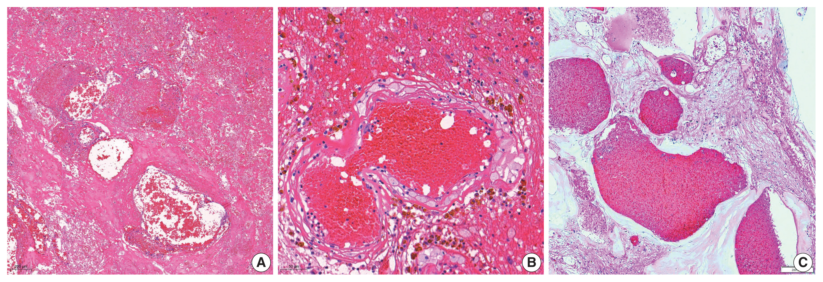
Radiation-induced organizing hematoma (RIOH) is a sporadic form of cavernous hemangioma (CH) that occurs after cerebral radiation. RIOH lesions are distinct histologically from de novo CH; however, detailed research on this subject is lacking. In the present study, the clinical and histological features of RIOHs were evaluated based on causative lesions.
Methods
The present study included 37 RIOHs confirmed by surgical excision from January 2009, to May 2020, in Yonsei Severance Hospital. All cases were divided into subgroups based on type of radiation treatment (gamma knife surgery [GKS], n = 24 vs. conventional radiation therapy [RT], n = 13) and pathology of the original lesion (arteriovenous malformation, n = 14; glioma, n = 12; metastasis, n = 4; other tumors, n = 7). The clinicopathological results were compared between the groups.
Results
Clinical data of multiplicity, latency, and size and wall thickness of the original tumors and RIOHs were analyzed. The GKS group showed shorter latency (5.85 ± 4.06 years vs. 11.15 ± 8.27 years, p = .046) and thicker tumor wall (693.7 ± 565.7 μm vs. 406.9 ± 519.7 μm, p = .049) than the conventional RT group. Significant difference was not found based on original pathology.
Conclusions
RIOH is more likely to occur earlier with thick tumor wall in subjects who underwent GKS than in patients who underwent conventional RT. These results indicate the clinical course of RIOH differs based on type of treatment and might help determine the duration of follow-up. -
Citations
Citations to this article as recorded by
- Impact of cranial irradiation on the clinical presentation of cerebral cavernous malformations
Neerav Kumar, Jeffrey Shi, Carlos Alcocer, Sara Luck, Andrew Garton, Maricruz Rivera, Mark M. Souweidane, Philip E. Stieg
Clinical Neurology and Neurosurgery.2026; 265: 109386. CrossRef - Radiation-Induced Cavernous Malformation in the Cerebellum: Clinical Features of Two Cases
Hyoung Soo Choi, Chae-Yong Kim, Byung Se Choi, Seung Hyuck Jeon, In Ah Kim, Joo-Young Kim, Kyu Sang Lee, Gheeyoung Choe
Brain Tumor Research and Treatment.2025; 13(2): 58. CrossRef - End-stage ADPKD with a low-frequency PKD1 mosaic variant accelerated by chemoradiotherapy
Hiroaki Hanafusa, Hiroshi Yamaguchi, Naoya Morisada, Ming Juan YE, Riki Matsumoto, Hiroaki Nagase, Kandai Nozu
Human Genome Variation.2024;[Epub] CrossRef - Recapitulating the Key Advances in the Diagnosis and Prognosis of High-Grade Gliomas: Second Half of 2021 Update
Guido Frosina
International Journal of Molecular Sciences.2023; 24(7): 6375. CrossRef - Earlier Age at Surgery for Brain Cavernous Angioma-Related Epilepsy May Achieve Complete Seizure Freedom without Aid of Anti-Seizure Medication
Ayataka Fujimoto, Hideo Enoki, Keisuke Hatano, Keishiro Sato, Tohru Okanishi
Brain Sciences.2022; 12(3): 403. CrossRef
- Impact of cranial irradiation on the clinical presentation of cerebral cavernous malformations
Case Reports
- Cavernous Hemangioma of the Uterus in a Postmenopausal Woman: A Case Report.
- Hye Ra Jung, Chi Hum Cho, Sang Hun Kwon, Sun Young Kwon
- Korean J Pathol. 2011;45(5):520-522.
- DOI: https://doi.org/10.4132/KoreanJPathol.2011.45.5.520
- 4,650 View
- 30 Download
- 4 Crossref
-
Abstract
PDF
- Cavernous hemangioma of the uterus is an uncommon mesenchymal tumor. Most cases have been reported in young, pregnant women and the condition is very rare in a postmenopausal patient. An 81-year-old woman presented with a huge pelvic mass. Abdominal computed tomography and magnetic resonance imaging results suggested a leiomyoma with degenerative change and hemorrhage. Microscopically, large, thick-walled and variable-sized vascular channels were evident in the majority part of myometrium; the lining cells were immunohistochemically reactive for CD31. Vascular tumors of the female genital tract should be cautiously excised due to the profuse intra-operative bleeding. The pathological examination of a hysterectomy specimen is the only method to confirm the diagnosis of this tumor.
-
Citations
Citations to this article as recorded by- A rare case report of cervical hemangioma and a comprehensive literature review of 137 cases of cervical and uterine hemangiomas
Maasomeh Farahani, Seyyed‐Ali Hashemi, Sogand Goodarzi, Bardia Hajikarimloo, Farzad Pour‐Ghazi, Shahrzad Noori, Saba Alijani, Armin Khavandegar
International Journal of Gynecology & Obstetrics.2024; 164(2): 421. CrossRef - Cavernous hemangioma of corpus imitating endometrial polyp in a young non‐pregnant woman: A case report study
Ali Emami, Ensiyeh Bahadoran, Fatemeh SamieeRad
Clinical Case Reports.2024;[Epub] CrossRef - Diffuse cavernous hemangioma of the uterus mimicking adenomyosis- A rare case report
Saloni Naresh Shah, N Geetha
Indian Journal of Obstetrics and Gynecology Research.2020; 7(2): 283. CrossRef - Haemangioma- Common Neoplasm in an Unusual Location - A Case Report
Dahlia Joseph, Elizabeth Joseph, Ajitha K
Journal of Evidence Based Medicine and Healthcare.2019; 6(51): 3216. CrossRef
- A rare case report of cervical hemangioma and a comprehensive literature review of 137 cases of cervical and uterine hemangiomas
- Solitary Pulmonary Lymphangioma in an Adult: A Brief Case Report.
- Hye Jong Song, Joungho Han, Kwhanmien Kim, Kyung Soo Lee, Jinwon Seo
- Korean J Pathol. 2008;42(2):125-127.
- 2,254 View
- 26 Download
-
Abstract
PDF
- Solitary pulmonary lymphangiomas are extremely rare. We report here on an unique case of solitary pulmonary lymphangioma in an adult. A well-circumscribed, 6 cm-sized, pleural based lesion with fluid attenuation was found in a 50-year-old Korean male. He had no previous history of disease or trauma. The wedge-resected lung revealed an ill-demarcated lesion with multiple microscopic cysts and the cystic walls had loose intervening stroma.
- Pulmonary Cavernous Hemangioma: A case report.
- Seung Yeon Ha, Sang Ae Yoon, Yang Seok Chae
- Korean J Pathol. 1994;28(2):203-205.
- 2,294 View
- 33 Download
-
Abstract
PDF
- The pulmonary cavernous hemangioma is usually from birth and there may be without symptoms until adulthood. Larger or multiple pulmonary angiomata with considerable pulmonary arteriovenous shunts may cause cyanosis, finger clubbing, dyspnea and frequently accompanyingbruit. Recently, we experienced a case of cavernous hemangioma of the lung. A 34-year-old woman was admitted to our hospital for surgical evaluation of a 4 cm solitary, round nodule in the right upper lobe on the chest X-ray and CT scan. She had no symptoms. Laboratory findings are within normal limits except for elevated glucose levels. At surgery, the mass was well encapsulated and easily excised from the peripheral portion of the posterior segment of the right upper lobe. Grossly, it consisted of a 4 cm in diameter, round, soft, sponge-like, hemorrhagic, slightly lobulated mass with a smooth external surface. Microscopically, the mass was composed of vessels, which were thin walled, dilated and filled with blood. The wall of the abnormal vessels was thin and composed of endothelium and fibrous connective tissue with only a little smooth muscle. Immunohistochemically, the wall of the dilated abnormal vessesls showed negative reaction for cytokeratin(low and high) and epithelial membrane antigen but weakly positive reaction for UEA-1 in focal areas.
- Cavernous Hemangioma of the kidney: Report of a case.
- Won Sang Park, Young Dae Kim, Ki Hwa Yang, Sun Moo Kim
- Korean J Pathol. 1991;25(4):363-366.
- 1,939 View
- 13 Download
-
Abstract
PDF
- Hemangioma of the kidney is a relatively uncommon tumor, which is most commonly located in the tip of the papilla. This lesion is usually small and has been found incidentally at postmorten examination. About 200 cases of renal hemangioma have been reported since Virchow's original report in 1876. In renal hemangioma, cavernous hemangioma is the most common type. They can create diagnostic problem for the clinician and the radiologist. We experienced a case of renal cavernous hemangioma in the medulla of the upper pole. The patient was a twenty-seven-year-old male who had gross hematuria and right flank pain. A nephrectomy was performed. An ill-defined mass, 4.5x3.0x1.5 cm, was observed around the pelvis. Microscopically, the tumor mass was hemangioma of the cavernous type.
Original Article
- Cavernous Hemangioma of the Ovary.
- Jin Hee Sohn, Hye Rim Park, Young Euy Park, Young Woo Lee
- Korean J Pathol. 1996;30(6):554-556.
- 2,041 View
- 16 Download
-
Abstract
PDF
- Hemangioma of the ovary is a very rare lesion, although the ovary itself is a highly vascularized organ. In the literature review, about 40 cases were reported all of which were small in size and they were usually identified incidentally. The age range spanned from 4 months to 81 years. Cavernous hemangioma was the most common histologic type. We experienced a case of cavernous hemangioma of the left ovary in a 26 year-old pregnant woman. The lesion was 8x6x2cm in size with well demarcated margin. The cut surface was purple to bluish red in color and had a spongy-like appearance. Microscopically, it was composed of dilated vascular spaces with a common wall.
 First
First Prev
Prev


