 E-submission
Search
- Page Path
- HOME > Search
Original Articles
- Inflammation and tissue remodeling contribute to fibrogenesis in stricturing Crohn’s disease: image processing and analysis study
- Mustafa Erdem Arslan, Rupinder Brar, Lianna Goetz, Dipti Karamchandani, Michael W. Mikula, Kyle Hodge, Hua Li, Sangtae Ahn, Hwajeong Lee
- J Pathol Transl Med. 2022;56(5):239-248. Published online July 4, 2022
- DOI: https://doi.org/10.4132/jptm.2022.05.18
- 6,018 View
- 144 Download
- 4 Web of Science
- 4 Crossref
-
 Abstract
 PDF
PDF Supplementary Material
 - Background
Inflammation and structural remodeling may contribute to fibrogenesis in Crohn’s disease (CD). We quantified the immunoexpression of calretinin, CD34, and calprotectin as a surrogate for mucosal innervation, telocytes (interstitial cells playing a role in networking), and inflammation, respectively, and correlated them with bowel alterations in stricturing CD.
Methods
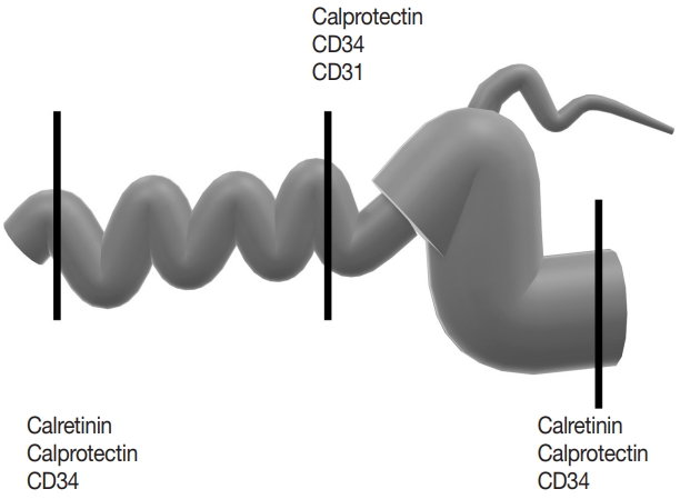
Primary resection specimens for ileal CD (n = 44, 31 stricturing CD, 13 inflammatory CD) were identified. Left-sided ulcerative colitis and trauma cases were used as controls. Proximal and distal margin and middle (diseased) sections were stained for calretinin, CD34, and calprotectin. Microscopic images were captured from the mucosa (calretinin), submucosa (calprotectin), and myenteric plexus (CD34), and the immunostaining was quantified using image processing and analysis. Bowel thickness at the corresponding sections were measured and correlated with the amount of immunoexpression.
Results
A total of 2,037 images were analyzed. In stricturing CD, submucosal alteration/thickening at the stricture site correlated with calprotectin staining and inversely correlated with calretinin staining at the proximal margin. Muscularis propria alteration/thickening at the stricture site correlated with mucosal calretinin staining at the proximal margin. Submucosal alteration/thickening at the proximal margin correlated with calretinin and CD34 staining at the proximal margin and inversely correlated with CD34 staining at the stricture site. Calretinin immunostaining at the distal margin was significantly higher in stricturing CD than the controls.
Conclusions
Inflammation and tissue remodeling appear to contribute to fibrogenesis in stricturing CD. Increased mucosal calretinin immunostaining distal to the diseased segment could be helpful in diagnosing CD in the right clinical context. -
Citations
Citations to this article as recorded by
- Pathophysiologic implications and therapeutic potentials of telocytes in multiorgan fibrosis
Irene Rosa, Eloisa Romano, Bianca Saveria Fioretto, Mirko Manetti
Current Opinion in Rheumatology.2026; 38(1): 26. CrossRef - Serum S100A8/A9 Correlates to Surgery‐Free Interval in Idiopathic Subglottic Stenosis
Laura M. Mafla, Raymond J. So, Ibrahim Abd‐Elazem, Samuel L. Collins, Yee Chan‐Li, Gabriela Lilly, Ioan A. Lina, Alexander H. Gelbard, Alexander T. Hillel, Kevin M. Motz
The Laryngoscope.2025; 135(5): 1724. CrossRef - Telocytes in inflammatory bowel diseases: contributions to pathology and therapeutic potentials
Ronaldo Paolo Panganiban, Christina McAninch, Marina Chulkina, Irina V. Pinchuk
Frontiers in Cell and Developmental Biology.2025;[Epub] CrossRef - Synergistic effects of vedolizumab and JAK 1,2,3 inhibitors in Crohn’s disease: insights from a systems biology and artificial intelligence-based approach
Ignacio Marín-Jiménez, Mónica Sierra-Ausín, Teresa Letosa-Abián, Jesús Aparicio, Carmen Montoto-Otero, Silvia Sánchez-Ramón
Frontiers in Immunology.2025;[Epub] CrossRef
- Pathophysiologic implications and therapeutic potentials of telocytes in multiorgan fibrosis
- Utility of Calretinin in Distinction between Benign Reactive Mesothelial and Carcinoma Cells in Serous Effusions.
- Byung Heon Kim
- J Pathol Transl Med. 2001;12(2):89-96.
- 1,929 View
- 16 Download
-
Abstract
PDF
- The cytological distinction of carcinoma cells from reactive mesothelial cells in serous effusions may be difficult or impossible based on morphology alone, especially in specimens containing reactive mesothelial cells which form glandular or ball- or papillary-shaped conglomerates or which mimic malignant nuclear features. Calretinin is a newly reported immunocytochemical marker for mesothelial cells, which can potentially be utilized for facilitating this distinction. This study evaluated the usefulness of calretinin for the discrimination between reactive mesothelial and metastatic carcinoma cells in serous effusion. Immunocytochemical staining was undertaken on 33 benign reactive and 87 malignant serous effusion specimens with histologically confirmed diagnoses. The specimens including smears and cell blocks were stained with polyclonal antibody to calretinin by labelled streptavidin-biotin method. The positive expression of calretinin was noted in 32(97.0%) of 33 benign reactive effusions and 9(10.3%) of 87 malignant effusions. The sensitivity and specificity of the calretinin immunostaining for reactive mesothelial cells was 97.0% and 89.7%, respectively. In conclusion, calretinin is a useful marker for distinguishing between reactive mesothelial cells and carcinoma cells in serous effusions.
- The Diagnostic Utility of Mesothelial Markers in Distinguishing between Reactive Mesothelial Cell and Adenocarcinoma Cells in Serous Effusions with Cytospin Preparation.
- Sooim Choi, Mi Sun Kang
- J Pathol Transl Med. 2006;17(2):108-115.
- 2,673 View
- 31 Download
-
Abstract
PDF
- Evaluation of serous effusions can include immunocytochemical stains that differentiate reactive mesothelial cell from adenocarcinoma cell. Among several positive mesothelial cell markers, we used desmin, CK5/6, WT1 and calretinin all known to have high sensitivity and specificity as selective mesothelial cell markers. We studied smears obtained with cytospin from 15 malignant and eight benign effusions. The mesothelial cells were positively stained by desmin, CK5/6, WT1 and calretinin in 60.9%, 29.1%, 26.7% and 56.5%, respectively among 8 benign and 15 malignant effusions; the adenocarcinoma cells were positively stained 6.7%, 13.3%, 1.0% and 0.0%, respectively among 15 malignant effusions. The percentage of positively stained mesothelial cells were somewhat lower for all antibodies compared to the results of previous studies. This was likely due to the differences in preparation methods and fixatives among studies. In conclusion, the use of desmin and calretinin were more valuable than CK5/6 and WT1 for distinguishing between reactive mesothelial cell and adenocarcinoma cells in serous effusion; however, choice of the proper preparation methods and fixatives are also important
 First
First Prev
Prev


