 E-submission
Search
- Page Path
- HOME > Search
Original Article
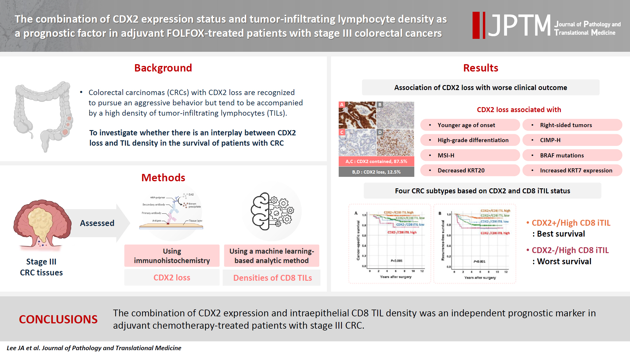
- The combination of CDX2 expression status and tumor-infiltrating lymphocyte density as a prognostic factor in adjuvant FOLFOX-treated patients with stage III colorectal cancers
- Ji-Ae Lee, Hye Eun Park, Hye-Yeong Jin, Lingyan Jin, Seung Yeon Yoo, Nam-Yun Cho, Jeong Mo Bae, Jung Ho Kim, Gyeong Hoon Kang
- J Pathol Transl Med. 2025;59(1):50-59. Published online October 24, 2024
- DOI: https://doi.org/10.4132/jptm.2024.09.26
- 4,128 View
- 286 Download
-
 Abstract
 PDF
PDF Supplementary Material
 - Background
Colorectal carcinomas (CRCs) with caudal-type homeobox 2 (CDX2) loss are recognized to pursue an aggressive behavior but tend to be accompanied by a high density of tumor-infiltrating lymphocytes (TILs). However, little is known about whether there is an interplay between CDX2 loss and TIL density in the survival of patients with CRC.
Methods
Stage III CRC tissues were assessed for CDX2 loss using immunohistochemistry and analyzed for their densities of CD8 TILs in both intraepithelial (iTILs) and stromal areas using a machine learning-based analytic method.
Results
CDX2 loss was significantly associated with a higher density of CD8 TILs in both intraepithelial and stromal areas. Both CDX2 loss and a high CD8 iTIL density were found to be prognostic parameters and showed hazard ratios of 2.314 (1.050–5.100) and 0.378 (0.175–0.817), respectively, for cancer-specific survival. A subset of CRCs with retained CDX2 expression and a high density of CD8 iTILs showed the best clinical outcome (hazard ratio of 0.138 [0.023–0.826]), whereas a subset with CDX2 loss and a high density of CD8 iTILs exhibited the worst clinical outcome (15.781 [3.939–63.230]).
Conclusions
Altogether, a high density of CD8 iTILs did not make a difference in the survival of patients with CRC with CDX2 loss. The combination of CDX2 expression and intraepithelial CD8 TIL density was an independent prognostic marker in adjuvant chemotherapy-treated patients with stage III CRC.
Case Report
- Sinonasal Low-Grade Adenocarcinoma: Report of Three Cases with the Clinicopathologic and Immunohistochemical Findings.
- Joon Seon Song, Shin Kwang Khang, Jooryung Huh, Bong Jae Lee, Kyung Ja Cho
- Korean J Pathol. 2006;40(3):235-240.
- 2,317 View
- 23 Download
-
Abstract
PDF
- Low-grade adenocarcinomas that primarily arise within the sinonasal tract are uncommon tumors. We report here on three cases of primary sinonasal low-grade adenocarcinomas. The patients were 2 females and 1 male with ages of 48, 57 and 64, respectively. Microscopically, the tumors had a well developed tubulopapillary growth pattern that consisted of columnar or pseudostratified cells with eosinophilic cytoplasm, round to oval nuclei and rare mitotic activity. On immunohistochemistry, the tumor cells were strongly positive for cytokeratin 7, but they were negative for cytokeratin 20, CDX-2 and p53. The Ki-67 labeling index was very low (mean: 1.9%). Two patients developed recurrent tumors at the primary site after the initial surgery, but all the patients are presently alive without metastasis 6 years 8 months, 8 years 8 months, and 11 months after the initial diagnosis. When considering the progress of these tumors, we think that it's important to understand the pathology of this entity to avoid underdiagnosis because a complete excision is required for effective treatment.
Original Article
- Mucin Phenotype and CDX2 Expression as Prognostic Factors in Gastric Carcinomas.
- Chan Kwon Jung, Kyo Young Song, Gyeongsin Park, Cho Hyun Park, Myeong Gyu Choi, Young Seon Hong, Kyo Young Lee
- Korean J Pathol. 2007;41(3):139-148.
- 2,228 View
- 27 Download
-
Abstract
PDF
- Background
: Mucin phenotypic markers and CDX2 are widely expressed in gastric carcinomas, however, recent studies have produced conflicting results regarding whether the expression patterns of these markers have clinicopathologic significance.
Methods
: We examined samples from 217 gastric carcinoma patients immunohistochemically to determine if the expression of mucin phenotypic markers and CDX2 was correlated with postoperative survival and other clinicopathologic factors.
Results
: All tumors were phenotypically classified as gastric (type G, 81 cases), gastric and intestinal mixed (type GI, 55 cases), intestinal (type I, 43 cases), or unclassified (type U, 38 cases). The occurrence of type G and GI tumors was positively correlated with tumor progression whereas that of type U tumors was negatively correlated with tumor progression. CDX2 expression was correlated with type I tumors. Tumors that expressed MUC5AC or MUC6 had a better prognosis than those that did not. When the relationship between phenotype and prognosis was considered, type GI had the best prognosis, followed by type G, then type U.
Conclusions
: The mucin phenotypic markers may be useful for predicting tumor progression and survival in patients with gastric carcinomas. Additionally, CDX2 may play an important role in gastric carcinogenesis of type I tumors.
 First
First Prev
Prev


